ابزار وبمستر

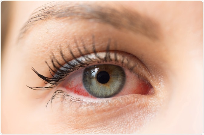
The pupil is a hole located in the center of the iris of the eye that allows light to strike the retina.It appears black because light rays entering the pupil are either absorbed by the tissues inside the eye directly, or absorbed after diffuse reflections within the eye that mostly miss exiting the narrow pupil. Anatomical term created by Gerard of Cremona.

In humans the pupil is round, but other species, such as some cats, have vertical slit pupils, goats have horizontally oriented pupils, and some catfish have annular types. In optical terms, the anatomical pupil is the eye's aperture and the iris is the aperture stop. The image of the pupil as seen from outside the eye is the entrance pupil, which does not exactly correspond to the location and size of the physical pupil because it is magnified by the cornea. On the inner edge lies a prominent structure, the collarette, marking the junction of the embryonic pupillary membrane covering the embryonic pupil.[9]

image

Functions of pupil

  • It regulates the amount of light entering the eye.Dynamic process of muscle reaction within the iris controls how much light enters the eye through the pupil.
  • It improves the visual acuity because it prevents the irregular refraction by the periphery of the cornea and lens, and increases the depth of focus.
  • It allows the passage of aqueous humour from the posterior chamber to the anterior chamber.

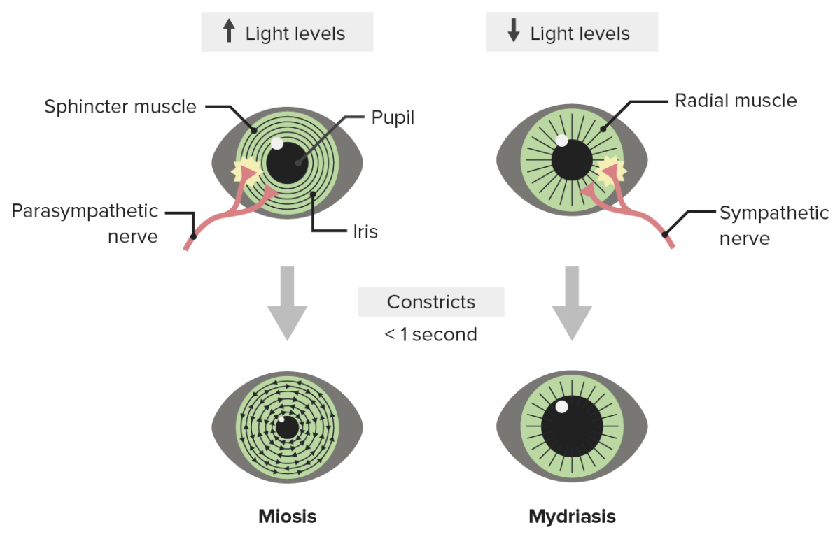
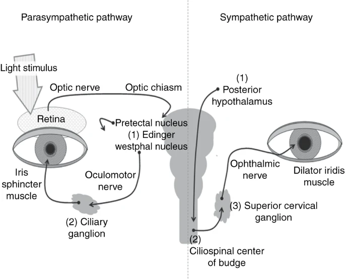
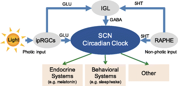
The pupillary light reflex (PLR) or photopupillary reflex is a reflex that controls the diameter of the pupil, in response to the intensity (luminance) of light that falls on the retinal ganglion cells of the retina in the back of the eye, thereby assisting in adaptation to various levels of lightness/darkness.

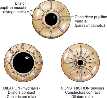
A greater intensity of light causes the pupil to constrict (miosis/myosis; thereby allowing less light in), whereas alower intensity of light causes the pupil to dilate (mydriasis, expansion; thereby allowing more light in)[1]

Pupillary pathways

Sympathetic stimulation of the adrenergic receptors causes the contraction of the radial muscle and subsequent dilation of the pupil. Conversely, parasympathetic stimulation causes contraction of the circular muscle and constriction of the pupil. The mechanism of mydriasis depends on the agent being used.[2]

Thus, the pupillary light reflex regulates the intensity of light entering the eye. Light shone into one eye will cause both pupils to constrict.[1]

Pupil size is determined by the interaction of the parasympathetic and the sympathetic nervous system. The parasympathetic system conducts the light reaction with its major center in the dorsal midbrain. The sympathetic nervous system acts either directly on the dilator muscle (peripherally) or centrally by inhibiting the Edinger-Westphal nucleus. Psychosensory reactions are transmitted via the sympathetic system.

The afferent input of the light reflex system in humans is characteristically wired, allowing a detailed analysis of a lesion of the afferent input. Even in humans a subgroup of ganglion cells containing melansopsin plays an important role as a light sensor for the pupillary system. To diagnose normal pupillary function, pupils need to be isocoric and react bilaterally equally to light. Anisocoria indicates a problem of the efferent pupillary pathway. Pupillary disorders may involve the afferent pathways (relative afferent pupillary defect) or the efferent pathways. Physiological anisocoria is a harmless condition that has to be distinguished from Horner's syndrome. In this case pharmacological testing with cocaine eye-drops is helpful. Disorders of the parasympathetic system will impair the light response. They include dorsal midbrain syndrome, third-nerve palsy, and tonic pupil.

Kids Eye Problems Parents Should Never Ignore

Tonic pupils are mainly idiopathic and do not need imaging.

People with Adie's pupil usually develop several distinct symptoms. The pupil of the affected eye first appears larger or more dilated than the normal eye and reacts abnormally to light. Initially, the pupil reacts slowly or irregularly during close tasks such as reading because the eye begins to lose its close-range focusing power. After extended near focusing or accommodation, the involved pupil may actually become tonic, remaining constricted long after discontinuing accommodative effort. Occasionally, the iris becomes depigmented, losing most or all of its color. Deep tendon reflexes, such as the classic hammer-to-knee reflex, may also be diminished in those patients that have systemic dysautonomia. Blurred vision, especially at close range, is another common symptom of the disorder, as well as excessive sweating.[14] eResearch by Navid Ajamin -- spring 2019

Disorders of the iris, including application of cholinergic agents, need also to be considered in impaired pupillary light reaction.[5]

Pupil reflections in photographs could help investigators solve crimes -- theverge.com

A variety of factors can influence pupil size, and not all of them have to do with light and distance. Some of these other factors include: [12]

  • your health
  • your emotions
  • medicines and drugs

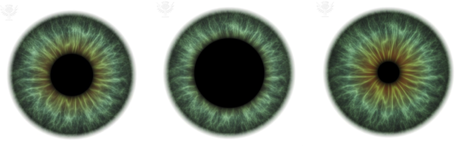
The normal pupil size in adults varies from 2 to 4 mm in diameter in bright light to 4 to 8 mm in the dark. The pupils are generally equal in size. They constrict to direct illumination (direct response) and to illumination of the opposite eye (consensual response). The pupil dilates in the dark. Both pupils constrict when the eye is focused on a near object (accommodative response). The pupil is abnormal if it fails to dilate to the dark or fails to constrict to light or accommodation.[3]

Adie syndrome, or Holmes-Adie syndrome, is a rare neurological disorder affecting the pupil of the eye. In most patients the pupil is larger than normal (dilated) and slow to react in response to direct light. Absent or poor tendon reflexes are also associated with this disorder.[16]

Amaurotic. This is seen when one eye has no perception of light. The pupil of this eye only constricts when light is shone into the other eye. When the light is shone back into the eye with no perception of light the pupil rapidly enlarges against the light.[15]

Anisocoria is a condition characterized by an unequal size of the eyes' pupils. Affecting 20% of the population, it can be an entirely harmless condition or a symptom of more serious medical problems.[4]

Everyone knows that your pupils will change size according to the amount of light you are experiencing. With more light, the pupil constricts and becomes smaller. Less light and your pupil dilates, letting more light into the back of the eye. It is the muscles of the iris working with your autonomic nervous system (ANS) to adjust the iris so the right amount of light enters the eye – like the aperture of a camera.

The iris is made up of two types of muscle:

Sphincter muscles that are like concentric rings that constrict the pupil to as small as two millimeters across

Dilator muscles that are laid out like the spokes of a bicycle wheel and can expand the pupil up to eight millimeters across dilated pupils respond

But the ANS is not only concerned with light reflex, it also reveals emotional and mental responses. The sympathetic branch of the ANS responds to a person being under stress, triggering the “fight or flight” response, which will cause the pupil to dilate. On the other hand, the parasympathetic branch known for “rest and digest” will cause pupil constriction. At any given time, your pupil is balancing between both the light and emotional reactions.

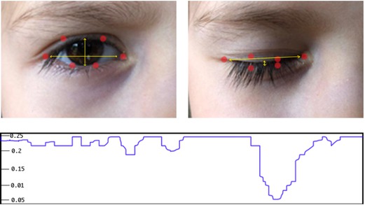
Even memory recall creates a pupil response. When subjects were instructed to remember and recite a series of seven digits, their pupils would grow steadily as they learned each number, but reduce as steadily when they recited back each of the numbers. Wolfgang Einhauser-Treyer, a neurophysicist at Philipps University Marburg in Germany, found that “pupil dilation can betray an individual’s decision before it is openly revealed.” He asked people to push a button at any point during a span of 10 seconds. Dilation began about one second before they pressed the button and continued to peak one to two seconds after the push.

Pupillometry, the measurement of pupil size and reactivity, is a key part of the clinical neurological exam for patients with a wide variety of neurological injuries. It is also used in psychology.[8]

Hutchinson's pupil is a clinical sign in which the pupil on the side of an intracranial mass lesion is dilated and unreactive to light, due to compression of the oculomotor nerve on that side. The sign is named after Sir Jonathan Hutchinson. These can be due to concussion injury to the brain and is associated with subdural haemorrhage and unconsciousness. The parasympathetic fibers to the pupil are responsible for pupillary constriction. The fibers pass through the periphery of the oculomotor nerve, and hence are the first to be affected in case of compression of the nerve. In Stage 1, the parasympathetic fibers on the side of injury are irritated, leading to constriction of pupil on that side. In stage 2, the parasympathetic fibers on the side of injury are paralysed, leading to dilatation of pupil. The fibers on the opposite oculomotor nerve are irritated, leading to constriction on opposite side. In stage 3, the parasympathetic fibers on both sides are paralysed - leading to bilateral pupillary dilatation. Pupils become fixed. This indicates grave prognosis.[18]

Cat eye 
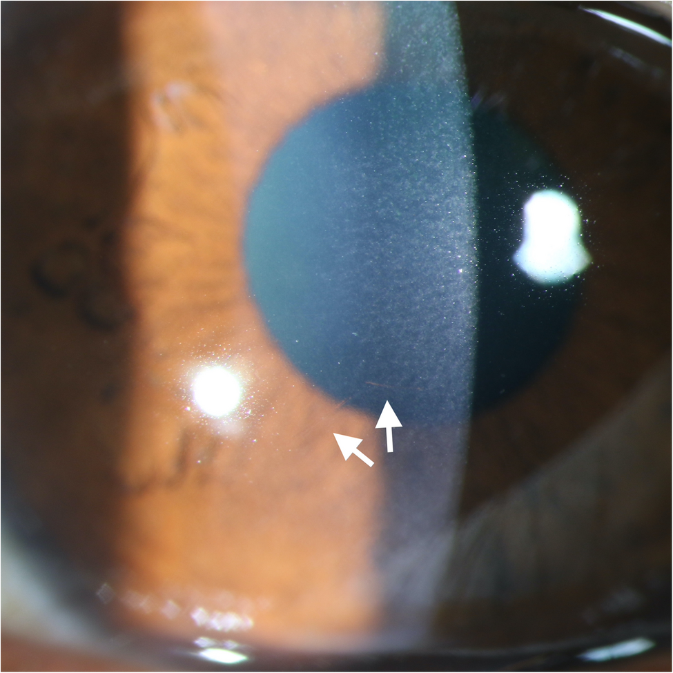
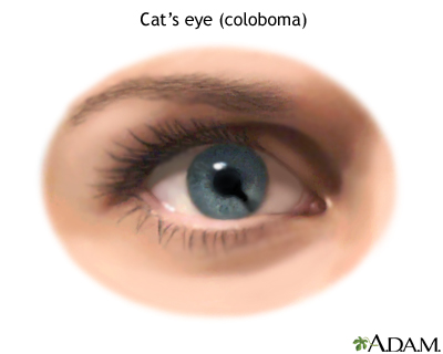
A cat eye is a type of coloboma. Any defect in the iris that allows light to enter the eye, other than through the pupil, is called a coloboma. An extra hole or slit may be present from birth, or may result from trauma. Colobomas may also exist in the eyelid, a defect which interrupts the border of the eyelid.[10]

This study of pupil size is known as pupillometry and is used to investigate a wide range of psychological phenomena including sleepiness, introversion, sexual interest, racial bias, schizophrenia, moral judgment, autism and depression. Kahneman said he has “never done any work in which the measurement is so precise.” And while “nobody really knows for sure what these changes do,” according Stuart Steinhauer, director of Biometric Research Lab at the University of Pittsburgh, pupillometry is a valuable tool for psychological research.

So the next time you look into someone’s eyes, know that you have the potential to see more than just their eye color. You might have a clue as to what is going on in their mind.[6]

Reverse Relative Afferent Pupillary Defect (RAPD) [24]

A relative afferent pupillary defect (RAPD) also known as a Marcus Gunn pupil, is a critically important ophthalmological examination finding that defines a defect ( pathology) in the pupil pathway on the afferent side. An RAPD is relative to the fellow eye and occurs because of the bilateral and equal innervation of the pupils in normal individuals. The RAPD manifests as a difference in pupillary light reaction between the two eyes. The test requires two eyes but only one working pupil. Patients do not have anisocoria.

What does a RRR pupil mean? The pupil is normally rounded, regular and reactive to light (RRR).[23]

PERRLA is an acronym for “pupils are equal, round and reactive/responsive to light and accommodation.”

Healthcare providers use the PERRLA eye test to check if your pupils look and function as they should.

Abnormal PERRLA results may show the following:

  • Unequal pupil size (anisocoria). It may indicate damaged neck blood vessels, brain aneurysms, or cranial nerve damage. It can also be a side effect of medications like anti-nausea or motion sickness.
  • Sluggish pupil reaction (Adie’s pupil syndrome). It may indicate the presence of an infection, trauma, eye surgery, tumors, or migraine.
  • Pinpoint pupil and drooping eyelid (Horner’s syndrome). This may indicate a damaged carotid artery, lymphoproliferative disorders, or tumors (neck and lungs)

The PERRLA test cannot detect a specific underlying condition. It also does not measure pupil size, shape, or reaction speed to light and motion. However, it can be a good starting point since it gives your doctor clues about any possible disorders to be investigated further for more accurate results.

Common Treatments for Pupil Defects

If your PERRLA test indicated pupillary abnormalities, your eye doctor may prescribe treatment depending on the underlying condition.

Common treatment options include: [21]

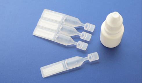
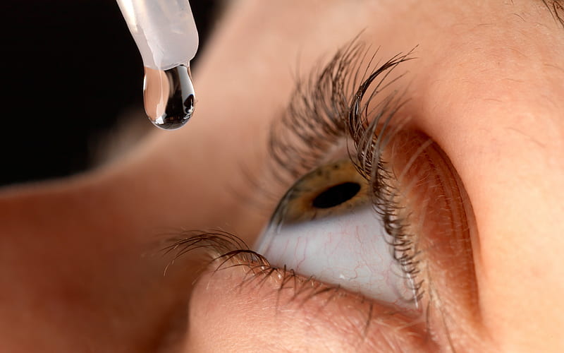
  • Eye drops. Pilocarpine eye drops can support pupil constriction in Adie’s tonic pupil.
  • Prism eyeglasses. These may help relieve diplopia (double vision) symptoms due to a brain tumor, aneurysm, or a stroke. An eye patch can also achieve this.
  • Sunglasses. These are recommended for use outdoors to deal with light sensitivity.
  • Pupil cerclage. This surgical technique involves running a suture around the margin of the affected pupil to shrink it. This procedure minimizes stretching of the iris (front colored part of the eye), thus maintaining the desired pupil size and round shape. It can correct abnormally dilated pupils like in traumatic mydriasis or Adie’s tonic pupil.
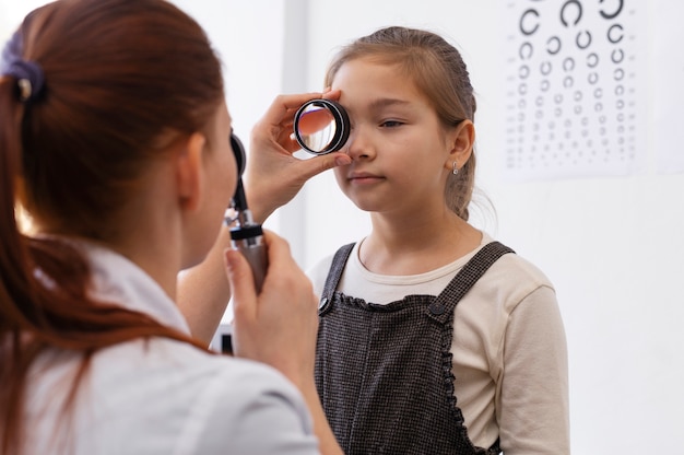
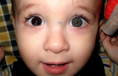
    Childhood Eye Examination

Eye doctors use three procedures to test pupil reflexes.[7]

1.Light Response Pupil Test
The light response pupil test assesses the reflex that controls the size of the pupil in response to light. Your doctor will first dim the lights, then ask you to look at an object in the distance. A light will be shone into your eyes from each side. Your doctor will watch your pupils closely to determine whether or not your pupils constrict in response to the light, making note of the size and shape of your pupils.

2.Swinging Flashlight Pupil Test
The swinging flashlight pupil test is used to compare your pupils' response to light. The lights in the room will be dimmed, and you will again be asked to look at a distant object. Your doctor will "swing" the light rhythmically from one eye to the other, noting the response of each pupil. Your pupils should constrict or stay the same size when the light is shone on them. Dilating pupils may alert your doctor to a possible optic nerve problem.

3.Near Response Pupil Test
The near response pupil test measures the pupil's response to a near target. This test will be performed in a room with normal lighting. Your doctor will ask you to look at a distant object, then move a small object or card in front of your eyes. As you fixate your eyes on the near object, your doctor will watch your pupils closely to make sure they constrict quickly as your fixation changes from far to near.

Pupillometry: Physiology, and Function

A few medicines can affect the muscles that control your pupils and prevent them from getting smaller when light shines in.

These meds include: [11]

  • Atropine (Atropen), which treats problems with heart rhythm, stomach issues, and some types of poisoning
  • Antihistamines, like diphenhydramine
  • Decongestants, like pseudoephedrine
  • Motion sickness and anti-nausea medicines such as dimenhydrinate
  • Parkinson's medications such as amantadine (Symmetrel) and carbidopa-levodopa (Sinemet)
  • Tricyclic antidepressants like amitriptyline (Elavil) and desipramine (Norpramin)
  • Botulinum toxin (Botox, Myobloc)
  • Anti-seizure drugs, such as phenobarbital (Luminal) and topiramate (Topamax)

Summary [13]

  • The many structures of the eye work together to allow for best possible vision.
  • The iris is composed of two layers of smooth muscle that dilate or constrict the pupil.
  • Dilation and constriction is often to regulate light entering the eye.
  • The pupillary light reflex controls this regulation and is triggered by the autonomic nervous system.
  • Dilation of the pupils often occurs when we see someone we find attractive , and we also deem people with more attractive when their pupils are dilated. This is because it indicates both sexual arousal and mutual interest. Our pupils have also been found to dilate in response to aesthetically pleasing artworks.
  • Constriction of the pupils influences as to see someone as more sad, and when we empathise with them our pupils also constrict in response. This is known as motor mimicry. The perception-action model and the autonomic nervous system trigger this constriction.
  • The perception-action model is about perceptions driving actions, and actions developing perceptions.
  • Contrary to popular belief, liars do not always avoid eye contact.
  • Due to the high cognitive load and stress of creating a lie, our pupils will often dilate.
  • Pupillometry is a reliable method of lie detection as, for the most part, dilation of the pupils is not a voluntary action.

The main types of pupillary abnormalities include: [19]

  • Anisocoria: unequal pupil sizes
  • Horner’s syndrome: disruption of a nerve pathway from the brain to the one side of the face and that eye
  • Third nerve palsy: one eyelid is completely closed, and that eye has moved outward and downward
  • Adie’s tonic pupil: one pupil is permanently dilated and unresponsive to light and other stimulants

Symptoms of a pupillary abnormality include:

  • Decreased or increased size of one pupil
  • Difficulty focusing on objects in near visual field
  • Diplopia (double vision)
  • Drooping eyelids (ptosis)
  • Headache
  • Light sensitivity
  • Problems moving your eye

Reference:

  1. en.wikipedia.org/wiki/Pupillary_light_reflex
  2. en.wikipedia.org/wiki/Mydriasis
  3. ncbi.nlm.nih.gov/books/NBK381
  4. en.wikipedia.org/wiki/Anisocoria
  5. ncbi.nlm.nih.gov/pubmed/21601076
  6. discoveryeye.org/pupils-respond-to-more-than-light
  7. verywellhealth.com/pupil-testing-3421844
  8. en.wikipedia.org/wiki/Pupillometry
  9. en.wikipedia.org/wiki/Pupil
  10. medlineplus.gov/ency/imagepages/1130.htm
  11. webmd.com/eye-health/why-are-pupils-dilated-mydriasis#1
  12. healthline.com/health/normal-pupil-size#changes
  13. en.wikiversity.org/wiki/Motivation_and_emotion/Book/2014
  14. verywellhealth.com/adies-pupil-causes-3421980
  15. pmc.ncbi.nlm.nih.gov/articles/PMC3588138
  16. rarediseases.org/rare-diseases/adie-syndrome
  17. tedmontgomery.com/the_eye/reflex.html
  18. Dr. Aruj Khurana. "Concussion injuries to the brain". Comprehensive Ophthalmology (fourth ed.). New Age International (P): 311.
  19. umiamihealth.org/en/bascom-palmer-eye-institute/specialties/neuro-ophthalmology/pupillary-abnormalities
  20. researchgate.net
  21. visioncenter.org/eye-health/perrla-exam
  22. lecturio.com/concepts/physiology-and-abnormalities-of-the-pupil
  23. ophc.mans.edu.eg/images/basic-ophthalmology.pdf
  24. eyewiki.org/Reverse_Relative_Afferent_Pupillary_Defect_(RAPD)

See also:

  • Enlarging the Pupil for Eye Examination verywellhealth.com
  • How to Check Pupil Reflexes Response aparat.com
  • Abnormalities of the pupil college-optometrists.org
  • How to examine the pupil eyeguru.org

You know what the sun can do to skin, right? Many a parent has learned the hard way. A few carefree hours in the sun – without sunscreen – can wreak havoc on the tender skin of children.

Well, their eyes are just as delicate. But while many parents religiously slather on the sunscreen, very few are just as careful with their kids’ eyes.

So, if you’re ready to go out and buy your kids sunglasses, read on to find out what to look for, and what to avoid. [1]

Sources of UV The main source of UVR is sunlight. Artificial lighting contributes to a lesser extent but may increase with the advent of energy efficient light sources.

Ambient UV: direct radiation, scatter, and reflection
Direct sunlight only partly contributes to ambient UV. Under average conditions, more than 50% of ocular exposure comes from scattering and reflection from clouds and the ground.

The World Health Organisation’s solar ultraviolet index (UVI), an international index of UV burden assesses risk of UV damage to the skin. Several studies have shown that this is not a valid indicator of eye protection and potentially misleading.

Identifying absorption and transmission of UVR within structures of the eye is key to understanding potential damage.

UV transmission is strongly dependant on age. Below 9 years of age, a larger portion (2-5%) of UVA is transmitted by the cornea and the lens. Significant inter-individual differences have also been shown.

Acute and chronic damage to the eye by UV and visible light has been extensively studied, including epidemiological studies, with greater significance on chronic exposure.

Cornea
The cornea is most exposed, with the greatest level of UVR absorption from direct irradiation. In addition oblique rays are reflected across the cornea and anterior chamber into the limbal area leading to elevated pathologies in this area. Most common diseases: Pterygium, pinguecula, climatic droplet keratopathy.

Cortical cataract
It is known that UV light induces cataracts with a damage threshold at 350 nm of 60 mJ/cm2. With growing and aging populations and other changing demographic factors the incidence and prevalence of cataracts will increase. Reducing the risks that can lead to cataracts is therefore important.

Dry eye, premature presbyopia, AMD
Decreasing tear film production linked to ageing, reduces UV absorption and antioxidant production by tears.

The association between UVR and AMD remains controversial. Blue light is a more significant contributor to development of AMD.

UV related skin aging and diseases of periorbital skin
The acute response of the skin to UV is inflammation (sunburn). Clinical symptoms include erythema, swelling, pain and pruritus.

Chronic effects include photoaging and photocarcinogenesis. Some clinical signs of photoaged skin include dryness, irregular pigmentation, lentigines, wrinkling and inelasticity. The delicate periorbital skin is particularly susceptible to effects of photoaging.

Mitochondrial DNA is a chromophore for UVA and UVB and subject to damage by UVR. DNA deletions are increased by up to 10-fold in photoaged skin compared to sun-protected skin of the same individual.

Photocarcinogenesis includes the development of actinic keratosis, squamous cell carcinoma, basal cell carcinoma, and malignant melanoma. 5% to 10% of skin cancers are appearing on the eyelids.

SPF measures sunscreen protection from UVB rays, the kind that cause sunburn and contribute to skin cancer. SPF does not measure how well a sunscreen will protect from UVA rays, which are also damaging and dangerous. Dermatologists recommend using a SPF15 or SPF30 sunscreen.

Higher SPFs don't give much more protection.[2]

SPF is an abbreviation for "sun protection factor." A sunscreen's protection factor (SPF) is figured by comparing how long it takes sunscreen-protected skin to burn to the length of time it takes unprotected skin to burn. The higher the SPF, the more protection you get against UVB rays.[3]

Image result for sunglasses uv factors kids

Sunglasses should be the first thing we reach for after applying sunscreen then, maybe a hat. With concerns over possible thinning of the ozone layer, the need to protect our bodies from UV exposure is becoming a growing concern.

It's more than a concern over sunburn. Our eyes as well as our skin need protection from UVA and UVB rays. These harmful invisible light rays are implicated as one of the leading causes of cataracts and macular degeneration. Children are of particular concern and should wear sunglasses for their protection as sun damage is cumulative with most ill effects occurring before age 25. Even on cloudy days, UV rays can be just as damaging to the eyes. Don’t save sunglasses for only the brightest days. Wear them when spending any time outdoors. eResearch by Navid Ajamin -- spring 2016

Quality ophthalmic sunglasses will give 100% protection against UVA and UVB. There are different types of ophthalmic lenses one should consider when purchasing sunglasses. Glass lenses are more scratch resistant but they are heavier and can shatter if hit with an object. Plastic lenses are lighter and available in more colors and coatings. Polycarbonate lenses offer the most protection from breaking and are recommended for sports and should be considered for active children.[4]

skin around our eyes is ten times thinner than the skin on our face and Sunscreens are not tested to be used around eye area ,So it is better not to use sunscreens for around the eyes.

The heat can be very harsh on your eyewear specifically if you have a plastic frame. What ever you do, remember not to leave your sunglasses in the car! If it’s nearing 100 degrees outside your car, it’s probably approaching 200 inside the car.

Excessive heat can cause your prescription lenses to peel or permanently smudge your anti-reflective coating. This is true even for the more durable forms of anti-reflective coating like Kodak brand Clean & Clear.

The heat can cause your plastic sunglass frame to actually warp, essentially melting the plastic. Now this does not look like some bad B-rated horror movie where your glasses literally liquify, usually it makes the frame flatten out and get wider. This of course produces some fitting problems for the wearer as the glasses will then be consistently falling off.

Take extra care of your eyewear in the harsh summer heat. Keep yourself (and your glasses) cool!

Choose a sunscreen labelled broad spectrum or high protection against UVA and UVB so it offers balanced UVA and UVB protection.

Do not stay in the sun too long, even whilst using sunscreen, as no sunscreen can provide 100% protection.

Use a high protection sunscreen and re-apply frequently and generously, especially after perspiring, swimming or towelling.

In sunny weather, seek shade between 11am and 3pm when UV is at its strongest.

Cover up with clothing and don’t forget to wear a hat that protects your face, neck and ears, and wear good quality UV protective sunglasses.

Never let your skin burn and remember, a tan is a sign of sun damage to the skin.

Children have more sensitive skin and need extra care – use sunscreen, clothing and shade. Keep babies and young children out of direct sunlight.

Reference:

1.vsp.com 2.badgerbalm.com 3.sharecare.com 4.eyecareassociatesnc.com 5.vivawoman.net 6.pointsdevue.com 7.drtavel.com 8.uvistat.com

مصرف لوازم آرایش به ویژه لوازم آرایش چشم و ابرو، از قدیم الایام در کشور ما رایج بوده و به ویژه طی سال های اخیر بین جوانان و نوجوانان رواج زیادی پیدا کرده است. این درست برخلاف چیزی است که در کشورهای غربی مشاهده می شود.


باید توجه داشته باشید که هریک از این مواد آرایشی، از ده ها ماده شیمیایی مختلف ساخته شده اند. این مواد شیمیایی به تنهایی یا در ترکیب با یکدیگر ممکن است باعث آسیب به لایه های محافظ پوست و ایجاد حساسیت های پوستی خفیف تا شدید به ویژه در نواحی اطراف چشم شوند. مصرف انواع سایه های رنگی، به ویژه انواع ضدآب آنها، می تواند علاوه بر قرمزی، تورم و خارش، فرد را دچار خشکی و پوسته ریزی و تغییر رنگ بارز در ناحیه اطراف چشم کند. البته آسیب به لایه های محافظ پوست می تواند زمینه ساز ایجاد عفونت های میکروبی و قارچی نیز باشد. این واکنش به ویژه در فصول گرم تشدید می شود.


متاسفانه استفاده لنزهای رنگی و مژه های مصنوعی به ویژه با روش کاشت مصنوعی نیز زمینه را برای بروز انواع واکنش های پوستی و حتی ریزش مژه های فرد و عفونت داخل و اطراف چشم فراهم می کند و اگر همزمان از خط چشم و ریمل هم استفاده شود، این واکنش ها تشدید خواهند شد. برخی از سایه ها به دلیل وجود مواد رنگی یا معطر در آنها، بعد از قرار گرفتن فرد در معرض اشعه آفتاب، لک های پوستی ایجاد می کنند. این لک ها و چروک زودرس و خشکی ایجادشده در ناحیه اطراف چشم، می تواند نمای ناخوشایندی به وجود آورد.


متاسفانه کنترل سریع این واکنش ها مقدور نیست و ممکن است در صورت مصرف دوباره این لوازم آرایش چه از همان برند و چه از برندهای دیگر، واکنش ها با شدت بیشتر تکرار شوند که در این موارد درمان دوباره ضروری خواهد بود. انواع تقلبی و غیراستاندارد این سایه ها که به وفور در بیشتر فروشگاه ها یافت می شود، از عوامل شایع واکنش های اطراف چشمی است که ما هر روز در مراکز درمانی با آنها مواجه می شویم.[1]


در واقع آرایش بیش از حد دختران جوان به دلیل کمبود اعتماد به نفس در آنها و هم چنین برگرفته از فرهنگ های واردتی است و به اعتقاد متخصصان این روزها جوانان و نوجوانان به این نتیجه رسیده‌اند که ابراز زیبایی‌های شخصی یكی از مهمترین شیوه‌های جلب توجه است، [2]

این روزها آرایش کردن بیش از گذشته میان زنان و دختران جوان رایج شده است. اگر در کشورهای غربی پیرزن های ۷۰ سال به بالا، خود را با انواع ترفندها می خواهند زیبا و جوان نشان دهند، در کشور ما بسیاری از دختران نوجوان ۱۴ سال به بالا، هر روز وقت زیادی از خود را روبروی آینه می گذرانند تا خوشگل شوند، غافل از این که پزشکان متخصص پوست نسبت به عوارض آرایش های غلط هشدار می دهند.


علل گرایش زنان به آرایش افراطی
کرم پودرها، لاک ناخن، رژ لب، ریمل چشم و سایه ها در رنگ های متنوع از جمله مواد آرایشی هستند که مورد استفاده قرار می گیرند.

 متاسفانه اغلب خانم ها بدون توجه به ویژگی های پوست خود از این مواد استفاده می کنند. مثلا کسی که پوست خشک دارد، به هیچ وجه نباید از کرم پودرهای خشک استفاده کند، در حالی که دارندگان پوست های چرب می توانند از این پودرها استفاده کنند.
بیشتر عوارض مواد آرایشی پوست، به مواد چرب مربوط می شود و در نهایت موجب بروز آکنه روی پوست خواهد شد.
مواد آرایشی چرب، منافذ پوست را می بندند و موجب بروز جوش های سرسیاه روی پوست می شوند.
دختران جوان از مصرف مواد آرایشی بپرهیزند. این مواد به پیر شدن زودرس پوست آنها کمک می کند.
، خانم ها پیش از استفاده از مواد آرایشی، آن را روی دست خود امتحان کنند و در صورت نداشتن حساسیت، از آن استفاده کنند. ضمن این که، مدت زمان استفاده از مواد آرایشی بین ۶ ماه تا یک سال است. بنابراین باید به تاریخ تولید و مصرف این مواد توجه شود و از خرید مواد آرایشی گرانقیمت خودداری کرد، چون ممکن است در مدت زمان کوتاه مورد استفاده قرار نگیرند.


چند توصیه:
- اگر بخواهید پوست خود را به انواع مواد آرایشی عادت دهید، باید هر روز ۲ ساعت جلوی آینه بایستید و دست کم ۵۰ لایه متفاوت را روی پوست خود قرار دهید. پس از انجام این کار منصرف شوید.
- از مواد آرایشی مختلف استفاده نکنید و سعی کنید برای استفاده از انواع کرم ها، با پزشک متخصص مشورت کنید.
- تحت تاثیر تبلیغات مواد آرایشی قرار نگیرید. بسیاری از این تبلیغات دروغین هستند و ممکن است روی پوست شما تاثیر مخرب بگذارند.
- لوازم آرایش خود را تمیز نگه دارید و پس از هر بار مصرف، برس آن ها را بشویید.
- از لوازم آرایشی دیگران به هیچ وجه استفاده نکنید.
- از دستفروش ها که مواد آرایشی و عطرهای تقلبی و تاریخ گذشته می فروشند، خرید نکنید.
- توجه داشته باشید قسمت بیشتر شیشه عطرهایی که در کشور تولید و پُر می شوند، الکل است. بنابراین از پاشیدن آن روی پوست خود خودداری کنید.
- دختران جوان از مصرف مواد آرایشی بپرهیزند. این مواد به پیر شدن زودرس پوست آنها کمک می کند.[3]


Good eye hygiene is essential to help keep your eyes healthy and free from infections.

  • Always use a clean face towel or flannel
  • If you wear make-up, make sure you keep your tools (brushes, etc.) clean
  • Don't share your towel or bed linen with anyone who has an eye infection


If you wear contact lenses:

  • Always wash and dry your hands before handling lenses
  • Clean and disinfect your lenses regularly according to professional instructions
  • Use fresh solution every time you clean or store your lenses - throw it away once it's used
  • Keep your lens case clean and dry and never use tap water to rinse it
  • Replace your lenses regularly and have regular eye check-ups [4]

Infection Control

There are many infections that relate to the eye, the most common being conjunctivitis. They are all very contagious and strict hygiene is needed to prevent the spread of infection.

If you are a wearer of contact lenses, it is very important to wash and rinse hands thoroughly before handling the lenses. Eyes should be free of make-up when inserting the lens to avoid particles of mascara etc from being trapped underneath the lens and causing irritation or even infection.

 little girl face paint tiger   

How toxic is Halloween face paint on your child’s face?

Always use cleaning solutions as directed and do not allow them to expire. These solutions are vital for removing the build-up of protein from the lens.

Eye-make-up should be removed daily to allow the skin to breathe. Use a gentle removal solution and dab the eye, do not rub it. Never share make-up or the brushes/sponges with others as this significantly increases the risk of cross infection.

Regular eye examinations (every two years) are important for keeping prescription lenses correct and for early diagnosis of any developing eye disorders.[5]

To help you keep your eyes healthy and beautiful, we’ve assembled some useful Dos and Don’ts for wearing makeup with contact lenses. Read more.

Do:

  • Put your lenses in before you put your makeup on
  • Wash your hands thoroughly before you apply your lenses
  • Apply eye shadow and liner gently, so you don't jostle or damage your lenses
  • Use oil-free and fragrance-free eye makeup
  • Use water-resistant mascara and eyeliner to prevent flaking and smudging
  • Replace your mascara every month
  • Replace your eyeliner every three months
  • Replace your eye shadows every six months
  • Remove your lenses before you remove your eye make-up
  • Remove your eye makeup every day with a hypoallergenic, oil-free remover
  • Call your optician if you have any redness, pain, swelling or irritation 

Don't:

  • Wear makeup (or put in your lenses) if your eyes are swollen, red or infected
  • Use saliva to apply eye shadow, liquid eyeliner or to try to get one more use out of old mascara
  • Apply eyeliner to your inner eye lid (inside the lashes)
  • Apply mascara at the base of your lashes - start from the midpoint and extend to the tips
  • Use mascara with 'lash-building fibres' as they can damage your lenses [6]

 eResearch by Navid Ajamin -- spring 2012


Image: www.eyecareopinion.com ewg.org berkeleyside.com

Reference: 1.PersianPersia.com 2.Tebyan.net 3.JameJamOnline.ir 4.GoldenEyecare.co.uk 

5.HygieneExpert.co.uk 6. acuvue.co.uk

Related To: Contact Lenses – A Beginner's Guide

آیا تا کنون فکر کردید که چرا گریه می کنید؟ دلایل بسیاری در گریه کردن وجود دارد

گریه بخشی از طبیعت انسان است چه دوست داشته باشید یا نداشته باشید. هنگامی که دل انسان دچار درد و عذاب می شود، گریه باعث می شود که احساس بهتری پیدا کنید و اخلاق و رفتار تغییر کند.اشک در غده اشکی، نهفته است که بین پلک و کره چشم تولید می شود.

سیستم بیولوژیکی زنان به گریه بیشتر حساس است . گریه زنان 5.3 بار در ماه در حالی که گریه مردان تنها حدود 1.4 است. طول متوسط گریه برای زنان 6 دقیقه و برای مردان 2 تا 4 دقیقه است

در واقع، همه اشک یکسان دارند. انواع مختلفی از اشک است که به دلایل مختلف تولید می شود.

۱-اشك پايه (basal tears): درچشمان پستانداران سالم قرنيه پيوسته (24 ساعته) توسط اشك هاي پايه خيس و تغذيه ميگردد. اين اشك باعث لغزنده شدن چشم ها و شستشوي چشمها از ذرات خارجي ميگردد. مايع اشك حاوي آب+موسين+چربي ها+ليزوزيم+لاكتوفرين+ليپوكالين+ايمنوگلوبولين ها+گلوكز+سديم كلرايد+كلرايد پتاسيم+آنتي اكسيدان ها+برخي ويتامينها ميباشد.

2-اشك رفلكس (reflex tears): هر چزي كه باعث تحريك و يا خشك شدن چشم شود، ميتواند توليد اين نوع اشك را افزايش دهد.اين نوع اشك بطورغيرارادي و در پاسخ به مواد محرك چشم نظير ذرات خارجي،گردوغبار،آلرژن ها،پاتوژن ها و مواد محرك ديگري همچون بخارات پياز،گاز اشك آور و افشانه فلفل ترشح ميگردد.اين اشك ها سعي ميكنند تا مواد محرك و آزاردهنده را از چشم ها پاك كنند.باد شديد و نور شديد خورشيد نيز ميتواند باعث توليد اشك رفلكس گردند.اين اشك بيشتر آبكي بوده و از ليزوزويم و ايمنوگلوبولين بيشتري برخورداراست.

3-اشك احساسي و يا گريه (emotional tears/crying): اين اشك هنگام احساساتي شدن انسان ها (استرس هيجاني ) و يا احساس درد فيزيكي ترشح ميگردد. آغازگرهاي هيجاني اين نوع اشك معمولا خشم، اندوه، شادي، ترس، شوخ طبعي و ناكامي ميباشند. تركيبات شيميايي اين نوع اشك با تركيبات دو اشك قبلي متفاوت است. اشك احساسي حاوي هورمونها و پروتئين هاي بيشتري ميباشد. اين اشك حاوي هورمونهاي پرولاكتين، هورمون رشد، آدرنوكورتيكوتروپين، آندروفين و فلز منگنز ميباشد.

نكات جالب درباره اشك و گريه :

1-به غير از انسانها برخي معتقدند فيل ها و گوريل ها نيز ميتوانند گريه كنند اما نه به پيچيدگي گريه انسانها. حيوانات ديگر اغلب هنگام درد كشيدن، وارد شدن اجسام خارجي در چشمها و يا خشك شدن چشمها دچار اشك ريزش ميشوند. حيوانات (به ويژه نوزادان آنها) بصورت آوايي و مانند نوزادان تازه متولد شده گريه ميكنند بدون توليد اشك.

2-غدد اشكي روزانه در حدود 2 قاشق پر چايخوري اشك توليد ميكنند.

3-هر پلك زدن (باز و بسته كردن چشم) در حدود 100-150 ميلي ثانيه بطول مي انجامد. انسانها بطور متوسط هر 2 تا 10 ثانيه يكبار و 10 بار در دقيقه پلك ميزنند.

4-شور بودن اشك عمدتا به علت وجود كلرايد سديم و پتاسيم در اشك ميباشد. ميزان نمك موجود در اشك با ميزان نمك پلاسماي خون برابراست.

5-از آنجايي كه جنين نيز در درون رحم قادر به گريه كردن است بنابراين گريه كردن نميتواند يك رفتارآموختني باشد. اما فرهنگ، سن، جنسيت، فرآيند اجتماعي شدن، شخصيت، روابط و بافت اجتماعي همگي ميتوانند در شكل بيان گريه تعيين كننده باشند.

6-PH اشك اندكي قليايي و خود اشك ترش و شور مزه ميباشد.

7-سرفه شديد، استفراغ، احتقان بيني، آلرژي بيني، التهاب يا عفونت بيني و يا چشم و اختلالات عصبي نيز ميتواند باعث اشك ريزش گردند.

8-گريه كردن در برخي فرهنگها عملي كودكانه و غير موقر محسوب شده و گريستن در انظارعمومي تقبيح ميگردد.

9-علت اشك ريزش هنگام خميازه كشيدن و يا خنديدن وارد شدن فشار به غدد و كيسه اشكي ميباشد. علت ديگر آن ميتواند اين مسئله باشد كه غدد اشكي تحت كنترل سيستم عصبي خود مختار ميباشند. اين سيستم عصبي تمام كاركردهاي غيرارادي را كنترل مي كند. هنگامي كه اين اعصاب تحريك ميشوند (هنگام خميازه) غدد اشكي نيز اندكي تحريك ميگردند.

10-بسياري از افراد به علت مبتلا بودن به خشكي چشم قادر به گريه كردن نميباشند. اما دراختلال ديگري كه در پي آسيب به مغزايجاد ميگردد فرد مبتلا ممكن است بطور مداوم براي ساعتها بدون علت گريه كند.

11-علت سوزش و اشك ريزش چشمها هنگام خرد كردن پياز به علت وجود آنزيمها و آمينو اسيد سولفوكسايد موجود در سلولهاي پياز ميباشد. آمينو اسيد سولفوكسايد توسط آنزيمهاي پياز به سولفينيك اسيد تبديل ميشود، كه بسيار ناپايدار بوده و فورا به گازي فرار تبديل ميگردد. هنگامي كه اين گاز با آب درون چشم (اشك) تركيب ميگردد توليد اسيد سولفوريك ملايمي را ميكند كه باعث سوزش و اشك ريزش ميشود. با افزايش توليد اشك بدن سعي ميكند تا اسيد سولفوريك را رقيق و از چشمها بشويد. براي جلوگيري از سوزش و اشك ريزش چشمها هنگام خرد كردن پياز به توصيه هاي زير عمل كنيد:

* دستان خود وپياز را قبل از خرد كردن با آب خيس كنيد تا گاز متصاعد شده با، آب روي دستها و پياز واكنش نشان دهد نه با اشك چشمها.

* تنها با دهان خود تنفس كنيد. عمل دم گازها را به درون ريه كشانده و عمل بازدم نيز باعث پراكنده شدن گاز ميشود.

* قبل از خرد كردن پياز آن را نيم ساعت درون يخچال قرار دهيد. چراكه كاهش دما فعاليت آنزيمهاي پياز و انتشار گاز را كاهش ميدهد.

علل گريه كردن: Reasons for crying

1-نوزادان از گريه به عنوان ابزار بقا استفاده ميكنند. گريه كودكان يك وسيله ارتباطي با مادر بوده و از طريق آن طلب كمك ميكنند. علل اصلي گريه نوزادان گرسنگي، پوشك خيس، كثيف و يا نامناسب، نياز به تماس بدني و نوازش، درد ناشي از گاز معده و روده، سرما و گرماي شديد، كسالت و بي حوصلگي، استرس، محيط پر ازدحام، درد و جلب توجه ميباشد.

2-علل گريه در افراد بزرگسال عمدتا شامل غم و اندوه(49%)، شادي و خوشي(21%)، خشم(10%)، همدردي(7%)، اضطراب(5%) و ترس(4%) ميباشد.

3-انسان ها براي تحت تاثير قرار دادن و برانگيختن احساسات و ترحم ديگران نيز متوسل به گريه ميشوند.

The Benefits of Crying for Eye HealthTears also clean your eyes, flushing out particles of dust and debris that could harm your vision. Plus, the antibodies in your tears help fight off a multitude of bacteria and viruses that can cause infections. A good cry is like a mini-cleanse for your eyes!

Crying may support both the body and mind by restoring emotional balance, dulling pain, and activating your parasympathetic nervous system to help you self-soothe, among other benefits.

How Your Body Makes Tears

Lacrimal glands above each eye produce your tears. As you blink, tears spread across the surface of the eye. Then the tears drain into puncta, tiny holes in the corners of your upper and lower eyelids. Your tears then travel through small canals in the lids and down a duct before emptying into your nose. There, tears will either evaporate or be reabsorbed.

Sometimes babies are born with a blocked tear duct, a condition that usually resolves on its own. An eye infection, swelling, injury or a tumor can cause a blocked tear duct in adults.

When a lot of emotional or reflex tears are made, they overwhelm the lacrimal drainage system. That’s why these tears can spill out of your eyes, run down your cheeks and sometimes dribble out of your nose.

Humans produce three types of tears:

  • Basal: The tear ducts constantly secrete basal tears, which are a protein-rich antibacterial liquid that help to keep the eyes moist every time a person blinks.
  • Reflex: These are tears triggered by irritants such as wind, smoke, or onions. They are released to flush out these irritants and protect the eye.
  • Emotional: Humans shed tears in response to a range of emotions. These tears contain a higher level of stress hormones than other types of tears.

فوايد گريه كردن: Benefits of crying

1-هنگامي كه ما دچار استرس هيجاني مي شويم، مغز وبدن ما شروع به توليد تركيبات شيميايي و هورمونهاي خاصي ميكند. گريه كردن كمك ميكند تا اين تركيبات شيميايي زائد كه نيازي به آنها نيست از بدن حذف گردند.

2-اشكهاي احساسي در واقع مواد سمي اي كه در پي استرس هيجاني در خون تجمع مي يابند را از بدن دفع ميكند.

3-اشكهاي احساسي سطح منگنز بدن را كاهش ميدهند. اين مواد معدني بروي خلق و خو تاثير مستقيم ميگذارد. غلظت منگنز اشك احساسي 30 برابر بيشتر از منگنز موجود در سرم خون است.

4-اشك احساسي نسبت به اشك پايه و رفلكس حاوي 24 درصد پروتئين آلبومين بيشتري ميباشد.

5-گريه مانند ادرار و تعريق باعث حذف مواد سمي و محصولات زائد بدن ميشود.

'Things to Say Instead of 'Stop Crying

6-گريه يك مكانيسم رايگان، طبيعي و قدرتمند براي كنار آمدن با درد، استرس و اندوه ميباشد.

7- هورمونهايي كه پس از استرس در بدن انباشته ميشوند به حد سطح سمي رسيده و باعث تضعيف سيستم ايمني بدن و ديگر فرآيندهاي بيولوژيكي ميشوند. اشك هاي احساسي به عنوان دريچه اطمينان براي قلب عمل ميكنند.

8-تركيباتي كه حين استرس در بدن تجمع مي يابد با اشك خارج ميگردند. همين امر باعث كاهش استرس ميگردد. اين مواد شامل آندورفين (lucine-enkephaline)، كه در كنترل درد موثر است، پرولاكتين كه در تنظيم توليد شير نقش دارد، و آدرنوكورتيكوتروپين (ACTH) كه يك نشانگر مهم استرس است، ميباشند. سركوب گريه و اشك باعث افزايش استرس ميگردد.

9-نه تنها گريه باعث ارتقاي سطح سلامتي فرد ميشود بلكه در افزايش حس تعلق در گروه نيز تاثير گذاربوده و موجب افزايش حضور و دخالت ديگران در تامين رفاه فرد ميشود. اشك ها يك روش موثر در ايجاد ارتباط بوده و در برانگيختن حس همدردي و دلسوزي سريعتر ازهر ابزار ديگري عمل ميكنند. گريه بطور موثري بازگو كننده آنست كه شما در مورد يك دلمشغولي خاص صادق و بي ريا بوده و در كنار آمدن با آن مشكل مضطرب ميباشيد.

10-گريه عملي طبيعي، سالم و شفا بخش است.

? What happens when you cry, exactly

11-گريه در كاهش و جلوگيري از بيماريهاي مرتبط با استرس موثر ميباشد.

12-خنده و گريه دو ابزار كارآمد دركاهش استرس و رهايي ازاحساسات منفي ميباشند.

13-هنگامي كه گريه سركوب ميشود، هيجانات رها نمي شوند و خصايص شخصيتي مخرب همچون پرخاشگري بروز ميكنند.

14- به باور دانشمندان سركوب و امتناع از گريه كار معقولي نيست. اما برخي افراد كه پس از مورد انتقاد قرار گرفتن، مشاجره با دوستان، مواجهه با ناكاميهاي كوچك فورا شروع به گريستن ميكنند، بايد به روانشناس مراجعه كنند. چراكه معمولا علت اصلي اين نوع گريه ها اعتماد بنفس پايين و يا مشكلات رواني مزمن در فرد بوده و بايستي فرد تحت درمان قرار گيرد.

15-انسان ها پس از گريه كردن از لحاظ جسمي و رواني احساس بهتري ميكنند و در واقع سبك و خالي ميشوند. (به علت دفع مواد انباشته شده كه توضيح داده شد)

16-گريه يك ابزار برقراري ارتباط است يك زبان جهاني براي درخواست كمك و حمايت اجتماعي.

17-گريه كمك ميكند به آزاد سازي آندورفين ها به جريان خون. آندورفين ها تركيبات شيميايي اي هستند كه باعث بهبود خلق و خو و تسكين درد ميشوند.

Crying, especially a "good cry," can offer a sense of liberation and emotional release, often leading to a feeling of lightness, calm, and clarity. This is due to the release of endorphins and oxytocin, which ease physical and emotional pain and promote well-being.

Here's a more detailed explanation:

Emotional Release: Crying can be a way to release pent-up emotions and trauma, allowing individuals to process difficult experiences and feel more in control of their feelings.

Physical Release: Crying also helps the body release tension and stress. After a good cry, the parasympathetic nervous system (responsible for "rest and digest") can take over, shifting the body out of a "fight or flight" response, leading to a sense of relief and clarity.

Chemical Release: Crying triggers the release of oxytocin and endorphins, which have mood-boosting and pain-relieving effects.

Psychological Relief: Crying can provide a sense of catharsis, allowing individuals to feel like a weight has been lifted and that they are moving forward.

Self-Soothing: Crying can be a way to self-soothe and manage difficult emotions, providing comfort and a sense of safety.

تفاوت گريه در مردان و زنان: The difference between crying in men and women

1-زنان 4 تا 5 برابر بيشتر از مردان گريه ميكنند.5.4 بار در ماه.

2-دختران و پسران تا قبل از بلوغ به يك ميزان گريه ميكنند، چراكه ترشح هورمون پرولاكتين كه باعث تحريك توليد اشك ميشود به يك ميزان در هر دو جنس توليد ميشود.

3-پس از بلوغ ميزان ترشح پرولاكتين كه در توليد و تناوب گريه موثر است در زنان 60 درصد بيشتر از مردان ميشود.

4- غدد اشكي مردان از لحاظ ساختاري متفاوت و كوچكتر از غدد اشكي زنان ميباشد. شايد يكي از دلايل بيشتر گريه كردن زنان نيزهمين مسئله باشد. مردان و زنان نميتوانند يكديگر را بخاطر چگونه گريه كردنشان مورد سرزنش قرار دهند.

5-يكي ديگر از علل بيشتر گريه كردن زنان باورهاي فرهنگي ميباشند، كه اجتماع گريه كردن زنان را بيشتر مورد پذيرش قرار ميدهد.

6-مردان آرام، بدون صدا و با اندكي اشك گريه ميكنند. بطوري كه معمولا كاسه چشمان آنها از اشك پر ميشود، اما بندرت اشك به گونه ها سرازير ميگردد.

7-اما زنان معمولا پر سرو صدا و با اشك هاي فراوان گريه ميكنند. eResearch by Navid Ajamin -- spring 2012

8-مردان اغلب در نتيجه احساسات مثبت و زنان پس از درگيري و مشاجره با ديگران و يا در نتيجه احساس بي كفايتي گريه ميكنند.

همچنان علت و فرآيند اشك هاي احساسي مبهم و رازآلود باقي مانده است. كماكان پرسشهاي بدون پاسخ مانده ي بسياري وجود دارند نظير: چنانچه گريه كردن به منظور كاهش استرس طراحي گرديده پس فلسفه اشك شوق و شادي چيست؟ و يا اگر گريه يك وسيله ارتباطي با ديگر همنوعان است پس چرا انسانها در خلوت وتنهايي خود نيز گريه ميكنند؟

Reference:

  • aao.org/eye-health/tips-prevention/facts-about-tears
  • arp.org/health/conditions-treatments/tears
  • medicalnewstoday.com/articles/319631
  • healthline.com/health/benefits-of-crying

See also:

  • ناگفته های اشک چشم اشک سه حرف ندارد کلی حرف دارد
  • Overflow Tearing and Chronic Eye Infections in Infants

در هوای آلوده عینک بزنید

چشم‌ها اعضای حساس و آسیب پذیری هستند. با این‌که آلودگی هوا به‌طور مستقیم به چشم آسیب نمی‌رساند، ولی در هوای آلوده بیماری‌های سطح چشم مانند خشکی چشم، حساسیت و عفونت‌های مختلف که ممکن است در سطح پلک اتفاق بیفتند، تشدید می‌شوند و در این شرایط افراد احساس ناراحتی بیشتری می‌کنند.

علاوه بر این تحریکات سطحی مثل خارش، سوزش، اشک ریزش و قرمزی چشم را نیز به‌دنبال خواهد داشت.

اگر فرد دچار یکی از بیماری‌های چشمی باشد، در این شرایط علائم آن افزایش پیدا می‌کند. بنابراین بهتر است ابتدا بیماری زمینه‌ای خود را درمان کند.

کسانی که آلودگی هوا بیشتر آن‌ها را اذیت می‌کند می‌توانند از عینک‌های محافظ مثل عینک آفتابی یا عینک بدون شماره با فریم بزرگ که چشم را در مقابل باد، خاک و چیزهای محرک دیگر محافظت می‌کند استفاده کنند.

How can we protect our eyes from air pollution?

Doctors suggests wearing sunglasses, using lubricating eye drops, washing hands often and avoiding rubbing your eyes on days when air pollution levels are high, so as not to exacerbate any irritation.

What You Can Do to Protect Your Eyes From Bad Air Quality

Exposure to pollution and other airborne irritants increases the risk of many diseases and health conditions, including heart disease, asthma, stroke, respiratory illnesses, and lung cancer. Unfortunately, your eyes may also suffer when the air quality is bad. Taking a few of these steps will improve your eye comfort and reduce the risk of vision problems.

How Air Quality Affects Your Eyes

The moist tissues in your eyes easily absorb pollutants in the air. Short-term exposure can cause irritation and inflammation, while long-term exposure can lead to age-related macular degeneration. Your risk of eye problems increases if you spend a significant amount of time outdoors when the air quality is poor.

Airborne Pollutants Can Trigger Painful Conjunctivitis

Conjunctivitis, or “pink eye,” affects the conjunctiva, a layer of tissue that covers the whites of your eye and the insides of the eyelids. Redness, itching, burning, foreign body sensation, and light sensitivity are common symptoms. Although conjunctivitis can be caused by viruses, bacteria, or chemicals, air quality is also a factor, particularly among people who have allergies.

Cases of allergic conjunctivitis among Japanese ophthalmologists and their families increased when nitrogen dioxide, a common pollutant was high. Results of the survey of Japanese eye doctors appeared in the December 3, 2019 issue of Scientific Reports.

In a study published in Investigative Ophthalmology & Visual Science in January 2012, high levels of ozone, nitrogen dioxide, sulfur dioxide, and particulate matter increased outpatient visits for non-specific conjunctivitis in Taiwan.

Pollution Could Be the Reason Your Eyes Feel So Dry

Do your eyes feel itchy and uncomfortable after spending time outdoors? You may be suffering from dry eye. The condition may also be to blame for blurry vision and redness. Pollution dries out the tear film that lubricates the sensitive tissues of your eyes, which causes the symptoms. Korean researchers discovered that higher ozone levels and lower humidity levels increased dry eye. Their conclusions were based on results obtained from the Korea National Health and Nutrition Examination Survey.

Poor Air Quality Increases Your Chance of Age-Related Macular Degeneration

Age-related macular degeneration (AMD) causes blurry vision or loss of vision in the center of your eye. AMD affects the macula, the center part of the retina responsible for color and central vision. You’re more likely to develop AMD if you’re over age 60 or have a family history of the disease, although pollution may also play a part in AMD, according to a recent research study.

Exposure to fine particle air pollution increased the risk of AMD in a study published in the British Journal of Ophthalmology. Some study participants also experienced changes in the thickness of their retinas due to pollution.

How You Can Reduce Your Risk of Pollution-Related Eye Issues

Although you may not be able to avoid pollution completely, these tips can help you protect your eyes:

  • Check Air Quality Forecasts. Take a look at the air quality forecast if you plan to spend time outdoors. If the air quality is unhealthy, it may be best to reschedule your activities if possible. Air quality forecasts are available on many weather websites.
  • Cover Your Eyes. Sunglasses or eyeglasses prevent pollutants from reaching your eyes. Wrap-around sunglasses offer the most protection.
  • Use Eye Drops. Lubricating eye drops keep your eyes moist and comfortable while reducing dry eye symptoms.
  • Increase Humidity. Use a humidifier in your home to increase moisture and prevent your dry eyes from feeling even worse.
  • Wear Your Glasses Instead of Contact Lenses. Contact lenses trap pollutants against your eye and increase your risk of developing dry eye and conjunctivitis. If you do wear your contacts on a day when the air quality is unhealthy, remove the lenses as soon as you return home and clean them immediately.
  • Contact Your eyeCare. Let your optometrist know if eye irritation or inflammation doesn’t improve after a day or two. Your eye doctor can prescribe eye drops that will reduce irritation, redness, and other symptoms.

عینک آفتابی در هوای ابری

خوب است بدانید که در هوای ابری هم مقادیر زیادی از اشعه خورشید از میان ابرها عبور می‌کند و پخش نور در قسمت‌های مختلف جو، UV (اشعه ماورای بنفش) قابل توجهی را در محیط ایجاد می‌کند، ولی مسلما کمتر از زمانی است که آفتاب به‌صورت کامل در آسمان وجود دارد. اگر هوا ابری ولی روشن است، طوری که نور چشمان‌تان را اذیت می‌کند، برای راحتی بیشتر از عینک آفتابی استفاده کنید. در این موارد، بسته به راحتی افراد میزان فتوکرومیک بودن عینک تنظیم می‌شود، مثلا در حالاتی مانند هوای ابری، عینک‌هایی که فتوکرومیک آنها خیلی بالا نیست، مناسبند، چون شیشه عینک کمتر تیره شده و دید فرد را مختل نمی‌کند. eResearch by Navid Ajamin

​How would sunglasses help from air pollution?

We must always wear sunglasses or eye protection when outdoors. As the face masks protect our lungs from inhaling pollution, the glasses or shades will help protect our eyes. Our cornea can be protected from dryness, itching, pathogenic bacteria, and hazardous pollutants by a layer of moisture.

How can we protect our eyes from air pollution?

Sunglasses or eyeglasses prevent pollutants from reaching your eyes. Wrap-around sunglasses offer the most protection. Use Eye Drops. Lubricating eye drops keep your eyes moist and comfortable while reducing dry eye symptoms.

Does bad air quality affect contact lenses?

Doctors have said that air pollution can damage your contact lenses, causing harm to the eyes. For this reason many are now opting for daily disposable contact lens brands rather than monthlies.

What do sunglasses protect?

Eyes filter out most, but not all, UV rays. Anything that limits UV exposure to the eye alleviates risk. Sunglasses protect your eyes and also can protect the skin around the eyes.

How does air pollution affect eyes?

Effect of long-term exposure to air pollution

Redness, soreness, and inflammation appear, and the eyes become sensitive to light, wind, and smoke. Sometimes mucus forms around the eyeballs too. The most dangerous air pollution and eye health effects include cataracts and cancer.

What causes eye pollution?

On the other hand, indoor air pollution from environmental tobacco smoking, heating, cooking, or poor indoor ventilation is also related to several eye diseases, including conjunctivitis, glaucoma, cataracts, and age-related macular degeneration (AMD).

Does light pollution affect eyesight?

Constant exposure to different wavelengths and intensities of light promoted by light pollution may produce retinal degeneration as a consequence of photoreceptor or retinal pigment epithelium cells death.

We have been hearing the news of how bad conditions have become in the capital city as it succumbs to hazardous smog. Due to this rampantly rising air pollution conditions, life has become pretty difficult and health of people living there has taken a major hit. Air pollutants present in smog adversely affects the health, especially the eyes. Dust and harmful particulate matter enter our eyes causing unclear vision, redness, burning sensation, dry eyes, and itching. They are some of the common symptoms that happen due to these elements. You are exposed to air pollutants every time you step out of your home or office, and it’s virtually impossible to be completely protected. However, there are certain practices a person can adapt to the precautionary measures to minimize these symptoms as much as possible.

1) Minimize the exposure by staying inside Avoid walking or riding a bike along busy streets. Areas which are heavily industrialized should be refrained from going. One can choose to close the windows of their car during peak traffic hour commute to prevent further exposure. If possible, don’t go out of your home or office between noon and 4pm as the air quality is lowest when the temperature is highest.

2) Wear proper eye gear when going outside Wear sunglasses every time you step outside. Make sure your sunglasses are appropriately large and have adequate UV protection for better eye protection. Wrap-around zero powered eyewear provide maximum protection.

3) Avoid rubbing eyes in case of pollutants getting in the eye If tiny pollutants enter the eyes, don’t rub your eyes, instead, wash your eyes thoroughly with cold water or apply lubricating eye drops for relief.

4) Do proper Care of eyes when inside home Wash your eyes with cold water, when you are back from outdoors. Buy a home air filter that can block out particulate matter. Place cucumbers/cold compress on your eyes and rest, to help reduce inflammation. Use lubricating eye drops recommended by specialists as a daily eye care routine.

5) Eat healthy food for better wellbeing Most important of all, eat a healthy diet including lots of carrots, green leafy vegetables, almonds, walnuts,papaya, berries, fish etc. which are extremely good for the eyes.In conclusion, for the best vision care and overall well-being of eyes, best practice would be to avoid exposure to air pollution whenever possible. Frequent blinking and keeping the eyes moist by using lubricating eye drops is a good way to relieve the symptoms. In case that infections and allergies prolong for a long period of time, visit an eye specialist at Centre for Sight for an eye check-up.

Reference:

  • cityspidey.com/news/19385/7-ways-to-protect-your-eyes-from-air-pollution
  • centreforsight.net/blog/how-to-protect-your-eyes-from-smog
  • duckloeyegroup.com/836847-poor-air-quality-and-your-eyes
  • buffalo.edu/news/ub-in-the-news/2024/Febraury/001.html

سوزش، سرخ شدن، خارش و اشكي شدن چشم ميتواند در صورت عدم درمان منجر به مشكلات جدي در فرد شود.

پنج دليل عمده سوزش چشم عبارت است از: عوامل محيط زيستي، بيماري، عفونت، عوامل شيميايي و اسيدي سوزش آور و جراحت در چشم. عوامل محيط زيستي همچون آلودگي هوا، دود سيگار يا گرد و غبار است كه موجب سوزش و آلرژي ميشوند. عوامل سوزش آور چشم مواردي از قبيل مصرف مواد شيميايي، صابون، استفاده از لنز آلوده و يا استفاده دائم از لنز را شامل ميشود.

عفونت مانند pinkeye ميتواند ويروس، باكتري و يا از نوع قارچي باشد. شما ممكن است به طور تصادفي چشم خود را بخارانيد و موجب سوزش چشم شويد. چشم شما ميتواند حتي هنگامي كه بيش از حد در معرض نور خورشيد قرار ميگيرد نيز بسوزد، شما ممكن است يك عفونت ويروسي و يا باكتريايي، سرماخوردگي يا آلرژي داشته باشيد كه باعث سوزش چشم شود و خشكي مزمن و ادواري چشم نيز ممكن است منجر به سوزش چشم شود.

هنگامي كه يك سوزش مزمن يا مكرر را تجربه ميكنيد و هيچ چيز به التيام آن كمك نميكند، هنگامي كه دچار يك سوختگي با مواد شيميايي ميشويد و يا چشم شما قرمز ميشود و زماني كه چشم شما ترشحاتي را توليد كرده و يا خوني ميشود، بايد به پزشك مراجعه كنيد.

همچنين اگرمتوجه تغييراتي در بينايي و درد غيرقابل تحمل شديد، هنگامي كه شما يك آلرژي سختي را تجربه ميكنيد، اگر بيماريهايي چشمي همانند آب مرواريد يا آب سياه داريد، اگر سوزش چشم شما همراه با سر درد، تب و يا ميگرن است و يا اگر مبتلا به ديابت هستيد بايد حتما براي معاينه و درمان به چشم پزشك مراجعه كنيد.

تشخيص و درمان:

اگر چشمان شما به طور دورهاي يا دائم خشك ميشود شما ميتوانيد با نظر پزشك از قطره و پماد استفاده كنيد.

يك چشم پزشك ميتواند به طور دقيق آزمايشاتي را انجام دهد كه مشخص ميكند شما در خطر ابتلا به خشكي مزمن چشم هستيد و اين آزمايشات ميتواند نوع درمان مناسب با چشم شما را مشخص كنند.

هنگامي كه در معرض نور خورشيد قرار ميگيرد از عينك آفتابي استفاده كنيد و يا هنگامي كه در آب كلردار مشغول شنا هستيد حتما از عينكهاي مخصوص استفاده كنيد.

در هنگام كار با مواد شيميايي همچون شويندهها و سفيدكنندهها بايد از عينكهاي ايمني آزمايشگاهي استفاده كرد و نيز در هنگام شستشو مراقب باشيد كه صابون به داخل چشم نرود و از دست زدن و يا ماليدن چشم خودداري كنيد.

عمل گذاشتن لنز درچشم بايد با رعايت نكات بهداشتي و با توصيه چشم پزشك صورت گيرد، لنزهاي آلوده ميتواند باعث عفونت و سوزش چشم شود.(۱)

Sometimes it's easy to tell what's causing an eye to burn. For example, your eyes might burn if you get chemicals in them, such as shampoo ingredients, chlorine from a swimming pomoisturizersol, or sunscreen. Other common irritants that can make your eyes burn include makeup, skin moisturizers, soap and cleaning products.

Burning eyes can have many causes. A trip to the eye doctor is the best way to get relief.

Wearing contact lenses for long periods of time also can make your eyes burn.

Burning eyes also can stem from environmental irritants like smog, smoke, dust, mold, pollen or pet dander. If you are allergic to any of these substances, they are even more likely to make your eyes burn. However, even "clean" air can cause your eyes to burn, especially when it's particularly hot, cold or dry.

Although getting something in your eyes can cause them to burn, burning eyes sometimes signal a serious eye condition. For example, conditions such as ocular rosacea, dry eyes and blepharitis can cause symptoms of burning eyes.

In fact, anything that causes inflammation can create a burning sensation. Eye allergies, as well as bacterial and viral eye infections, can cause inflammation that leads to burning eyes. Even a common cold or the flu can cause eyes to burn.

In rare instances, burning eyes can be a sign of a serious sight- or life-threatening condition such as uveitis or orbital cellulitis.

Often, burning eyes occur alongside other symptoms that can give your eye doctor clues about the root cause of your discomfort. For example, when burning eyes occur with itching, it may signal allergies; or if you have burning and eye discharge, this could mean an infection.

How To Get Relief From Burning Eyes If a household product gets in your eyes and causes burning, the first thing you should do is check the product label for specific instructions. In many cases, you will be able to safely rinse your eyes to alleviate the burning sensation.

For example, children and adults often get sunscreen in their eyes during the warmer months. Though the burning or stinging may initially be significant, rinsing your eyes gently with clean water often will provide quick relief.

If you are an allergy sufferer, your doctor may prescribe specific eye drops that can minimize the burning you might usually experience during allergy season. These drops differ from oral allergy medicines, which can sometimes cause eyes to burn by drying them out.

If you are taking an allergy medication, or any other medication that you believe is causing your eyes to burn, make sure you discuss your concerns with your doctor before discontinuing use.

Burning eyes caused by a dry eye condition usually can be relieved with frequent use of lubricating eye drops (also called artificial tears). When selecting a brand of artificial tears, consider one that is preservative-free — particularly if you plan to use the drops frequently. If your discomfort continues, let your doctor know, since there are other dry eye treatments that may be more effective and also help relieve your burning eyes.[3]

Many underlying problems and conditions can cause itchy eyes.

These underlying causes include:

  • Eyestrain: Caused from staring at a computer screen for too long or driving long distances.
  • Allergies: Contact with a foreign substance that causes itchy, red eyes, such as makeup, pollen, and dander.
  • Dry eye: A condition where the eye does not get enough lubrication throughout the day.
  • Atopic dermatitis: A type of eczema that causes red, itchy skin as well as dry eyes.
  • Conjunctivitis: A highly contagious infection often referred to as pink eye that causes red, itchy, and burning eyes.
  • Blepharitis: Inflammation of the eyelid caused by the follicle becoming blocked.
  • Meibomian gland dysfunction: When the meibomian glands are blocked and do not produce enough liquid to lubricate the eyes.
  • Some medications: Common culprits that can cause dry eyes include antihistamines and blood pressure medication.

Diagnosis
People with persistent itchy eyes should see a doctor to diagnose the cause.

A doctor will likely start by reviewing the person's medical history and symptoms. The doctor will then probably do a physical exam, which includes checking the person's eyes and eyelids. If there is any discharge on the person's eyelids, the doctor may take a sample of the discharge with a swab and send it to a lab for testing.

If a doctor suspects that an allergy is causing the itchy eyes, they may do a patch test. They might also recommend a follow-up visit with an eye doctor.

Home remedies

Using a humidifier may help to prevent the eyes and skin from becoming dry and itchy.

Some home remedies can help treat and prevent itchy eyes including:

  • applying warm and cool compresses
  • keeping the eye area clean
  • using a humidifier[4]
  • avoiding allergens
  • using eye drops
  • following the 20-20-20 rule
  • Rinse your eyelids with lukewarm water. Rinsing can remove allergens and irritants from your eye, reducing inflammation and dryness.[5]
  • Mix a small amount of baby shampoo with warm water. Dip a cotton swab into the water, and then use it to clean the base of your eyelashes. This method unclogs oil glands and minimizes inflammation.
  • Drink more water to increase eye moisture and reduce dryness. Dry eyes can trigger stinging, burning, and irritation.
  • Step away from the computer and give your eyes a break. Staring at a bright computer screen for hours may contribute to irritation and burning.
  • Wear sunglasses to protect your eyes from the sun and wind.
  • Eat more omega-3 fatty acids to alleviate dry eyes and burning. Good sources of omega-3 include salmon, tuna, anchovies, and sardines. You can also receive omega-3s from flaxseeds if you’re a vegan or vegetarian. Talk to a doctor to see if supplements are right for you.
  • Apply cucumber slices over the affected eye to reduce inflammation, swelling, puffiness, and burning.

آلرژی یعنی این که بدن نسبت به برخی از عوامل خارجی واکنش بیش از حد نشان دهد. در حالت معمولی وقتی بدن در شرایط خاصی قرار می گیرد ممکن است پوست، بافت های مخاطی، بینی و چشم به عوامل محرک آن محیط واکنش نشان دهند که این امری طبیعی است، اما گاهی اوقات این واکنش بدن به آن عوامل خارجی از حد طبیعی بیشتر است که به آن افزایش حساسیت گفته می شود لذا در واقع آلرژی واکنش افزایش حساسیتی نسبت به عوامل محرک خارجی است. آلرژی دارای طیف وسیعی است و از خفیف شروع می شود و تا شکل شدید آن که به شکل آسم است ادامه پیدا می کند. آلرژی می تواند به شکل تحریکات و حساسیت های پوستی مانند قرمزی، خارش، سوزش، تغییر رنگ پوست، حتی کهیر تظاهر کند. eResearch by Navid Ajamin -- spring 2012

در آلرژی چشمی، چشم دچار قرمزی، خارش، سوزش و ریزش اشک می شود که باید آن ها را جدی گرفت. در چشم ملتحمه چشم در تماس مستقیم با مواد حساسیت زای موجود در هوا قرار می گیرد.

در ملتحمه تعداد زیادی رگ، سلول های ایمنی و مواد دفاعی وجود دارد که باعث حفاظت لایه های داخلی تر کره چشم در برابر عفونت ها و عوامل خارجی می شود. هرگاه کنترل طبیعی این واکنش های دفاعی به هم بخورد واکنش های حساسیتی ایجاد می شود لذا چشم ها به شدت می خارد، پلک ها متورم و سنگین می شوند و ممکن است به نور هم حساسیت ایجاد شود.

هنگامی که ذرات معلق در هوا پخش می شوند، در حالت معمولی چشم همه افراد قرمز می شود اما اگر چشم قدری بیشتر از حالت طبیعی واکنش نشان دهد این حالت آلرژی چشمی است. حال ممکن است در مرحله ای فرد هیچ گونه واکنش آلرژی نداشته باشد یا ممکن است در ابتدای طیف آلرژی باشد. لذا این افراد علاوه بر خارش و سوزش در بافت های مخاطی آبریزش بینی دارند و به دفعات زیاد عطسه می کنند، حتی ممکن است این افزایش حساسیت، پوست را هم درگیر کند و اختلالات تنفسی مانند آسم را ایجاد کند.

آلرژی چشمی منجر به آسم نمی شود اما ممکن است فردی که در ابتدای طیف واکنش های آلرژی زاست دچار آلرژی چشمی شود اما هیچ وقت آلرژی تنفسی پیدا نکند. به عبارت دیگر اگرچه آلرژی چشمی در ابتدای واکنش آلرژیک است اما ممکن است این عوامل محرک یا همان آلرژن ها برای هر فردی متفاوت باشد و هر فرد نسبت به یک ماده خاص حساسیت داشته باشد و عوامل محرک برای همه افراد یکسان نباشد.

برای بسیاری از بیماران این سوال پیش می آید آیا می توان عامل حساسیت زا را از بین برد تا بدن نسبت به آن واکنش نشان ندهد، جواب این سوال این است که خیر، تا زمانی که عامل محرک خارجی وجود داشته باشد می تواند روی چشم اثر بگذارد و علائم بالینی در فرد تظاهر کند. از طرفی چون نمی توانیم عامل محرک را در محیط حذف کنیم لذا به طور کامل نمی توانیم از ابتلاء به این بیماری پیش گیری کنیم اما می توانیم تا حدودی با عوامل ایجاد آلرژی چشمی مقابله کنیم.استفاده از عینک آفتابی یکی از مواردی است که به کمک آن می توان تا حدودی از تماس مستقیم عوامل محرک با چشم جلوگیری کرد. سلول هایی در داخل عروق خونی وجود دارند که در شرایط خاص مانند وجود عوامل محرک در محیط تحریک می شوند. این سلول ها پس از تحریک شدن، آزاد و از عروق خونی بدن خارج می شوند. خارج شدن این سلول ها از عروق خونی سبب ایجاد واکنش های آلرژی زا می شود که به صورت خارش، قرمزی و سوزش در بدن تظاهر می کند. هنگامی که این سلول ها در پوست آزاد شوند پوست دچار کهیر و هنگامی که در ناحیه تنفسی آزاد شوند به صورت آسم پدیدار می شود. یکی از علامت های بارز آلرژی، خارش است و وقتی نقطه ای از بدن که احساس خارش در آن جا وجود دارد خارانده می شود موجب می شود که عروق خونی گشادتر و تعداد بیشتری از سلول های عروق خونی که در بالا ذکر شد آزاد می شود وقتی هم که تعداد بیشتری از این سلول ها آزاد شود فرآیند خارش تشدید می شود.

آماری در زمینه مبتلایان به آلرژی ها وجود ندارد اما تعداد این مبتلایان بسیار زیاد است افرادی هم که زمینه آلرژی دارند بیشتر در معرض خطر هستند و واکنش آلرژی چشمی در این افراد بیشتر است.

از عوامل شیمیایی می توان به ذرات معلق پراکنده در محیط کارخانه ها و یا ذرات آلوده کننده هوا اشاره کرد که سبب تحریک چشم ها و مخاط می شود. عوامل فیزیکی هم عواملی مانند گرده گل ها و گیاهان، ذرات خاک، مو، کرک و پر حیوانات هستند که ممکن است یک چشم را و یا هر دو چشم را درگیر کنند.

ممکن است آلرژی از دوران کودکی شروع شود اما زمان اوج ابتلا به آلرژی در دوران نوجوانی و جوانی است که بدن بیشترین شدت واکنش آلرژی را دارد و سپس در سن میانسالی خودبه خود کاهش پیدا می کند.

همیشه پیش گیری آسان تر از درمان است اما چون نمی توانیم عوامل حساسیت زا را از محیط حذف کنیم می توانیم با استفاده از عینک آفتابی تا حدودی از ورود عوامل محرک به چشم جلوگیری کنیم. در صورت شدت بیماری نیز می توان با مراجعه به پزشک متخصص چشم و طبق نظر ایشان داروهایی را استفاده کرد که پاسخ بدن به عوامل محرک را کاهش داد. در صورتی که آلرژی چشمی درمان نشود فرد دچار مشکل و ناراحتی می شود و کیفیت زندگی وی پایین می آید ضمن این که در صورت خاراندن چشم، ممکن است عوامل عفونی و آلودگی نیز وارد چشم شود و مشکلات بیشتری را برای بیمار ایجاد کند. اگرچه آلرژی معمولا روی دید افراد تاثیر ندارد اما در حال حاضر در مقالات جدید عنوان شده است افرادی که در سن اوج ابتلا به آلرژی چشمی هستند ممکن است در اثر خاراندن چشم دچار قوز قرنیه چشم نیز شوند.(۲)

Reference:

  1. shahmir.org
  2. salamatnews.com
  3. allaboutvision.com
  4. medicalnewstoday.com
  5. healthline.com

پوست اطراف چشم، نازک ترین و ظریف ترین قسمت از پوست بدن است که باید از ۲۰ سالگی به بعد، حسابی مراقب اش باشیم تا چین و چروک زودهنگام روی آن ننشیند و پیری زودرس را نصیب مان نکند...

مصرف مشروبات الکلی، خندیدن بیش از حد و استفاده نکردن از کِرِم های دور چشم از جمله عواملی هستند که می توانند روند چروک افتادن پوست اطراف پلک را تسریع کنند و از آنجایی که چشم، اولین نقطه ای از صورت است که نظر دیگران به آن جلب می شود، بهتر است آنچنان از آن مراقبت کنیم که هرچه دیرتر، پیری و چین و چروک به سراغش بیایند. اگر با این حرف ها موافقید، ۴ راهکار زیر را برای رهایی از پیری زودرس چشم هایتان به خاطر بسپارید.

۱)عینک آفتابی بزنید

اگر خوب به چروک های دور چشم اطرافیان تان دقت کرده باشید، حتما دیده اید که برخی چروک های دور چشم، درست شبیه اثر پای پرندگان است! مسبب اصلی این نوع از چین و چروک ها، نور آفتاب است. شما معمولا به توصیه متخصصان پوست، پیش از خارج شدن از منزل، کلی ضدآفتاب به تمام صورت تان (جز اطراف چشم هایتان) می زنید. حالا حسابش را بکنید که این طوری پوست دور چشمتان بدون هیچ گونه محافظتی در برابر نور آفتاب قرار می گیرد و بیش از پیش، خشک و شکننده می شود و شما تازه این شکنندگی و چروک ها را اواخر ۲۰ سالگی و اوایل ۳۰ سالگی می بینید و به فکر درمان می افتید. این می شود که به دنبال هزار جور کِرِم مختلف می گردید! بنابراین بهتر است که همه افراد، حتی بچه ها، از عینک آفتابی (مخصوصا در روزهای گرم و آفتابی) استفاده کنند و همیشه اطراف چشمشان را با کمی کِرِم دور چشم یا حتی مرطوب کننده، نرم و مرطوب نگه دارند. هیچ گاه فراموش نکنید که نور آفتاب می تواند به سرعت باعث چروک خوردن پوست های خشک شود. ضمنا می توانید از پماد ویتامین A هم برای نرم و لطیف نگه داشتن پوست اطراف چشم و خط لبخندتان کمک بگیرید. پماد ویتامین A، کم خرج ترین پمادی است که می توانید دور چشم هایتان بمالید و با این کار باعث افزایش تولید کلاژن در زیر پوست این ناحیه شوید. این را هم باید گفت که استفاده از کِرِم، پماد و عینک های آفتابی غیراستاندارد و نامناسب، نه تنها از چروکیدگی چشم هایتان جلوگیری نمی کند؛ بلکه باعث تسریع این امر هم می شود.

۲) حواستان به آلرژی زاها باشد

دکتر رانلا هیرش، استاد پوست و موی دانشگاه کمبریج، می گوید: «آلرژی های فصلی و آلرژی به مواد غذایی مختلف، از مهم ترین دلایل پف کردن چشم است.» بنابراین اگر دوست ندارید که صبح ها با چشم هایی پف آلود به محل کارتان بروید یا دوست ندارید که در یک میهمانی دوستانه با چشم هایی قرمز و متورم حاضر شوید، عوامل آلرژی زا را در خودتان پیدا کنید و اگر می توانید با آنها روبه رو نشوید (مثلا اگر به فلفل حساسیت دارید، آن را نخورید) و اگر آلرژی تان فصلی است و گریز از آن برایتان امکان ندارد، به مدت ۲ تا ۳ هفته، روزی ۱ قرص آنتی هیستامین مصرف کنید. استفاده از رول های ضدپف آلودگی موجود در داروخانه ها هم می تواند ورم چشم هایتان را از بین ببرد. فقط حواستان باشد که این رول ها را درون فریزر نگهداری کنید تا هم مدت زمان ماندگاری شان بیشتر شود و هم کارایی شان بهتر شود! در ضمن، یادتان نرود که برای جلوگیری از قرمزی و ورم کردن چشم، هیچ گاه نباید آن را با دستان تان محکم بمالید. به جای این کار می توانید صورتتان را با آب خنک بشویید و ۱۰ دقیقه چشمان تان را ببندید و یک حلقه خیار روی آنها قرار دهید تا خستگی شان از بین برود و بیش از پیش شاداب به نظر برسند.

۳) همیشه پوست اطراف چشمتان را روشن نگه دارید


چند دلیل مختلف برای تیرگی پوست اطراف چشم وجود دارد. اولین و مهم ترین دلیل، وراثت و عوامل ژنتیکی است. دومین دلیل، آسیب های ناشی از تابش مستقیم نور خورشید به این بخش از پوست است و آخرین دلیل هم خودتان هستید! شما می توانید با یک آرایش نامناسب و غلط، کاری کنید که پوست اطراف چشم هایتان تیره تر از آنچه که هستند، دیده شوند.

۴) مراقب افتادگی پلک هایتان باشید

هرچه سن بالاتر می رود، روند تولید کلاژن در پوست اطراف چشم کندتر می شود و چشم ها نرمی و انعطاف پذیری سابق شان را از دست می دهند. این می شود که شما با افزایش سن با پدیده افتادگی پلک ها و پیر به نظر رسیدن چشم هایتان مواجه می شوید. به همین خاطر باید پیش از اینکه این اتفاق برایتان بیفتد، با یک متخصص پوست مشورت کرده و از کِرِم های کلاژن ساز برای پلک های بالایی تان استفاده کنید تا روند افتادگی پلک را در خودتان هرچه بیشتر عقب بیندازید.

پوست اطراف چشم نازک‌ترین و شکننده ‌ترین پوست در تمام سطح بدن است و با توجه به این که هنگام صحبت‌کردن با دیگران،چشم‌ها کانون توجه محسوب می‌شوند،چین و چروک‌های اطراف آن به‌عنوان بزرگ ‌ترین علت پیری چهره شناخته شده‌اند.
کیست‌های زیر چشمی نیز صدمه زیادی به چهره فرد وارد می‌کند و باعث ایجاد قیافه‌ای آشفته و پریشان می‌شود.
ورزش‌های مخصوص چشم می‌تواند کمک زیادی به چشم‌های پف‌آلود کند.افتادگی پلک‌ها نیز با تمرین‌های مؤثر و ورزش‌های ضد پیری چشم،تا حد زیادی برطرف می‌شود.

تمرین شماره ۱
دو طرف سر خود را – در قسمت گیجگاه – به آرامی با دو انگشت فشار دهید تا عضلات آن کشیده شوند و در همین حال،مرتبا چشم‌ها را باز و بسته کنید.این کار را ۵ بار تکرار کنید.

تمرین شماره ۲
در حالی‌که چشم‌هایتان بسته و آرام است،به صورت قائم بنشینید.همان‌طور که پلک چشم‌ها در تمام طول مدت ورزش بسته است،اول به پایین نگاه کرده و بعد تا جایی که ممکن است به بالا نگاه کنید.این حرکت را ۱۰ بار انجام دهید.

تمرین شماره ۳
با چشم‌های بسته و آرام،راست بنشینید.همان‌طور که چشم‌ها را در تمام طول تمرین بسته نگه داشته‌اید،ابروها را تا جایی که می‌توانید،بالا ببرید و پلک چشم‌ها را تا حد ممکن به سمت پایین بکشید.در همین حالت بمانید و تا ۵ بشمارید.استراحت کنید و این ورزش را۵ بار تکرار کنید.

تمرین شماره ۴
با چشم‌های باز و آرام،به صورت راست بنشینید.پلک بالای چشم‌ها را تا نیمه ببندید و در همین حال، ابروهایتان را به سمت بالا ببرید.بعد چشم‌ها را کاملا باز کنید تا سفیدی چشم‌ها در قسمت بالایی عنبیه دیده شود.

تمرین شماره ۵
مثل سه حرکت قبلی،راست بنشینید و در حالی‌که چشم‌هایتان باز است،سرتان را صاف نگه دارید. همان‌طور که سرتان را مستقیم نگه داشته‌اید،اول به بالا و بعد به پایین نگاه کنید.این حرکت را ۱۰بار تکرار کنید.حالا بدن و سرتان را در حالت قبلی حفظ کنید و این بار،به چپ و راست نگاه کنید.این حرکت را نیز ۱۰ بار انجام دهید. eResearch by Navid Ajamin -- spring 2012

منبع: www.tebyan.net 

اشعه ماوراء بنفش یا همان (UV) دارای سه نوع طول موج UVA ،UVB و UVC است که اشعه «UVC» در لایه ازن جذب شده و به زمین نمی‌رسد و آن قسمتی که برای چشم مضر است UVB است که موجب سوختگی پوست و اثرات مضر روی چشم است.

Image result for sun

مثلا وقتی UVB به روی برف تابیده می‌شود، می‌تواند موجب کوری برگشت‌پذیر شود و برای ۱۲ تا ۴۸ ساعت بینایی خود را از دست دهد.

دانشمندان بر این اعتقادند که تماس زیاد با اشعه UVB می‌تواند در مدت زیاد بیماری‌های برگشت‌ناپذیر، مانند: آب مروارید، تغییر در مرکز دید و ناخنک چشم را فراهم آورد.

اشعه «UVA» بیشتر توسط عدسی چشم جذب می‌شود و مدرکی برای ضرر چشم وجود ندارد. اشعه‌ای که باید چشم را از آن مصون نگه داشت UVB است.

عینک‌ها باید حداقل ۹۸ درصد از UVA و UVB را جذب کنند چون عینکی که استاندارد نیست، بسیار مضرتر از نزدنش است، چراکه عینک آفتابی باعث می‌شود مردمک چشم باز شود و حال اگر UV را جذب نکند، تمام اشعه‌های مضر به داخل چشم نفوذ می‌کنند.

استفاده از عینک‌های آفتابی  به عنوان یک ضرورت و حفظ سلامت چشم‌ها مطرح شده است . محافظت چشم در مقابل نور شدید آفتاب و اشعه فرابنفش موجود در آن بسیار مهم است. استفاده از عینک آفتابی مناسب در شرایطی که نور آفتاب شدید است، باعث بهتر شدن دید و پیشگیری از صدمه به عدسی و شبکیه چشم می‌شود. استفاده از عینکهای تقلبی نه تنها این خاصیت را ندارند بلکه سبب بسیاری از عوارض چشمی‌می‌شوند که از آن جمله می‌توان، کاهش بینایی، آب مروارید و بیماری شبکیه را نام برد.

در نور شدید، چشم احساس خستگی و کاهش دید رنگی می‌کند. در صورتی که چشم‌ها بدون محافظ هر روز برای ساعات طولانی در نور آفتاب به سر برد احتمال ابتلا به آب مروارید بسیار افزایش می‌یابد.

Image result for Sun damage to eyes

توصیه های لازم در مورد انتخاب عینک آفتابی

۱- افرادی که در حال اسکی کردن، حمام آفتاب گرفتن و کوهنوردی در ارتفاعات هستند باید حتماً از عینک آفتابی استفاده کنند. برای اینگونه محیط‌ها عینک‌هایی لازمند که فقط ۱۲ ـ ۸ درصد نور را از خود عبور دهند.

۲ ـ عینکهای رنگی متمایل به سبز و قهوه ای به علت مختل کردن طیف رنگ ، مضر هستند . البته برای افرادی که می‌خواهند به مدت طولانی رانندگی کنند ، توصیه می‌شود از عینکهای قهوه ای استفاده کنند ، چرا که این عینکها نور آبی آسمان را کاهش وحدت بینایی را افزایش می‌دهند، لذا برای مصارف روزمره توصیه به استفاده از این رنگ نمی‌شود .

 ۳ -اگر پشت فرمان می‌نشینید توصیه می‌شود که از عینکهای آفتابی سبز رنگ استفاده نکنید چرا که تشخیص نور قرمز و زرد را مختل می‌کند و از این نظر مناسب نیستند. به عنوان یک اصل بدانید که بهترین رنگ برای عینکهای آفتابی در وهله اول خاکستری و پس از آن قهوه ای است.

۴ـ  شیشه‌های عینک آفتابی باید مقاومت کافی داشته باشد. پلی کربنات از همه مواد مقاوم تر است.

۵ ـ قاب عینک باید بزرگ باشد تا محافظت کامل در این خصوص صورت گیرد .

۶ – یک عینک آفتابی استاندارد عینکی است که وقتی آن را در مقابل یک صفحه طرح دار تکان می‌دهیم نباید صفحه موجدار تار و کج و معوج دیده شود .

۷ – اگر می‌خواهید بدانید که تیرگی عینک آفتابی شما مناسب است یا نه، در یک اتاق با نور معمولی با عینک آفتابی به آینه نگاه کنید اگر عینک آفتابی شما مناسب باشد نباید چشمان خود را ببینید .

۸ – کودکانی که برای ساعتهای طولانی در زیر نور آفتاب در حال بازی کردن هستند باید از عینکهای مخصوص خودشان استفاده کنند .

۹ – اگر در محل کار خود با اشعه‌های زیاد در تماس هستید، عینک آفتابی جوابگو نخواهد بود لذا توصیه می‌شود عینکهای متفاوت و مخصوص استفاده نمایید (برای مثال در جوشکاری ویا کار با وسائلی که تولید اشعه میکند عینک ویا محافظ های خاصی لازم است )

۱۰ – افرادی که تحت عمل آب مروارید یا لیزیک یا لازک قرار گرفته‌اند باید در مواجهه با آفتاب از عینکهای آفتابی مجاز استفاده کنند .

۱۱- بعضی داروهای پوستی یا چشمی‌حساسیت چشم‌ها را به نور آفتاب بیشتر می‌کند در صورت استفاده از این داروها ( به عنوان مثال کسانی که از قطره های چشمی‌یا قرص داکسی سیکلین استفاده می‌کنند ) باید حتماً از عینک آفتابی استفاده کنند .

۱۲- عینکهای آفتابی با شیشه‌های رفلکس که دارای پوشش آینه‌ای هستند برای مصارف اسکی یا کوهنوردی مناسب می‌باشند .

۱۳- با توجه به هزینه‌های مختلف که روزمره متحمل می‌شوید خرید یک عینک آفتابی استاندارد نه تنها عملی درست و به جا بلکه ضروری می‌باشد . بهای استفاده از عینکهای تقلبی به خطر انداختن سلامتی چشم هایتان می‌باشد .

۱۴ – برای تهیه عینک آفتابی بهتر است با یک چشم پزشک مشورت کنید و یا از یک اپتومتریست کمک بگیرید . و جهت خرید آن به عینک فروشیهای معتبر مراجعه نمایید . با خرید یک عینک خوب به راحتی می‌توانید تا چند سال از آن استفاده کنید .

۱۵- اگر دچار اختلال در بینایی هستید و از عینک طبی استفاده می‌کنید چنانچه مایل بودید می‌توانید از عینکهای آفتابی مخصوص که بر روی عینک طبی قرار می‌گیرند استفاده نمایید و یا برای عینک طبی خود شیشه های فتوکروم و یا شیشه های رنگی سفارش دهید .

نحوه شناخت عینک استاندارد

«در لنز عینک‌های ارزان ماده‌ای به نام (Triace tate) است که فقط ۴۰ درصد ازUV را جذب می‌کند و برای چشم بسیار مضر هستند بنابراین بهترین راه شناخت عینک‌های استاندارد، استفاده از دستگاه UV متر است که اغلب در عینک‌فروشی‌های معتبر یافت می‌شود.»

بدون استفاده از دستگاه «یووی متر» جهت شناخت استاندارد بودن عینک‌های آفتابی، تشخیص یک عینک آفتابی مناسب، حتی برای چشم پزشکان نیز سخت و غیرممکن خواهد بود.

گفتنی است این دستگاه باید UV عینک را از ۹۸‌درصد به بالا نشان دهد. همچنین گاهی علامت «۴۰۰ UV» در کنار برخی از عینک‌ها درج شده است که نشان‌دهنده قابلیت حفاظت بر علیه UVB است.

نمی‌توان هر عینک گران‌قیمتی را استاندارد قلمداد کرد و باید تمامی آنها توسط دستگاه (یووی‌متر)‌ تست شوند.

خاصیت عینک‌های شیشه‌ای بدون نمره

در حال حاضر اکثر عینک‌های طبی و آفتابی به‌دلیل هزینه‌های بالای آن از شیشه‌ای تبدیل به تلق شده‌اند، ولی جالب است که بدانید اگر شیشه بدون شماره‌ای را جلوی چشمان خود بگذارید، باعث می‌شود ۹۸ درصد از UV توسط آن جذب شود. آنچه در انتخاب عینک آفتابی مهم می‌باشد، این است که باید کل چشم توسط آن گرفته شود، بنابراین استفاده از عینک‌های کوچک که بیشتر برای زیبایی طراحی شده‌اند و هیچ پوششی ندارند، توصیه نمی‌شود.

آیا کودکان هم به عینک آفتابی نیاز دارند ؟

کودکان به دلیل این که ممکن است بیش از سایرین در معرض آفتاب قرار گیرند، احتیاج به عینک‌های آفتابی دارند.  هر قدر افراد جوانتر باشند UV بیشتر به چشم آنها صدمه خواهد زد، چرا که قرنیه و عدسی چشم کودکان بسیار شفاف است وUV بدون برگشت داخل چشم می‌رود در صورتی که در افراد سن بالاتر به دلیل کدر بودن عدسی و قرنیه، مقداری ازUV بازتاب خواهد داشت، پس بهتر است کودکان و بزرگسالان در معرض آفتاب از ۱۰ صبح تا ساعت‌۱۴ زدن عینک را به هیچ وجه فراموش نکنند؛ چرا که در این مدت زمان، حتی اگر شخص در سایه نیز قرار داشته باشد، خطر صدمه اشعهUV چشمان را تهدید می‌کند.

فیلتر پلاریزه

اغلب اشعه‌های مضر از استوا می‌آیند و به‌شکل افقی‌اند. عینک‌های پلاریزه می‌توانند جلوی این اشعه‌ها را به خوبی بگیرند، چرا که این عینک‌ها اجازه می‌دهند فقط اشعه‌های عمودی، داخل چشم شوند و تنها مشکل این عینک‌ها این است که اگر خودرویتان (ال‌سی‌دی)‌ دارد شما به هنگام نگاه کردن به این صفحه، کمی دچار مشکل شده و ممکن است این صفحه را واضح نبینید. بنابراین، این نوع از عینک‌ها بیشتر برای افرادی که دارای دید بسیار خوب و شغل خلبانی یا تیراندازی هستند توصیه می‌شود.

آیا رنگ رنگ شیشه عینک هم مهم است ؟

رنگ و لایه‌های مختلف موجود در شیشه هر‌یک با توجه به کاربرد آن طراحی می‌شود مثلا رنگ خاکستری به دلیل این که شدت نور را در تمامی رنگ‌ها به طور یکسان کم می‌کنند و کمترین اختلالی را در دید رنگی ایجاد می‌کنند، برای رانندگی و سایر کاربردهای روزمره مناسبند. دکتر اساسی درخصوص عینک‌های شیشه زرد می‌افزاید: این عینک‌ها نور آبی را تا حد زیادی جذب می‌کنند، بنابراین بیشتر سطح‌ها می‌توانند آن را منعکس و منتشر کنند و به همین دلیل عینک‌های اسکی بیشتر زرد هستند.

همچنین عینک‌های قهوه‌ای علاوه بر جذب بهتر نور آبی و فرکانس‌های بالا، اشعه‌ ماوراءبنفش را هم بیشتر جذب می‌کنند و عینک‌های سبز بخشی از نور آبی را فیلتر کرده و درخشندگی سطوح را کاهش می‌دهد و در پایان عینک‌های قرمز و ارغوانی که بیشتر برای شکار و اسکی روی آب مناسبند در محیط‌های سبز و آبی استفاده می‌شود.

Related image

آیا در زمستان هم احتیاج به عینک آفتابی داریم ؟

استفاده از عینک آفتابی در زمستان، بخصوص در برف را توصیه می شود . از آنجا که برف بازتاب ۸۰ درصد ازUV را دارد و زمین و آب فقط ۱۰ درصد ازUV را منعکس می‌کنند، اهمیت استفاده از عینک آفتابی در زمستان کمتر از استفاده از آن در فصل تابستان نیست.

عیـنـک آفتابی خوب باید شرایط زیر را داشته باشد :

۱- چشمان شما را در برابر اشعه ماوراء بنـقش محافظت کند.

۲- چشمان شما را در برابر نور شدید خورشید محافـظت کند.

۳- چـشمـان شما در برابر نورهای زننده و خـیـره کـنـنـده محافظت کند.

۴- وضوح و کنتراست را بهبود ببخشد.

یـک عیـنـک ارزان قیـمت و بی کیفیت مـعمـولا مـزایای فـوق را در اختیار شما قرار نمی دهـند بـلکه تنها از شدت نور میکاهد ولی اشعه ماوراء بنفش را حذف نمی کنـد. از ایـن رو عـنـبـیــه چـشمـان بـواسطـه کـاهش شـدت نـور منبسط تـر و بازتر شده و اجازه می دهد اشـعـه مـاوراء بنـفـش بیـشتری به آنها برسد که سبب آسیب به شبکیه چشم و در پی آن ابتلا به آب مروارید و حتی سرطان چشم می گردد. بنـابـرایـن حتما از عیـنـک هـای آفـتـابی دارای حداقل حفاظت UV-400 بـا مـارک هـای مـعـتبر استفاده کنید.

انواع جنس فریم عینک های آفتابی

۱- پلاستیکی

* CELLULOSE ACETATE – ZYL: مقرون به صرفه و خیلی سبک میباشد.
* PROPIONATE: پـلاسـتـیـک نـایـلونی که حساسـیـت زا نمیباشد و سبک وزن است.
* NYLON – GLIDAMIDE: گلیدامید مقـاوم در بـرابر گـرما و سـرمـا بــوده و انـعــطاف پذیر اما سخت و حساسـیـــت زا نمیباشد. فریم های پلاستیکی شکننده تر از فریم های فلزی بوده و در برابر تابش خورشید بمرور استحکامشان کاهش می یابد.

۲- فلزی
* MONEL: آلیاژی از چند فلز میباشد. ضد خـوردگی بوده و معمولا دارای روکش پالادیوم است.
* TITANUM: سبک و بادوام، با استحکام و مقاوم دربرابر خوردگی (نقره ای رنگ میباشد)
* BERYLLIUM: ارزان قیمـت، مـقـاوم در برابر خوردگی و کدر شدن، بسیار قابل انعطاف(خاکستری مات)
* STAINLESS STEEL: اسـتـیـل ضـد زنـگ سـبـک وزن و حساسیت زا نمی بـاشد، مـقـاوم در بـرابـر خــوردگــی و ساییدگی بواسطه داشتن فلز کرمیوم در ترکیبش
* FLEXON: نـوعـی آلـیـاژ تـیـتـانیـوم مـــی بـاشـد که بـه “فلز حافظه” موسوم است چون در صـورت آسـیـب دیـدن مـجـددا بـه شـکـل اولیه خود باز می گردد. حـتـی پـس از پیچاندن خم کردن و له کردن. سبک وزن، ضـد خـوردگی و حساسیت زا نمیباشد.
* ALUMINUM: سبک، بسـیـار مـقاوم در برابر خوردگی، برای استحام بیشتر با آهن و سیلیکون ترکیب میگردد. فریـم باید هم اندازه و مکمل شکل و رنگ پوست شما باشد. مـثـلا افـراد بـا صـورت گـرد بهتر است فریمهای مستطیل شکل را انتخاب کنند و افـرادی که فرم صورتشان مربعی و مستطیلی شکل است بهتر است فریمهای بیضی شکل را انتخاب کنند. نکته دیگر آنکه سطح بالای فریم نباید ابروهای شما را بپوشاند.
انواع فریم، جنس لنز…


انواع فریم


۱- WRAP-AROUND: به فریمهایی اطلاق میگردد که از قوس صورت تبعیت کرده و چشمها را کاملا میپوشاند. در مقابل برف و باد و باران نیز محافظند.

۲- CLIP-ON: به فریمهایی اطلاق میگردند که مستقیما روی عینکهای طبی قرار میگیرند.

جنس لنز (عدسی)                       eResearch by Navid Ajamin --- Winter 2012

۱- CR-39: نـوع پلاستیکی کـه از یـک نـوع رزیـن سـاخته میشود

۲- POLYCARBONATE: پلاستیک مصنوعی بسیار سبک وزن و دارای استحکام زیاد میباشد.

۳- ACRYLIC: لـنـزهـای آکــریلی ارزان قـیـمـت و سـخـت میباشد اما وضوحشان کمتر از پلی کربنات میباشد. ولی زود خراشیده میشوند.

۴- شیشه: سنگین وزنتر از لنزهای پلاستیکی بوده اما در برابر خراشیدگی مقاومت بیشتری دارند. خطر شکستن.
* لنزهای پلاستیکی سبکتر بوده و در برابر ضربه مقاومت بیشتری دارند.
* لنزهای قابل تعویض لنزهایی می بـاشنـد کـه دارای تـه رنـگهای متـفــاوت بوده و روی فریمهای ویژه قابل جایگزینی با یکدیگر میباشند.

چرا بعضی از لنزها از لنزهای دیگر تیره تر است ؟ 
میزان تیرگی لنز بستگی به محیطی دارد که می خواهـیـد از عیـنـک استـفـاده کنید. در محیطهایی که شدت نور شدید است مانند ورزش کـوهنـوردی و اسکی روی برف، شما نیاز به لنزی دارید که بیشتر نور را سد کند %۹۵ برای رانندگی و کنار دریا %۹۰-۷۰ نـــور را جذب کند و لنزهایی که %۲۰-۱۰ نور را کاهش میدهد صرفا برای نـمایـش مد و خوش نمایی مناسب میباشد. نکته مهم این است که میزان تیرگی لنز هیچ ارتباطی با میزان حفاظت کنندگی آن عینک در برابر اشعه ماوراء بنفش ندارد. رنگ ها لنز و کاربرد آنها، پوششهای لنز…

ته رنگ لنز
لنزهـا در تـه رنـگای متـفـاوتی مـوجود می باشند که هر کدام برای فعالیت های ویژه ای مناسب است.

۱- ته رنگ خاکستری: یک ته رنگ فوق العاده که سبـب کـاهش کـلـی روشنـایــی نور گشته و چشمها را در برابر تابشهای زننده (خیره کننده) محافظت کرده و بـرای رانندگی و استفاده عمومی مناسب میباشد.

۲- ته رنگ زرد و طلایی: میزان نور آبی را کاهش داده و در عـیـن حـال به فرکانسهای دیگر نور اجازه عبور می دهـد. تـه رنـگ زرد بـطور کـلی رنـگ آبـی را حـذف کرده و سبب میگردد همه چیز واضح تر بنظر برسد (چـون نور آبی تمایل به پخش و بازتاب فراوان دارد از این رو حذف آن در وضوح دید موثر است) و به همین خـاطر اسـت کـه بیشتر عینکهای ویژه اسکی روی برف زرد رنگ میباشند. در واقع این ته رنگ دید نور را مختل میکند برای رانندگی در شب، هوای ابری و مه آلود نیز مناسب است.

۳- ته رنگ قهوه ای و کهربایی: ته رنگهای عمومی میباشند و مزایای آن کاهش نور زننده و حذف فرکانسهای بالای نور مانند نـور آبـی و مـاوراء بنـفش را دارد. مانند ته رنگ زرد دید نور را مختل میکند اما کنتراست و وضوح را افزایش میدهد. برای دویدن، دوچرخه سواری و رانندگی مناسب است.

۴- ته رنگ سبز: کاهش نور زننده و حـذف نـور آبی از ویژگیهای آن میـباشد از آن رو که ته رنگ سبز بالاترین میزان کنتراست و بیشترین درجه تیزبینی را فراهم می آورد بسیار محبوب میباشد.

۵- ته رنگ صورتی و قرمز: کنتراست عالی را از اجسام بـا پـس زمیـنـه آبـی و سـبـز فراهم می آورد. برای شکار و اسکی روی آبی مناسب است.

۶- ته رنگ آبی: بیشترین نور آبی را از خود عبور می دهند. بـرای گـلف و تـنـیـس و یـا نشانه گیری به سوی هدفهای سبز رنگ.

پوشش های لنز

۱- ultraviolet: این پوشش اشعه ماوراء بنفش را بلوکه می کند. حـتــما هنگام خرید عـیـــنکی را انتخاب کنید که تا امواج ۴۰۰ نانومتر را حذف کرده و ۱۰۰ درصد امـواج مـاوراء بنفش را بلوکه کند. برخی از عینکهای آفتابی اشعه مادون قرمز را نیز حذف میکنند.

۲- scratch-resistant: ضـد خـش بــروی پلاستیکها اعمال می گـردد چـون لـنـزهـای شیشه ای خودشان تقریبا ضد خش می باشند.

۳- photochromic: لنزهای فتو کرومیک هنگامیکـه در معرض نور خورشید قرار میگیرند تیره می شوند. پوشش این لنـزهـا از تـرکیـبـات نـقره مـانـنـد کـلرید نـقـره و هـالـید نقره میباشد. توجه داشته باشید که فتوکرومیکها به نور مرئی واکنش نسان نمیدهند و تنها هنگامی که در معرض اشعه ماوراء بنفش واقع میگردند تیره میشوند از ایـن رو در داخـل اتومبیل بخاطر آنکه شیشه اتومبیل اشعه ماوراء بنفش را اجازه عبور به آن نمی دهـــد، بنابراین لنز فتوکرومیک تیره نمی شود. زمانی که نیاز است تا لنز تیره گردد گاهی اوقات به ۸ دقیقه میرسد.

۴- flash یا mirroring: ایـن پـوششها لنز را مـانـند آینه کرده و ۱۰ الی ۶۰ درصد بیشتر نور را کاهش داده و جذب میکند. میتواند از نقره -طلا و یا مس باشد. در ارتفاعات و برای برف وشن وآب مناسب است.

۵- ar یا anti-reflective: پوشـشـی اسـت کـه ضـریب شــکسـتــی مــابـــیـن هـوا و شـیـشـه داشـتـه و شدت نـور بـاز تـابیده از سطح داخلی و خارجی لنز یکسان میگردد. در واقــع این پوشش سبب میگـردد نـور بـیـشـتـری بــه چـشمـها رسیـده در عـیــن حال که نور زننده را بسـیار کاهش میدهـد و هــمچنین چشمـان شمـا نـمایـان تـر می گردد. بسیار مناسب برای رانندگی در شب.

۶- polarization: امـواج نــور خــورشـیـد و یـا مـنابع نور مصنوعی مـانند لامپ ارتعاش یافـتـه و در تـمـام جـهـات پخش می گـردد. در ایـن پـوششـها از مــواد استفاده شده که به طـور طـبـیـعی در یـک پـیـوند مـوازی در بـرابر هم ردیف شده اند. زمانی که نور به لنـز می تـابـد مانند یک فیلتر میکروسکوپی عمل کرده و امواج نوری را که با ردیفهای مـوازی مطابقت داشته را جذب کرده و به مابقی اجــــازه عـــبور میدهد. بیـشتـرین نـور زننده از سـطـوح افـقی حاصل می گردند بنابراین این لنزها طوری تنظیم می شــونـد کـه امــواج انعکاسهای یافته افقی را حذف میکنند و بـه شـمـا اجازه میدهد به سطوحی مانند آب، یخ و برف نگاه کنید...

Reference: pezeshk.us 

به طور كلی رنگ چشم بستگی به مقدار ماده ای به نام ملانین دارد. ملانین رنگدانه ی قهوه ای تیره است كه در عنبیه افراد وجود دارد. چشم آبی نشان دهنده میزان كم ملانین است، در حالی كه چشم قهوه ای حاوی مقدار زیادی از این ماده است. در نتیجه انسانهایی كه پوست و موی تیره دارند ، دارای مقدار زیادی ملانین هستند، بنابراین استعداد داشتن چشم قهوه ای را دارند ولی افرادی كه مو و پوست روشن دارند مقدار كمی از این رنگدانه دارند و رنگ چشم اكثر آنها روشن است.

به همین علت است كه بیشتر نوزادان با چشم آبی به دنیا می آیند ولی به مرور زمان وقتی بدن آنها شروع به تولید ملانین می كند ، رنگ چشم آنها هم عوض می شود.

وقتی كه یك انسان مقدار متفاوتی ملانین در هر یك از عنبیه هایش باشد، رنگ دو چشم او متفاوت خواهد بود. eResearch by Navid Ajamin -- winter 2011

"Heterochromia Iridium"( اصطلاح علمی برای داشتن رنگ چشم های متفاوت در یك جاندار) تقریباً در انسانها نادر ، ولی در بسیاری از حیوانات مثل اسب، گربه و بعضی از نژادهای سگ رایج است.

There are many types and causes of heterochromia. An infant can be born with it or develop it soon after birth. In these cases, it is called congenital heterochromia.

Understanding Genetics of Human Eye Color - ppt download

In most cases, children born with heterochromia will experience no other symptoms. They do not have any other problems with their eyes or general health. However, in some cases heterochromia can be a symptom of another condition.

Causes of heterochromia in infants can include:

  • Horner’s syndrome
  • Benign heterochromia
  • Sturge-Weber syndrome
  • Waardenburg syndrome
  • Piebaldism
  • Hirschsprung disease
  • Bloch-Sulzberger syndrome
  • von Recklinghausen disease
  • Bourneville disease
  • Parry-Romberg syndrome

When a person gets heterochromia later in life, this is called acquired heterochromia.

Causes of acquired heterochromia include:

  • Eye injury
  • Bleeding in the eye
  • Swelling, due to iritis or uveitis
  • Eye surgery
  • Fuchs' heterochromic cyclitis
  • Acquired Horner’s syndrome
  • Glaucoma and some medications used to treat it
  • Latisse, a repurposed glaucoma medication used cosmetically to thicken eyelashes
  • Pigment dispersion syndrome
  • Ocular melanosis
  • Posner-Schlossman syndrome
  • Iris ectropion syndrome
  • Benign and malignant tumors of the iris
  • Diabetes mellitus
  • Central retinal vein occlusion
  • Chediak-Higashi syndrome

این خصوصیت ( داشتن دو رنگ چشم متفاوت در یك فرد) نتیجه تغییرات در یكی از ژنهایی است كه رنگ چشم را كنترل می كنند كه می تواند ارثی باشد. البته صدمات و یا بعضی از داروهای خاص در افزایش و یا كاهش مقدار رنگدانه ها در یكی از عنبیه ها تاثیر می گذارد و یا حتی بعضی از سندرم ها مانند:Syndrome Waardendurg می تواند باعث بروز این خصوصیت درفرد شود. البته درحالیكه گروهی از این افراد از لنزهای رنگی برای تطابق دو چشم خود استفاده می كنند، عده ای دیگر به این خصوصیت بارز خود می بالند!

دلیل رنگی بودن چشم

Image result for heterochromia test

چشم شما به شکل کره است، که در جلو يک برآمدگی دارد و نور از همان جا وارد چشم می شود. این برجستگی را قرنيه نامند ، که بی رنگ است. پشت آن به ترتيب مردمک و عنبيه چشم قرار دارند ، که اولی بر حسب مقدار نور، فراخ يا تنگ ميشود. دومي به شکل عدسي است که شکست نور را سبب ميشود. اگر به چشم افراد مختلف نگاه کنيد، آنها را در رنگ‌های مختلفي خواهيد ديد . چرا در افراد مختلف رنگ چشم متفاوت است؟ در داخل عنبيه ملانين (رنگدانه‌ها) وجود دارند همان ماده‌ای که پوست را رنگ مي کند . سلول های کوچک ملانين قهوه ای پررنگ هستند. اگر تعداد آنها زياد باشد. رنگ چشم ها قهوه ای سير خواهد بود ، وبر عکس کم بودن آنها باعث ميشود،که چشم ها قهوه ای پررنگ باشند.

البته رنگ چشم نمي تواند به طور کامل سياه باشد ،اما دليل آبي بودن برخي چشم ها را مي توان چنين توضيح داد : چشم هايي ميتوانند آبي باشند ، که ملانين کمتری دارند . در اين گونه چشم ها توده هاي کوچکي از ماده خاصي وجود دارند، که قابل رويت نيستند آنها از هفت رنگ تشکيل دهنده نور سفيد (قرمز- نارنجي – زرد- سبز- آبي – نيلي – بنفش)،که دريافت مي کنند، همه را جذب کرده، فقط آبي را پخش مي کنند . پس رنگ آبي چشم ، بازتاب ملانين هايي است، که در عنبيه قرار دارند.

Types of eye heterochromia

The different types of heterochromia of the eye include:

Central heterochromia

Central heterochromia is characterized by having two different colors in the same iris. Usually, the outer ring of the iris is one color while the inner ring is another.

The inner ring often seems to have "spikes" of different colors that radiate from the pupil or the black circle at the center of the iris. Eyes that have this pattern may be referred to as "cat eyes." The outer color is considered to be the true iris color in people with central heterochromia.

Central heterochromia tends to occur in irises that have low levels of melanin.

Complete heterochromia

People with this condition have two different-colored eyes. For example, they may have one blue eye and one brown eye.

Sectoral heterochromia(Cat eye)

Related image

In people with sectoral heterochromia, also known as partial heterochromia, one part of the iris is a different color from the rest. Sectoral heterochromia often resembles an irregular spot on the iris of the eye and does not form a ring around the pupil.

Identifying heterochromia

Heterochromia of the eye is easy to identify. The person will have two different colored eyes or color differences within one or both eyes.

Color differences may be slight and may only become apparent under certain lighting conditions or in photographs.

Aside from variations in eye color, there are usually no other signs and symptoms of heterochromia. However, if a medical condition or trauma is responsible for the heterochromia, other signs and symptoms may be present.

Reference:

  • aao.org/eye-health/diseases/what-is-heterochromia
  • medicalnewstoday.com/articles/319389.php#identifying-heterochromia

Hyperopia, also known as farsightedness, longsightedness or hypermetropia, is a defect of vision caused by an imperfection in the eye (often when the eyeball is too short or the lens cannot become round enough), causing difficulty focusing on near objects, and in extreme cases causing a sufferer to be unable to focus on objects at any distance. As an object moves toward the eye, the eye must increase its optical power to keep the image in focus on the retina. If the power of the cornea and lens is insufficient, as in hyperopia, the image will appear blurred.[1]

What does it mean to show farsightedness?

People with hyperopia can experience 

  • blurred vision,
  • asthenopia
  • accommodative dysfunction
  • binocular dysfunction
  • amblyopia, and strabismus.

A color illustration of hyperopia highlighting the cornea, pupil and lens, and the way an image focuses behind the retina.

Classification of hyperopia

  • Simple hyperopia
  • Pathological hyperopia
  • Functional hyperopia
  • Ornithological hyperopia

Causes

Hyperopia can be caused by sinus infections, injuries, migraines, aging or genetics.

How is farsighted vision corrected?              eResearch by Navid Ajamin -- summer 2011

Farsightedness

Farsighted (also called hyperopia) is a term to describe an eye condition that lets you clearly see objects “far” or distant in your field of vision, while objects that are near appear blurry or hazy. Due to the nature of this type of vision problem, farsightedness can affect vision in different ways.

Farsightedness happens in eyes that are incorrectly focusing images behind the retina rather than directly on it. The retina is the light-sensitive tissue at the back of the human eye responsible for processing images.

Farsighted vision is treated with corrective lenses like eyeglasses or contact lenses, and can also be treated surgically with types of surgery. Farsighted vision can develop in children or adults, and between 5 and 10 percent of all Americans are considered to be farsighted.

Persons who are extremely nearsighted, have diabetes, or have had cataract surgery are also more likely to report eye floaters.

Farsightedness Symptoms

Symptoms of farsightedness include eyes that feel tired or strained, headaches, squinting and blurred vision, especially when viewing objects that are near. But symptoms can vary person to person based on the degree of farsighted vision; some may notice little visual impairment, while others may have blurred or hazy vision for objects at distance and nearby.

Farsighted vision can develop at any time, and happens in both children and adults.

Farsightedness develops when the eyeball becomes “shorter” than it should be, moving the “focal point” of the images we see from on top of the retina, to behind the retina. Abnormalities in the eye’s lens or cornea can also cause farsighted vision.

Reference:

1.en.wikipedia.org/wiki/Hyperopia 2.bausch.co.uk 3.eyeglassguide.com

Your child always seems to have tears streaming down their face. And they can’t stop rubbing their eyes. But they aren’t sad — so what’s going on?

Babies usually don’t make tears for at least a few weeks after birth.

After that time your child may have a blocked tear duct if they have:

  • Eye mucus or discharge with a yellowish color.
  • Redness or crusting around their eye.
  • Tears that build up near the inner corner of their eye but don’t drain.
  • Tears that run down your their cheek when they’re not crying (epiphora).
  • Crusting and discharge on their eyelashes when they wake up in the morning.

اصولا علل اشک ‌ریزش را می‌ توان این ‌طور دسته‌بندی کرد :

1) تولید اشک زیادتر از حد طبیعی باشد (وجود ایرادی در غدد اصلی سازنده اشک)

2) اشکال در مسیر خروج اشک باشد که اصطلاحا انسداد مجرا نامیده می‌شود (دلیل آن اختلالی در کار خروج اشک از چشم و هدایت آن به داخل بینی است.)

یکی از علل مهمی که شیرخواران را به مطب چشم پزشک می‌آورند، همین اشک‌ریزش است. حدود 10 درصد نوزادانی که متولد می‌شوند، مبتلا به انسداد مجرای اشکی‌ هستند، یعنی موقع تولد، هنوز مجرای اشکی آنها به داخل بینی باز نشده و این شیرخواران با اشک‌ریزش و گاهی با ترشحات چرکی مواجه می‌شوند.

تجمع مواد کنار مژه‌ها و گاه چسبیدن مژه‌ها به هم خصوصا هنگام خواب و لزوم دستکاری پلک‌ها در موقع بیدار شدن از مشکلات عمده‌ای است که موجب نگرانی والدین می‌شود و معمولا مجبورند پلک‌ها را تمیز کنند تا چشم‌ها از یکدیگر بازگردد.

Blocked Tear Duct in Children

The most common cause is tear duct (the tube that drains tears away from the eyes) blockage. This condition occurs in up to 20% of children in the first year of life, as the tear duct is slow to develop or the lower end of the tract is not fully opened.

Watery eyes can be due to many factors and conditions.

In infants, persistent watery eyes, often with some matter, are commonly the result of blocked tear ducts. The tear ducts don't produce tears, but rather carry away tears, similar to how a storm drain carries away rainwater. Tears normally drain into your nose through tiny openings (puncta) in the inner part of the lids near the nose. In babies, the tear duct may not be fully open and functioning for the first several months of life.

In older adults, persistent watery eyes may occur as the aging skin of the eyelids sags away from the eyeball, allowing tears to accumulate and flow out.

Sometimes, excess tear production may cause watery eyes as well.

Allergies or viral infections (conjunctivitis), as well as any kind of inflammation, may cause watery eyes for a few days or so.

Medication causes

  1. Chemotherapy drugs
  2. Epinephrine
  3. Eyedrops, especially echothiophate iodide and pilocarpine

Common causes

  1. Allergies
  2. Blepharitis (which is eyelid inflammation)
  3. Blocked tear duct
  4. Common cold
  5. Corneal abrasion (scratch): First aid
  6. Corneal ulcer
  7. Dry eyes (caused by decreased production of tears)
  8. Ectropion (a condition in which the eyelid turns outward)
  9. Entropion (a condition in which the eyelid turns inward)
  10. Foreign object in the eye: First aid
  11. Hay fever (allergic rhinitis)
  12. Ingrown eyelash (trichiasis)
  13. Keratitis (which is inflammation of the cornea)
  14. Pink eye (conjunctivitis)
  15. Stye (sty) (a red, painful lump near the edge of your eyelid)
  16. Tear duct infection
  17. Trachoma

Other causes

  1. Bell's palsy
  2. Blow to the eye or other eye injury
  3. Burns
  4. Chemical splash in the eye: First aid
  5. Chronic sinusitis
  6. Granulomatosis with polyangiitis
  7. Inflammatory diseases
  8. Radiation therapy
  9. Rheumatoid arthritis
  10. Sarcoidosis
  11. Sjogren's syndrome
  12. Stevens-Johnson syndrome
  13. Surgery of the eye or nose
  14. Tumors affecting the tear drainage system

به طور کلی انسداد مادرزادی مجرای اشکی در بچه‌ها به سه صورت دیده می‌شود:

- اولین حالت، اشک ریزش مداوم بدون همراه بودن ترشحات چرکی است.

- دوم، اشک‌ریزش مداوم همراه با ترشحات چرکی به خصوص هنگام ماساژ کیسه اشکی است که این بیانگر انسداد کامل مجرای اشکی می‌باشد (این فرم شایعی است).

- سوم، اشک‌ریزش متناوب به خصوص هنگام سرماخوردگی است که بیانگر مشکلات بینی می باشد که هنگام سرماخوردگی شدت یافته و به طور موقت مجرای اشکی را می‌بندد.

هریک از سه حالت فوق باید توسط والدین مورد پیگیری قرار گیرد و بچه‌ توسط چشم‌پزشک معاینه شود.

معمولا این بچه‌ها نسبت به نور حساس نیستند، ولی قرنیه، شفاف و کره چشم سالم است و تنها انسداد مجرا مطرح است.

حدود 90 درصد این بچه‌ها تا سن یک سالگی انسدادشان برطرف می‌شود و والدین نباید نگران باشند. تنها لازم است معاینه شوند و دستور ماساژ صحیح و درمان طبی توسط پزشک معالج به والدین داده شود.

هیچ اقدام خاصی برای باز شدن مجرا لازم نیست و نباید در درمان جراحی سونداژ بی‌مورد عجله شود و باید منتظر بمانیم تا مجرا باز شود. تنها در موارد عفونت حاد کیسه اشکی است که سونداژ را زودتر از موعد توصیه می‌کنیم.

سونداژ یک عمل کاملا ساده و موفقیت‌آمیز است که در بیمارستان تحت بی‌هوشی عمومی انجام می‌شود.

بهترین زمان انجام سونداژ حوالی یک سالگی است و تاخیر می‌تواند شانس موفقیت را کم کند، ولی تا پنج سالگی هم ، بهتر است اولین اقدام سونداژ (به تنهایی) و یا سونداژ همراه با لوله‌گذاری باشد. eResearch by Navid Ajamin

Teary eyes: other causes


Another common cause of tearing in children is epiblepharon, where an extra horizontal fold of muscle causes the lashes to turn inwards.

This is usually more common in the lower lids. The constant rubbing of the lashes against the cornea causes irritation, tearing, redness and glare, and increases the risk of scarring and infection of the cornea. Mild cases can resolve with age as the facial structures elongate and mature, while severe cases need to be corrected with surgery. Surgery is usually performed to direct the lashes outwards, away from the cornea.
Viral or bacterial infections of the eye can also cause tearing. These are usually transient and resolve once the acute infection resolves. Viral conjunctivitis can be preceded by a cold or there may be a positive contact history with someone who has a similar infection. However, if a newborn
has red and sticky eyes from birth, bacterial infections such as chlamydia have to suspected. This is usually passed from the mother to the baby through the birth canal. Hence patients with severe conjunctivitis should be evaluated by an eye specialist, who will advise on the appropriate treatment.
A rare but serious cause of tearing in infants that needs to be excluded is congenital glaucoma. It is commonly diagnosed between the ages of three and six months and typically presents with – apart from tearing – over-sensitivity to light, involuntary closure of the eyes, redness, cloudiness or enlargement of the eyes. A thorough examination of the eye pressure and other parts of the eye is needed to diagnose
the condition and its severity.
Delayed diagnosis or treatment of this condition would result in potentially irreversible blindness.
Generally, once the more serious causes have been excluded, tearing in children is a relatively harmless condition that can be managed conservatively. Unless there is a definite bacterial infection, usually just keeping the lids clean and wiping away any discharge without antibiotic eyedrops is sufficient.

Reference:

  • eas.clinic/pdf/teary-eyes-in-children.pdf
  • mayoclinic.org/symptoms/watery-eyes/basics/causes/sym-20050821
  • my.clevelandclinic.org/pediatrics/services/blocked-tear-duct-treatment

 خوراکی و ویتامین های مفید برای تقویت چشم

بسياري از مردم نمي توانند باور كنند كه چربي ها نيز براي سلامتي لازم هستند. اسيدهاي چرب در واقع زير بناي چربي ها هستند و بعضي از آنها از اين جهت " ضروري " خوانده مي شوند كه بدن نمي تواند آنها را بسازد و بايد حتماً از طريق مواد غذايي تأمين شوند.

بررسي ها نشان داده اند كه ميزان ويتامين A موجود در يك هويج متوسط دو برابر مقدار توصيه شده در روز است ، پس مصرف اين سبزي واقعاً براي تقويت چشم مؤثر است. اگر شما هويج دوست نداريد نگران نباشيد. مواد غذايي زياد ديگري وجود دارند كه حاوي مقدار زيادي ويتامين A و ديگر مواد لازم براي سلامت چشم هستند. در اين مقاله سعي کرده ايم ويتامين ها و ديگر تركيبات مفيد براي سلامتي چشم و مواد غذايي حاوي آنها را معرفي کنيم.

Vitamins For Eyes

1- آنتي اكسيدان ها

آنتي اكسيدان ها مولكول هايي هستند كه با خنثي كردن " راديكال هاي آزاد " مانع اثر مخرب آنها بر سلول هاي بدن مي شوند. راديكال هاي آزاد تركيباتي هستند كه بر اثر واكنش هاي شيميايي در داخل بدن توليد مي شوند. تحقيقات نشان داده است كه بعضي از آنتي كسيدان ها خطر ابتلا به بسياري از بيماري ها از جمله بيماري هاي چشم مثل كاتاراكت ( آب مرواريد ) را كاهش مي دهند.

الف ) ويتامين A و كاروتنوئيدها

ويتامين A آنتي اكسيداني است كه در مواد غذايي حيواني مثل شير و لبنيات، جگر ، تخم مرغ ، كره و مواد غذايي گياهي حاوي كاروتنوئيدها وجود دارد. كاروتنوئيدها رنگدانه هاي قرمز و زردي هستند كه در گياهان و حيوانات وجود دارند. اين مواد به عنوان آنتي اكسيدان عمل كرده و همچنين انواع خاصي از آنها ( كه بيشتر بتا-كاروتن است) به ويتامين A تبديل مي شوند. بعضي از كاروتنوئيدهاي ديگر نظير لوتئين و زيگزانتين نيز مفيد هستند.

ويتامين A مانع شب كوري مي شود. كمبود ويتامين كه سبب شب كوري شده باشد، در صورت عدم درمان مي تواند سبب گزروفتالمي ( خشكي چشم )، زخم هاي قرنيه و تورم پلك ها شود. خشكي چشم نيز در صورت عدم درمان مي تواند سبب كوري شود. بد نيست بدانيد كه كمبود ويتامين A يكي از علل اصلي كوري در كشورهاي در حال توسعه است. تحقيقات همچنين نشان داده است كه ويتامين A مانع شكل گيري كاتاراكت شده است.

ميزان لازم ويتامين A براي مردان بالاتر از 11 سال 1000RE1 و در زنان بالاي 11 سال 800RE است. RE يا RETINAL EQUIVALENT ؛ واحد اندازه گيري ويتامين A و به معناي " معادل رتينول " است. ويتامين A با واحد بين المللي (INTERNATIONAL UNIT) نيز سنجيده مي شود. در مواد غذايي با منشاء گياهي هر RE معادل 10 واحد بين المللي و در مواد غذايي با منشاء حيواني هر RE معادل 3.3 واحد بين المللي است.

افراد سيگاري و افرادي كه الكل مصرف مي كنند، بايد ويتامين A بيشتري دريافت كنند، زيرا توتون مانع جذب ويتامين A مي شود. الكل نيز ذخيره ويتامين A بدن را از بين مي برد. در عين حال بايد مراقب قرص هاي ويتامين حاوي بتا- كاروتن نيز بود. مطالعات نشان داده اند كه اين قرص ها شيوع سرطان ريه را در افراد سيگاري بالا مي برند. در اين موارد بهتر است ويتامين A لازم از طريق مواد خواركي تأمين شود. جدول زير مواد غذايي غني از ويتامين A و مقدار اين ويتامين را در آنها نشان مي دهد:

ماده غذايي RE در 100 گرم RE در مصرف معمول:

  • روغن كبد ماهي 30,003 4,080 (يك قاشق غذاخوري)
  • جگر سياه 10,503 11,868 (113 گرم)
  • جگر مرغ 6,165 1,973 (1 عدد)
  • هويج 2,813 2,025 (1 عدد)
  • سيب زميني شيرين (نوع خاصي از سيب زميني) 2,006 2,668 (1 عدد)
  • کلم 890 596 (يك فنجان، خرد شده)
  • کدو 870 1,092 (يك فنجان، خرد شده)
  • فلفل دلمه اي قرمز 570 849 (يك فنجان، خرد شده)
  • انبه 389 806 (1 عدد)
  • طالبي 322 515 (يك فنجان، خرد شده)

مقادير بالا در مورد مواد غذايي خام صدق مي كند. پختن مواد غذايي محتواي ويتاميني آن را تغيير مي دهد، زيرا گرما سبب تخريب ويتامين A و بتا- كاروتن موجود در مواد غذايي مي شود. همچنين، در صورتي كه از مواد غذايي تازه استفاده نمي كنيد ، مواد يخ زده بهتر از مواد كنسرو شده هستند.

ب ) لوتيئين و گزانتين

لوتيئين و گزانتين رنگدانه ها ي گياهي زرد رنگ هستند. مطالعات مختلف نشان داده اند لوتئين و گزانتين ممكن است در كاهش خطر ابتلا به كاتاراكت مؤثر باشند. لوتئين و گزانتين ارتباط تنگاتنگي با هم دارند. اين دو ماده اغلب همراه با يكديگر در سبزيجات و ميوه جات يافت شده و بدن مي تواند لوتئين را به گزانتين تبديل كند. در حال حاضر مقدار مصرف روزانه اي اين دو ماده مشخص نشده است ولي بهتر است روزانه کمي مصرف شوند زيرا بدن نمي تواند اين دو ماده را بسازد. بهترين منبع تأمين اين مواد، سبزيجات سبز و پربرگ بويژه اسفناج است. كلم و سبزيجات سبز رنگ نيز منابع خوبي هستند. لوتئين و گزانتين را مي توان در سبزيجات و ميوه جات زرد و نارنجي رنگ نظير ذرت نيز يافت. سبزيجات پخته مقادير بيشتري لوتئين دارند، زيرا پختن سبب شكسته شدن ديواره سلول ها و آزاد شدن لوتئين مي شود.

ج ) ويتامين C و بيوفلاونوئيدها (BIOFLAVONOIDS)

هر چند که اين مطلب با ويتامين A شروع شد، ولي اين ويتامين تنها ماده غذايي مفيد براي چشم نيست. يكي ديگر از مواد بسيار مفيد براي بينايي ويتامين C است. بيوفلاونوئيد ها نيز مواد آنتي اکسيداني هستند که علاوه بر کمک به اثر گذاري ويتامين C ، خود نيز فوايدي دارد. مطالعات نشان داده اند كه مقادير بالاي ويتامين C مي تواند باعث كاهش خطر ابتلا به كاتاراكت شود. همچنين مي تواند با كاهش فشار چشم ، مانع گلوكوم ( آب سياه ) شده و يا بيماري را در مبتلايان تسكين دهد.

ميزان نياز روزانه به ويتامين C 60 ميلي گرم در روز است. نكته قابل توجه اين است كه اين مقدار بايد به صورت روزانه مصرف شود، زيرا بدن انسان نه تنها قادر به ساخت اين ويتامين نيست، حتي نمي تواند آن را براي مدت طولاني ذخيره كند. افراد سيگاري ، كساني كه الكل مصرف مي كنند و مبتلايان به ديابت بايد مقادير بيشتري مصرف كنند.

مركبات مثل پرتقال و ليمو ، فلفل دلمه اي، کيوي ، گوجه فرنگي ، توت فرنگي ، سيب زميني و سبزيجات سبز و پر برگ منابع غني ويتامين C هستند. جدول زير مواد غذايي غني از ويتامين C و مقدار اين ويتامين را در آنها نشان مي دهد:

ماده غذايي ميلي گرم در 100 گرم ميلي گرم در مصرف معمول:

  • فلفل دلمه اي قرمز 190 283 (يك فنجان، خرد شده)
  • كلم 120 80 (يك فنجان، خرد شده)
  • كلم بروكلي 93 82 (يك فنجان، خرد شده)
  • فلفل دلمه اي سبز 89 133 (يك فنجان، خرد شده)
  • توت فرنگي 57 86 (يك فنجان، نصف شده)
  • پرتقال 53 70 (1 عدد)
  • طالبي 42 68 (يك فنجان، خرد شده)
  • گريپ فروت 34 44 (نصف گريپ فروت)
  • انبه 28 57 (1 عدد)
  • تمشك 25 (يك فنجان)

مقادير بالا در مورد مواد خام صدق مي كند. پختن مواد غذايي اين مقادير را تغيير مي دهد، زيرا گرما سبب كاهش ويتامين C موجود در مواد غذايي مي شود. نور نيز سبب تخريب ويتامين C مي شود. از اين رو بهتر است مثلا آب پرتقال در بسته بندي هاي پلاستيكي يا مقوايي خريداري شود تا شيشه هاي شفاف. همچنين، مواد غذايي تازه ، ويتامين C بيشتري دارند تا مواد غذايي يخ زده يا كنسرو شده.

د ) ويتامين E و مواد معدني

ويتامين E نيز يکي از آنتي اکسيدان هاي قوي مي باشد. مطالعات زيادي نشان داده كه ويتامين E به پيشگيري از كاتاراكت مي کند . مقدار توصيه شده ويتامين E در مردان بالاي 11 سال 10 ميلي گرم و در زنان بالاي 11 سال 8 ميلي گرم در روز است. سيگار كشيدن نياز به ويتامين E را نيز زياد مي كند.

بهترين منبع ويتامين E آجيل و روغن هاي گياهي است. جدول زير مواد غذايي غني از ويتامين E و مقدار اين ويتامين را در آنها نشان مي دهد:

ماده غذايي ميلي گرم در 100 گرم ميلي گرم در مصرف معمول:

  • تخمه آفتاب گردان 50 36 (نصف فنجان)
  • بادام 24 17 (نصف فنجان)
  • فندق 24 16 (نصف فنجان)
  • بادام زميني 9 7 (نصف فنجان)
  • انبه 1 2 (1 عدد)

چشم براي سلامتي خود به بعضي از مواد معدني نيز نياز دارد. براي مثال سلنيوم به جذب ويتامين E و ساخته شدن آنتي اكسيدان ها در بدن كمك مي كند. غذاهاي دريايي از جمله مواد غذايي حاوي مقادير زياد سلنيوم هستند. روي نيز به جذب ويتامين A كمك كرده و همچنين بخشي از يك آنزيم است كه تعداد راديكال هاي آزاد را كم مي كند. مطالعات نشان داده اند كه روي، چشم را در برابر شب كوري محافظت مي كند. روي در غذاهاي دريايي، جوانه گندم و آجيل يافت مي شود.

2- اسيدهاي چرب ضروري

بسياري از مردم نمي توانند باور كنند كه چربي ها نيز براي سلامتي لازم هستند. اسيدهاي چرب در واقع زير بناي چربي ها هستند و بعضي از آنها از اين جهت " ضروري " خوانده مي شوند كه بدن نمي تواند آنها را بسازد و بايد حتماً از طريق مواد غذايي تأمين شوند.

اسيدهاي چرب ضروري شامل چربي هاي اشباع نشده اُمگا-3 و اُمگا-6 هستند اُمگا-3 اصلي، آلفا-لينولنيك اسيد است و مشتقات شامل ايكوزاپنتانوئيك اسيد (EICOSAPENTAEONIC ACID)، داكوزاهگزانوئيك اسيد (DACOSAHEXAENOIC ACID) و چند تركيب ديگر است. اُمگا-6 اصلي نيز لينولئيك اسيد است كه مشتقات آن تاثيري در سلامت چشم و بينايي ندارند.
اسيدهاي چرب ضروري در تكامل بينايي در نوزادان نقش دارند. كمبود اين مواد ( به ويژه اُمگا-3 ) در بزرگسالان ممكن است سبب كاهش ديد شود. مطالعات نشان داده كه كمبود طولاني مدت ممكن است سبب تخريب شبكيه شود. بدن هر دو نوع اسيد هاي چرب ضروري را به پروستاگلاندين ها تبديل مي كند كه يكي از اثرات آنها كمك به خروج مايع چشم و تنظيم فشار داخل چشم است.

مقدار توصيه شده مصرف اسيدهاي چرب ضروري براي اُمگا-3، 65/0 گرم و براي اُمگا-6، 44/4 گرم است، ولي بسياري از محققين نسبت مصرف اُمگا-6 به اُمگا-3 را مهم مي دانند. مقدار مناسب اين نسبت 1 به 1 تا 4 به 1 است، در حاليكه در بيشتر رژيم هاي غذايي اين نسبت بسيار بيشتر از اين است. بنابراين مقدار مصرف اُمگا-6 در مقايسه با اُمگا-3 در بسياري از رژيم هاي غذايي بايد كاهش يابد.

بهترين منبع تأمين اُمگا-3 ماهي است و مقدار توصيه شده مصرف دو بار در هفته است.
نكته اي كه بايد به آن توجه داشت اين است كه روغن ماهي راديكال هاي آزاد توليد مي كند ، به همين دليل كساني كه زياد ماهي يا روغن ماهي مصرف مي كنند، بايد به ميزان كافي آنتي اكسيدان به ويژه ويتامين E مصرف كنند.

نكته قابل توجه ديگر اين است كه بيشتر اُمگا-6 مصرفي از روغن نباتي حاصل مي شود، به همين دليل استفاده از روغن مايع مي تواند ميزان اُمگا-6 مصرفي را تا حد زيادي كاهش دهد.

تحقيقات نشان داده است كه آنتي اكسيدان ها خطر ابتلا به كاتاراكت را كاهش مي دهند. بعضي از آنتي اكسيدان ها فوايد ديگري نيز دارند. بطور مثال ويتامين A چشم را در برابر كوري محافظت كرده و ويتامين C ممكن است در جلوگيري يا تسكين گلوكوم نقش داشته باشد. همچنين اسيد هاي چرب ضروري علائم خشكي چشم را كاهش داده و از چشم محافظت مي كنند.

مواد غذايي زير براي داشتن چشمي سالم توصيه مي شوند:

ويتامين A : روغن كبد ماهي ، جگر، هويج
لوتئين و گزانتين : اسفناج، كلم
ويتامين C: فلفل دلمه اي ، كلم ، توت فرنگي ، پرتقال
بيوفلاونوئيدها : مركبات، انگور
ويتامين E : تخمه آفتاب گردان، بادام ، فندق
سلنيوم : آجيل، غذاهاي دريايي
روي : جوانه گندم ، آجيل
اسيدهاي چرب ضروري : ماهي

Reference: asriran.com/fa/news/9908588

The eye depends on the flow of tears to provide constant moisture and lubrication to maintain vision and comfort. Tears are a combination of water, for moisture; oils, for lubrication; mucus, for even spreading; and antibodies and special proteins, for resistance to infection. These components are secreted by special glands located around the eye. When there is an imbalance in this tear system, a person may experience dry eyes.

When tears do not adequately lubricate the eye, a person may experience:

  • Pain

  • Light sensitivity

  • A gritty sensation

  • A feeling of a foreign body or sand in the eye

  • Itching

  • Redness

  • Blurring of vision

Sometimes, a person with a dry eye will have excess tears running down the cheeks, which may seem confusing. This happens when the eye isn't getting enough lubrication. The eye sends a distress signal through the nervous system for more lubrication. In response, the eye is flooded with tears to try to compensate for the underlying dryness. However, these tears are mostly water and do not have the lubricating qualities or the rich composition of normal tears. They will wash debris away, but they will not coat the eye surface properly.

Image result for Anatomy of lacrimation

Anatomy of lacrimation, showing
a) Lacrimal gland
b) Superior lacrimal punctum
c) Superior lacrimal canal
d) Lacrimal sac
e) Inferior lacrimal punctum
f) Inferior lacrimal canal
g) Nasolacrimal canal

What Causes Dry Eyes? eResearch by Navid Ajamin -- summer 2011

In addition to an imbalance in the tear-flow system of the eye, dry eyes can be caused by situations that dry out the tear film. This can be due to dry air from air conditioning, heat, or other environmental conditions. Other conditions that may cause dry eyes are:

  • The natural aging process, especially menopause.

  • Side effects of certain medications such as antihistamines and birth control pills.

  • Diseases that affect the ability to make tears, such as Sjogren's syndrome, rheumatoid arthritis, and collagen vascular diseases.

  • Structural problems with the eye lids that don't allow them to close properly.

How Are Dry Eyes Treated?

Though dry eyes cannot be cured, there are a number of steps that can be taken to treat them. You should discuss treatment options with an ophthalmologist.

Related image

Treatments for dry eyes may include:

  • Artificial tear drops and ointments. The use of artificial teardrops is the primary treatment for dry eye. Artificial teardrops are available over the counter. No one drop works for everyone, so you might have to experiment to find the drop that works for you. If you have chronic dry eye, it is important to use the drops even when your eyes feel fine, to keep them lubricated. If your eyes dry out while you sleep, you can use a thicker lubricant, such as an ointment, at night.

  • Temporary punctal occlusion. Sometimes it is necessary to close the ducts that drain tears out of the eye. This is first done via a painless test where a plug that will dissolve over a few days is inserted into the tear drain of the lower eyelid to determine whether permanent plugs can provide an adequate supply of tears.

  • Permanent punctal occlusion. If temporary plugging of the tear drains works well, then silicone plugs (punctal occlusion) may be used. The plugs will hold tears around the eyes as long as they are in place. They can be removed. Rarely, the plugs may come out spontaneously or migrate down the tear drain. Many patients find that the plugs improve comfort and reduce the need for artificial tears.

  • Restasis. In 2002, the FDA approved the prescription eye drop Restasis for the treatment of chronic dry eye. It is currently the only prescription eye drop that helps your eyes increase their own tear production with continued use.

  • Other medications. Other medications, including topical steroids, may also be beneficial in some cases.

  • Surgery. If needed, the ducts that drain tears into the nose can be permanently closed to allow more tears to remain around the eye. This is done with local anesthetic on an outpatient basis. There are no limitations in activity after having this surgery.

Dry eyes can be diagnosed through a comprehensive eye examination. Testing, with emphasis on the evaluation of the quantity and quality of tears produced by the eyes, may include:

  • Patient history to determine the patient's symptoms and to note any general health problems, medications or environmental factors that may be contributing to the dry eye problem.
  • External examination of the eye, including lid structure and blink dynamics.
  • Evaluation of the eyelids and cornea using bright light and magnification.
  • Measurement of the quantity and quality of tears for any abnormalities. Special dyes may be put in the eyes to better observe tear flow and to highlight any changes to the outer surface of the eye caused by insufficient tears.

You can take the following steps to reduce symptoms of dry eyes:

  • Remember to blink regularly when reading or staring at a computer screen for long periods of time.
  • Increase the humidity in the air at work and at home.
  • Wear sunglasses outdoors, particularly those with wraparound frames, to reduce exposure to drying winds and the sun.
  • Nutritional supplements containing essential fatty acids may help decrease dry eye symptoms in some people. Ask your optometrist if taking dietary supplements could help your dry eye problems.
  • Avoiding becoming dehydrated by drinking plenty of water (8 to 10 glasses) each day.

Related to:

  • Dry eyes due to smoking and radiations
  • Dry eyes due to hormone imbalance
  • Dry eyes due to alcohol usage
  • Dry eyes due to sun exposure
  • Dry eyes due to lasik
  • Dry eyes due to bell's palsy
  • Dry eyes due to allergies
  • Dry eyes due to sjogren's syndrome
  • Dry eyes due to contacts
  • Dry eyes due to low humidity
  • Dry eyes due to dry air
  • Dry eyes due to glacuma
  • Dry eyes due to hypothyroidism
  • Dry eyes due to lack of sleep
  • Dry eyes due to dehydration
  • Dry eyes due to thyroid
  • Dry eyes due to inflammation
  • Dry eyes due to sinus infection
  • Dry eyes due to vitamin D deficiency
  • Dry eyes due to computer , LCD/LED , Lamp , tablet , phoneset or any digital devices
  • Dry eyes due to some medications, including antihistamines, nasal decongestants, birth control pills, and antidepressants

Related image

Reference: webmd.com wikipedia.org aoa.org/patients-and-public/eye-and-vision-problem

See Also:

  • Getting Relief From Dry Eyes -- american optometric association
  • Why It’s Important to Treat Dry Eyes -- bettermedicine.com

    یوئیت(Uveitis) به التهاب بافت يووه‌آ(uvea) چشم گویند. يووه‌آ لایهٔ میانی چشم است و شامل عنبیه، جسم مژگانی و مشیمیه می‌باشد. بسیاری از انواع یووئیت مزمن هستند.

    Image result for uveitis

    uvea /ˈjuːvɪə/

    the pigmented layer of the eye, lying beneath the sclera and cornea, and comprising the iris, choroid, and ciliary body.[5]

    علائم یوئیت:

    علائم یووئیت عبارتند از:

    • تاري ديد
    • حساسيت به نور
    • ريزش اشك از چشم
    • اجسام شناور در ميدان بينايي
    • درد و قرمزي (قرمزي دور قرنيه)

    البته علائم یوئیت بستگي به محل درگيري التهاب در سيستم يووه‌آ دارد.

    اهميت يووه‌آ(uvea) چيست؟

    بيشتر عروق خوني تغذيه كننده چشم در يووه‌آ مي‌باشد. التهاب يووه‌آ مي‌تواند بر قرنيه,‌ شبكيه, صلبيه و قسمت‌هاي حياتي ديگر چشم تأثير گذارد. از آنجايي كه يووه‌آ در مجاورت بسياري از قسمت‌هاي مهم چشم قرار دارد لذا التهاب اين لايه از چشم ممكن است ديد را تهديد نموده و از التهاب‌هاي شايع‌تر لايه‌هاي بيروني چشم شديدتر باشد.

    یوئیت قدامی Anterior : به التهاب عنبیه به تنهایی و یا به التهاب عنبیه و جسم مژگانی یوئیت قدامی اطلاق می‌شود.

    یوئیت میانی Intermediate : به التهاب جسم مژگانی یوئیت میانی گفته می‌شود.

    یوئیت خلفی Posterior : به التهاب مشیمیه گفته می‌شود. یوئیت میانی و یوئیت خلفی معمولاً بدون درد هستند. نشانه‌های یوئیت خلفی و یوئیت میانی شامل، کاهش دید و مگس پران (دیدن سایه‌ای متحرک شبیه مگس در میدان بینایی) بوده و معمولاً دو طرفه‌است. بیشتر بیماران دچار یوئیت میانی، در سنین طفولیت یا جوانی مبتلا می‌شوند.

    یوئیت منتشر Panuveitis : به التهاب در همهٔ قسمت‌های یوه یوئیت منتشرگفته می‌شود. یوئیت قدامی شایع‌ترین نوع یووئیت است. نشانه‌های یوئیت قدامی ممکن است تنها در یک چشم بروز کند. بیمار ممکن است درد خفیف تا شدید، قرمزی چشم و حساسیت به نور داشته باشد. ممکن است دید بیمار نیز دچار کاهش شده باشد.

    بیماری یووئیت منتشر مجموعه‌ای از نشانه‌های همهٔ انواع را دارد.

    علل يوئيت بسيار گوناگون است از جمله:

    • ويروس (مانند شينگل، مامپس يا هرپس)
    • قارچ (مانند هيستوپلاسموزيس)
    • پارازيت (مانند توكسوپلاسموزيس)

    ممكن است باعث ايجاد يوئيت شوند. در بسياري از موارد علت ابتلا به بیماری یوئیت ناشناخته است. همچنين يووئيت ممكن است در نتيجه آسيب به چشم يا ناشي از بيماري باشد كه در نقاط ديگر بدن رخ داده است (مانند آرتريت). ايجاد التهاب در يك چشم ممكن است در نتيجه آسيب شديد چشم ديگر باشد(يووئيت همدرد Sympalhetic).

    عوارض بیماری یوئیت چشم:

    ممکن است بیماری یووئیت منجر به برخی عوارض از جمله آب مروارید، کاهش یا افزایش فشار چشم، آب سیاه، مشکلات شبکیه و کراتوپاتی باند شکل (Band keratopathy) شود. هر یک از این عوارض ممکن است منجر به کاهش دید شود. بیماری یوئیت چشم ممکن است در هر سنی بروز کند.

    چگونگی تشخیص بیماری یوئیت چشم:

    انجام معاينه دقيق چشم توسط چشم پزشك به هنگام بروز علائم از اهميت ويژه‌اي برخوردار است. التهاب درون چشمي ممكن است براي هميشه روي بينايي اثر بگذارد يا گاهي به نابينايي منجر شود. چشم پزشك از وسايل ويژه‌اي استفاده كرده، درون چشم را معاينه مي‌كند و غالباً بر همان اساس تشخيص بيماري را مي‌دهد.

    در برخي شرايط انجام آزمايش‌هاي خوني، آزمايش‌هاي پوستي عكس از سينه و گاهي حتي نمونه‌برداري به طريقه جراحي از چشم ممكن است در تشخيص مفيد باشند از آنجايي كه بیماری یوئیت مي‌تواند با وجود بيماري در قسمت‌ هاي ديگر بدن همراه باشد، لذا ارزيابي و درك سلامتي عمومي بيمار حائز اهميت است. اين كار ممكن است نيازمند انجام مشاوره با متخصصين پزشكي ديگر باشد.

    بیماری يوؤيت جدى است كه مى تواند عوارض دائمى روى چشم بگذارد، بنابراين بايد بلا فاصله بعد از تشخيص، درمان بیماری یوئیت چشم را شروع شود.

    معمولا استروئيد (كورتون) به شكل قطره به همراه قطره سيكلوپلژيك مثل تروپيكاميد جهت كاهش درد و التهاب تجويز مي‌شود. بسته به نوع و شدت بيماري ممكن است استروئيد به صورت تزريق اطراف چشم ويا قرص خوراكى نيز تجويز گردد. یوئیت قدامي بدليل گرفتاري قسمت هاي قدامي چشم، معمولا با قطره قابل درمان است.

    گاهي در درمان یوئیت از داروهاي ديگري كه باعث كنترل التهاب مي شوند و اصطلاحا به آنها ايمونوساپرسيو (سرکوبگر سیستم ایمنی) گفته مي شود، استفاده مي گردد. اين داروها و همچنين استروئيدها ممكن است عوارضي ايجاد كنند كه شامل گلوكوم، كاتاراكت، افزايش قند خون، افزايش فشار خون، مشكلات كليوي و پوكي استخوان مي باشند.

    بنابراين بسيار مهم است كه فردی که دارای بیماری یوئیت است دستورات دارويي پزشك را بدقت اجرا کند و بيمار بطور منظم تحت معاينه و پيگيري قرار گيرد، تا پيشرفت بيماري و يا عوارض ناشي از داروهای یوئیت تحت كنترل باشد. گاهي در يوئيت هاي مزمني كه لازم است سالها دارو مصرف كنند، دارو را به صورت مخازني در داخل چشم كاشته، و دارو به مدت چند ماه يا سال بتدريج در داخل چشم آزاد مي گردد.[4,5]

    Uveitis (pronounced you-vee-EYE-tis) is basically an inflammation of the eye. The condition involves all inflammatory processes of the middle layers of the eye, also called the uveal tract or uvea. The uvea includes the iris (colored part of the eye), choroid (a thin membrane containing many blood vessels) and ciliary body (the part of the eye that joins these together).

    The uvea is very important because its many veins and arteries transport blood to the parts of the eye that are critical for vision.

    Symptoms of uveitis may include: eResearch by Navid Ajamin -- summer 2011

    • Eye redness and irritation

    • Blurred vision

    • Eye pain

    • Increased sensitivity to light [1]

    • Floating spots before the eyes

    • Alteration of the color of the iris

    • Photophobia - abnormal sensitivity to light
    • Eye redness
    • General vision problems
    • Floaters - debris in the eye make you see tiny rods, chains of transparent bubbles floating around in your field of vision
    • Cloudy vision

    • Headaches
    • A small pupil [3]

    Uveitis may develop rapidly, and it is very important that you see your ophthalmologist for a complete eye exam if you develop these symptoms, especially if a painful, red eye does not clear up quickly.Left untreated, uveitis may permanently damage your vision.[1]

    About half of all uveitis cases — with most occurring in the anterior uvea — don't have an obvious cause. Symptoms of anterior uveitis include light sensitivity, decreased visual acuity and eye pain.

    Intermediate and posterior uveitis usually are painless. Symptoms of these types of uveitis include blurred vision and floaters, typically in both eyes. Most people who develop intermediate uveitis are in their teens, 20s or 30s.

    Diffuse uveitis has a combination of symptoms of all types of uveitis.[2]

    Uveitis has many potential causes, including infection with a virus, fungus, bacteria or parasite, inflammatory disease affecting other parts of the body, or injury to the eye.

    There are four types of uveitis:

    • Iritis is the most common form of uveitis. It affects the iris and is often associated with autoimmune disorders such as rheumatoid arthritis. Iritis may develop suddenly and may last up to eight weeks, even with treatment.

    • Cyclitis is an inflammation of the middle portion of the eye and may affect the muscle that focuses the lens. This also may develop suddenly and last several months.

    • Retinitis affects the back of the eye. It may be rapidly progressive, making it difficult to treat. Retinitis may be caused by viruses such as shingles or herpes and bacterial infections such as syphilis or toxoplasmosis.

    • Choroiditis is an inflammation of the layer beneath the retina. It may also be caused by an infection such as tuberculosis.

    Because uveitis is serious, treatment needs to begin right away. For uveitis not caused by an infection, your ophthalmologist may prescribe eye drops containing steroids to reduce swelling and drugs to relieve pain. Antibiotics are used in patients with infectious uveitis.

    Dark glasses will help with light sensitivity.

    Complications of uveitis may include glaucoma, cataracts, abnormal growth of blood vessels in the eyes that interfere with vision, fluid within the retina, and vision loss. Early diagnosis and treatment by your ophthalmologist is critical.[1]

    New evidence shows that smoking also appears linked to development of uveitis, according to a study reported in the March 2010 issue of Ophthalmology.

    "Cigarette smoke includes compounds that stimulate inflammation within the blood vessels, and this may contribute to immune system disruption and uveitis," said University of California San Francisco (UCSF) researcher and ophthalmologist Dr. Nisha Acharya.[2]

    Types of Uveitis

    There are four types of uveitis: anterior, intermediate, posterior and panuveitis.

    • Anterior uveitis (also called iritis) affects the front section of the uvea, including, the tissue between the iris and the cornea, the iris itself, and the ciliary body. This type is usually the easiest to treat.
    • Intermediate uveitis affects the middle of the uvea, including the retina, the blood vessels behind the lens and the vitreous (the transparent, gel-like substance in the center of the eye).
    • Posterior uveitis affects the back of the uvea, including the retina or the choroid (the tissues that make up the wall in the back of the eye. This type is difficult to treat and is most likely to cause severe complications.
    • Panuveitis affects all three layers of the uvea at once. This is the rarest type of uveitis.[6]

    What are the risk factors of uveitis? [7]

    Patients over 60, those with long-term uveitis, and some who have uveitis in the middle or back of the eye might be at increased risk of complications. If left untreated, uveitis can cause long-term problems such as:

    • Cataracts, which cloud the lens of the eye
    • Damage to the optic nerve that can lead to vision loss
    • Detachment of the retina
    • Glaucoma, when fluid can’t drain from the eye leading to increased pressure in the eye
    • Macular edema, or swelling of the retina
    • Retina scarring in spots, which can lead to vision loss
    • Permanent vision loss

    Reference:

    1. medicinenet.com
    2. allaboutvision.com
    3. medicalnewstoday.com
    4. lod.ir
    5. dictionary.com
    6. myvision.org/eye-conditions/uveitis
    7. utswmed.org/medblog/uveitis-eye-inflammation

    Nutrition plays a big role in your vision health and its function depends on the nutrients that you're getting from your foods. Deficiency of certain eye vitamins may cause eye disorders and impair your vision. So, let’s see what vitamins for eyes are important that you need to be sure you’re getting enough for your healthy vision. 

    Vitamin A is a real eye vitamin because one of its main functions to maintain eye health. Vitamin A deficiency leads to night blindness and “dry eye” disorder. To support eye health you need to make sure your diet is filled with vitamin A foods.

    Vitamin C deficiency increases the risk for the development of cataracts and other eye problems. Studies showed that in early stages this antioxidant helps to slow or stall the process of cataracts and improve vision. Also vitamin C appeared to be helpful in the treatment of glaucoma by reducing intraocular pressure that leads to this disease.

    Vitamin E plays a crucial role in eye protection from the development of cataracts. Vitamin E acts as an antioxidant protecting eye cells from free radical damage and it believed to retard the adding process of the lens of the eyes. Provided studies showed that vitamin E supplementation can reduce the risk of cataract development by 50 percent.

    Carotenoids – Powerful Antioxidants for Vision Health

    Beta-carotene helps to protect the eyes from oxidation and aging. So, you may help your vision by filling your diet with orange, yellow and dark green vegetables that are rich in this powerful antioxidant.

    Lutein and Zeaxonthin – carotenoids, that are found in high concentrations in the eye tissue. Studies have shown that people who get the most lutein and zeaxonthin in their diets are protected against common eye disease such as cataracts and macular degeneration.

    Unfortunately, most people do not consume enough foods that are rich in these carotenoids. It you will eat more of fruits and vegetables especially dark green, yellow and orange colors probably you’ll get enough of carotenoids necessary for eye health.

    Today most natural eye supplements contain extracts from the foods that are high in these carotenoids such as spinach, broccoli, bilberry, blubbery, black currant and cranberries. You may give extra support to your eyes with special supplements for eyes that usually contains all necessary nutrients that are vital for healthy vision.[1]

    B Complex vitaminsThese vitamins are necessary for nerve function. The retinal receptor cells send all their messages through nerve fibres into the optic nerve, and into the brain. These vitamins maintain many nerve and general body activities. B-12 is especially important, as it is the most common deficiency in elderly individuals. 1000 mg of B-12, sublingually (under the tongue) a day is recommended for people with optic nerve disease or glaucoma.Alpha Lipoic AcidThis is a very important nerve stabilizer and is helpful in diabetics, and in patients with glaucoma, to protect their remaining optic nerve fibres.
    DHA
    Dicosahexaenoic acid, with six unsaturated double bonds, comprises 30 percent of the good fat that is in the retina, brain and adrenal gland. The primary source for this is algae and cold water fish such as tuna, mackerel, salmon and sardines. DHA has been used as a supplement to infant formulas to improve visual performance in the first year of life, as well as emotional and physical development. People have found that following objects at a distance, driving and hand/eye coordination have all been improved with the use of this important fat, that stabilizes cell membranes throughout the body.
    N-Acetyl CysteineThis is the primary component in the production of glutathione. Glutathione is produced and released by the liver, and is the major antioxidant in the lens of the eye. Cellular enzymes -- glutathione reductase, super oxide dismutase and catalase, are the primary free radical-fighting potions inside our cells. Glutathione helps fortify these and protect many structures throughout the body while removing toxins from the body.

    Magnesium
    Magnesium is important in nerve conduction and it dilates blood vessels. Magnesium at bedtime, 400-500 mg, is important for maintaining blood flow to the eye and brain in elderly individuals with 
    macular degeneration or diabetes, at a time of decreased blood pressure because they are lying down. Magnesium and B-12 deficiencies are the two most common deficiencies in the elderly.

    Chromium, Selenium and Zinc
    This is important in regulation of blood sugar. Selenium is a cofactor for vitamin E, as well as glutathione reductase. Zinc plays a role in many enzymes present in the retina. Supplementation of more than 30 mg zinc daily requires adding 2 mg of copper.

    Gingko Biloba
    This herb has been used for millennia for eye and central nervous system problems. It is known to be a selective cerebro-vascular dilator and seems to increase circulation to the back of the eye. It is becoming an adjunct in the treatment of 
    macular degeneration and glaucoma. Although there are no critical studies that show a definite scientific value, there are many reports about its increasing blood flow to the eye.

    Coleus forskohlii, Pilocarpus jaborandi, and Triphala
    These have been recommended for patients with 
    glaucoma to lower intra ocular pressure via parasympathetic relaxation of the body. Triphala (composes of Emblica officinaliis, Terminalia belerica and Terminalis chebula) has long been known in Ayurvedic medicine for the treatment of glaucoma. It is interesting that its mild laxative effect brings about a generalizing calming feeling, which is so important for glaucoma individuals, to reduce stress. Magnesium and B-12 are also important for these individuals with chronic glaucoma.

    Silymarin
    This is the primary component of milk thistle. Silymarin is a major liver support and is the only known treatment for chronic active hepatitis and for alcoholic cirrhosis. The liver is the key organ for the eye, since all the fat soluble vitamins and glutathione are stored there. The B vitamins are activated in the liver. The eye is subjected to bright light throughout the day and the important ingredients for repair are stored in the liver. When the liver is overburdened, eyesight will be compromised.
    [2]

    Bilberry – Natural Support for Eye Health

    Bilberry extract is the leading herbal remedy for maintaining healthy eye vision. Bilberry is rich in flavonoids that proven to prevent eye disorders and especially in people with diabetes. Also it helps eliminate toxins from eye tissue. One study showed that bilberry combined with vitamin E stopped the progression of cataracts in 97 percent of people with early stage disease.

    Bilberry helps the eyes to adapt properly to dark and light. It has been widely used to treat nigh blindness and poor vision resulting from day-time glare. It plays important role in prevention and treatment of many eye disorders and diseases in early stages such as retinopathy, macular degeneration and cataracts (as you already know).[1]

    Reference:

    1.vitamin-deficiency-today.com 2.eyerobics.com.au

    Related To:

    پوست اطراف چشم نازک‌ترین پوستی است که در بدن وجود دارد و به همین دلیل بسیار آسیب‌پذیر است و در مقابل تحریک‌ها به سرعت واکنش نشان می‌دهد.

    حلقه‌های سیاهی که گاهی اطراف چشم مشاهده می کنیم، در واقع مویرگ‌های خونی هستند که هاله‌ای سیاه رنگ به وجود می‌آورند.

    علل این سیاهی:

    به گفته پزشکان، آلرژی و ناراحتی‌های کلیه می‌تواند از جمله دلایل تیره‌تر‌شدن این حلقه‌ها باشد.

    البته کمبود ویتامین K ، آهن و آنتی‌اکسیدان‌ها و نیز بی‌خوابی از دیگر عوامل شایع در ایجاد این حلقه‌هاست و هرچه سن بالاتر رود با توجه به نازک‌ترشدن پوست، حلقه‌های دور چشم تیره‌تر می‌شوند.
    راه های رفع سیاهی:

    کارشناسان توصیه می‌کنند که ۷ تا ۹ ساعت خواب شبانه و تغذیه سالم سرشار از میوه و سبزیجات تا حدود زیادی از ایجاد حلقه سیاه دور چشم جلوگیری می‌کند.
    همچنین در صورتی که شب، هنگام خواب یک دستمال یا حوله مرطوب روی چشم ها قرار داده شود، تاثیر شگفت‌انگیز آن پس از بیدارشدن از خواب قابل مشاهده است.

    گفته می‌شود که گلابی و اسفناج هم در درمان این حلقه‌ها مفیدند.

    برخی پزشکان مالیدن روغن بادام به اطراف چشم ها در دو نوبت صبح و بعد‌ از ظهر را توصیه می‌کنند.

    خیار حلقه حلقه شده و چای کیسه‌ای خنک هم می‌تواند پف و کبودی اطراف چشم‌ها را برطرف کند.

    جای تردیدی وجود ندارد که پرهیز از استعمال سیگار نیز نه تنها به نفع سلامتی بدن است، بلکه مانع از آن می‌شود که مویرگ های اطراف چشم تیره‌تر به نظر برسند.

    Dark circles under the lower eyelids are common in men and women. Often accompanied by bags, dark circles can make you appear older than you are. To make matters worse, they can be difficult to get rid of.

    Though they can affect anyone, dark circles are most common in people who:

    • are elderly
    • have a genetic predisposition to this condition (periorbital hyperpigmentationTrusted Source)
    • are from non-white ethnic groups (darker skin tones are more prone to hyperpigmentation around the eye area)

    While fatigue may seem like the most logical explanation for this condition, there are a number of factors that can contribute to dark circles under the eyes. In most cases, they are no cause for concern and do not require medical attention.

    What causes dark circles?

    There are a number of contributing factors for dark circles. Some common causes of include:

    Fatigue

    Oversleeping, extreme fatigue, or just staying up a few hours past your normal bedtime can cause dark circles to form under your eyes. Sleep deprivation can cause your skin to become dull and pale, allowing for dark tissues and blood vessels beneath your skin to show.

    Lack of sleep can also cause fluid to build underneath your eyes, causing them to appear puffy. As a result, the dark circles you see may actually be shadows cast by your puffy eyelids.

    Age

    Natural aging is another common cause of those dark circles beneath your eyes. As you get older, your skin becomes thinner. You also lose the fat and collagen needed to maintain your skin’s elasticity. As this occurs, the dark blood vessels beneath your skin become more visible causing the area below your eyes to darken.

    Eye strain

    Staring at your television or computer screen can cause significant strain on your eyes. This strain can cause blood vessels around your eyes to enlarge. As a result, the skin surrounding your eyes can darken.

    Allergies

    Allergic reactions and eye dryness can trigger dark circles. When you have an allergic reaction, your body release histamines as a response to harmful bacteria. Other than causing uncomfortable symptoms — including itchiness, redness, and puffy eyes — histamines also cause your blood vessels to dilate and become more visible beneath your skin.

    Allergies can also increase your urge to rub and scratch the itchy skin around your eyes. These actions can worsen your symptoms, causing inflammation, swelling, and broken blood vessels. This can result in dark shadows beneath your eyes.

    Dehydration

    Dehydration is a common cause of dark circles under your eyes. When your body is not receiving the proper amount of water, the skin beneath your eyes begins to look dull and your eyes look sunken. This is due to their close proximity to the underlying bone.

    Sun overexposure

    Overexposure to the sun can cause your body to produce an excess of melanin, the pigment that provides your skin with color. Too much sun — particularly for your eyes — can cause pigmentation in the surrounding skin to darken.

    Genetics

    Family history also plays a part in developing dark circles under your eyes. It can be an inherited trait seen early in childhood, and may worsen as you age or slowly disappear. Predispositions to other medical conditions — such as thyroid disease — can also result in dark circles beneath your eyes.

    Treatment for dark eye circles depends on the underlying cause. However, there are some home remedies that can help manage this condition.

    Some of the more common methods include:

    Apply a cold compress. A cold compress can help reduce swelling and shrink dilated blood vessels. This can reduce the appearance of puffiness and help eliminate dark circles. Wrap a few ice cubes in a clean washcloth and apply to your eyes. You can also dampen a washcloth with cold water and apply it to the skin under your eyes for 20 minutes for the same effect. Repeat this process if the cloth becomes warm or if the ice melts.

    Get extra sleep. Catching up on sleep can also help reduce the appearance of dark circles. Sleep deprivation can cause your skin to appear pale, making the dark circles more obvious. Allow yourself seven to eight hours of rest to prevent dark circles from appearing.

    Elevate your head. While sleep deprivation can play a part in producing those dark bags under your eyes, sometimes it’s how you sleep. Elevate your head with a few pillows to prevent fluid from pooling under your eyes which can make them look puffy and swollen.

    Soak with tea bags. Applying cold tea bags to your eyes can improve their appearance. Tea contains caffeine and antioxidants that can help stimulate blood circulation, shrink your blood vessels, and reduce liquid retention beneath your skin. Soak two black or green tea bags in hot water for five minutes. Let them chill in the refrigerator for 15 to 20 minutes. Once they’re cold, apply the teabags to your closed eyes for 10 to 20 minutes. After removing, rinse your eyes with cool water.

    Conceal with makeup. While makeup and cosmetics do not cure dark eye circles, they can help to camouflage them. Concealers can cover dark marks so they blend in with your normal skin color. However, as with any topical treatment or makeup product, use proper care. Some products can cause your symptoms to worsen and may trigger an allergic reaction. If you begin to experience irregular symptoms from any topical treatment, stop use immediately and schedule a visit with your doctor.

    For a more effective and permanent solution, some medical treatments are available to reduce the appearance of dark circles. Some of the more common methods include:

    or many people, dark circles are temporary and are often an indication of aging or lack of sleep. Though there are a number of at-home and medical treatments available to improve the appearance of your eyes, dark circles are typically no cause for alarm. However, if the discoloration or swelling worsens over time, schedule a visit with your doctor or dermatologist to ensure you have correctly diagnosed the issue and are receiving the best treatment.

    ۱۰ توصیه برای کاهش

    1. مخفي كنيد. افراد جوان‌تر كه با تيرگي دور چشم مواجهند، مي‌توانند با كرم‌هاي پوشاننده استاندارد و معتبر آن را مخفي نمايند.
    2. عينك بزنيد. در افرادي كه از عينك استفاده مي‌كنند، اين تيرگي كمتر به نظر مي‌آيد.
    3. خوب و به اندازه بخوابيد. توجه داشته باشيد كه دومين علت شايع كبودي زير چشم اختلالات خواب است.
    4. صورتتان را با آب خنك بشوييد.
    5. از ماسك‌هاي طبيعي شبانه استفاده كنيد. اين ماسك‌ها محيطي مغذي و مرطوب براي پوست صورت شما ايجاد مي‌كنند.
    6. سيگار نكشيد. توجه داشته باشيد سيگار بزرگ‌ترين دشمن پوست شماست.
    7. مصرف نمك را كاهش دهيد.
    8. سبزي‌ و ميوه تازه بخصوص سبزي‌هاي حاوي مواد آنتي‌اكسيدان مثل اسفناج بخوريد.
    9. ويتامين ث و آهن را به ميزان كافي مصرف كنيد.
    10. ورزش را هيچگاه فراموش نكنيد.

    Reference:

    • pezeshkan.org
    • healthline.com/health/dark-circle-under-eyes#causes

    See also: Causes of Dark Circles Under The Eyes

    Glaucoma is a multi-factorial, complex eye disease with specific characteristics such as optic nerve damage and visual field loss. While increased pressure inside the eye (called intraocular pressure or IOP) is usually present, even patients with normal range IOP can develop glaucoma.

    There is no specific level of elevated eye pressure that definitely leads to glaucoma; conversely, there is no lower level of IOP that will absolutely eliminate a person’s risk of developing glaucoma. That is why early diagnosis and treatment of glaucoma is the key to preventing vision loss.

    Image result for glaucoma

    Eye pressure is measured in millimeters of mercury (mm Hg). Normal eye pressure ranges from 12-22 mm Hg, and eye pressure of greater than 22 mm Hg is considered higher than normal. When the IOP is higher than normal but the person does not show signs of glaucoma, this is referred to as ocular hypertension.

    Glaucoma - The Lancet

    High eye pressure alone does not cause glaucoma. However, it is a significant risk factor. Individuals diagnosed with high eye pressure should have regular comprehensive eye examinations by an eyecare professional to check for signs of the onset of glaucoma.

    In the past, eye care professionals sometimes referred to a person with an elevated IOP as a glaucoma suspect, because of the concern that the elevated eye pressure might lead to glaucoma. However, the term glaucomasuspect is usually reserved today to describe a person who has suspicious optic nerve cupping in the setting of normal IOP and normal visual fields. 

    In the majority of cases, vision loss usually occurs when the eye pressure is too high for the specific individual and damages the optic nerve. Any resultant damage cannot be reversed. In eyes with glaucoma, peripheral (side) vision is affected first. The changes in vision may be so gradual that they are not noticed until a lot of vision loss has already occurred.

    In time, if the glaucoma is not treated, central vision will also be decreased and then lost — this is how blindness from glaucoma is most often noticed. The good news is that glaucoma can be managed if detected early, and with medical and/or surgical treatment, most people with glaucoma will not lose their sight.

    If you are at higher risk for glaucoma, you should have a thorough eye exam every one or two years after age 35. If you are diagnosed with glaucoma, it is important to work with your eye doctor and carefully follow your prescribed treatment. Glaucoma medications will only work if they are taken exactly as advised by your doctor, so it is important to follow your medication regimen carefully.

    Reference: glaucoma.org/learn/glaucoma_insigh_1.php

    Dry eyelids are an occurrence that affects many people, especially those who already have issues with dry skin. When it comes to dry, flaky skin it can sometimes be embarrassing, especially if you suffer from eczema or psoriasis. If you suffer from dry, scaly or flaky skin on your eyelids this could be the worse of these conditions, as it's extremely uncomfortable, irritating and unattractive. Before applying simple lubrications, moisturizers or lotions to your eyelids to relieve the dryness, it's important you talk to a dermatologist to find the underlying cause. There could be several different reasons or causes behind the dry skin.

    Certain symptoms you might suffer from could also be a tell-tell sign of an underlying skin condition such as Blepharitis. We will go over the symptoms, causes and treatment methods of dry eyelids, as well as discuss other skin conditions that could be causing the problem below.

    Eyelid: Parts of the Eyelid and How They Work

    Symptoms of Dry Eyelids

    It's not a guarantee that you will have or experience all of these symptoms. In fact, you may only experience the dryness of the eyelids and nothing else. However, there are symptoms to watch for that will let you know a certain condition might be lurking. The main ones of course are dry, flaking eyelids that can get itchy as the skin begins to peel.

    Dryness Scales Itching Swelling Soreness Discoloration of skin

    Causes of Dry Eyelids

    Dry eyelids can be caused by many underlying problems, some maybe very minor like facial cleansers to major ones such as Sjogren's syndrome. As with the symptoms, there could be several different causes as to why one is suffering or experiencing dry, itchy eyelids and if it is a constant problem the best solution is to consult a professional. Some of the most common causes are:

    • Cosmetics or Make-up (eyeliner, foundation, eye shadow)

    • Shampoo

    • Food

    • Cleansers

    • Hair Dye containing the ingredient p-Phenylenediamine

    • Eyelash Curlers, the nickel body could cause irritation

    • Underlying Skin Conditions

    • Eczema

    • Psoriasis

    Underlying Conditions that could be causing dry eyelids are:

    • Eyelid Dermatitis - It's known to manifest itself as an allergic reaction to something that has direct contact with ones eyelids.

    • Atopic Dermatitis - This is the result of food or airborne allergens.

    • Eborrheic Dermatitis - This is the result when the skin reacts to its own natural oils and bacteria.

    • Blepharitis - This condition is a chronic inflammation of the eyelids, caused by excess bacteria.

    Treatment for Dry Eyelids

    Unlike other parts of the body, the skin around your eyes, including your eyelids is much more sensitive. Simply applying lotions or moisturizers is unsafe and not recommended by most dermatologists. Companies make specific types of gels and creams for the skin around your eyes. Using these types of lubricants can help you achieve the moisturized skin you're looking for on your eyelids. Another good treatment option is to gently wash with baby shampoo and wipe the area clean. Talk with a dermatologist, possibly one referred by an ophthalmologist, or if possible, try talking to an ophthalmologist who specializes in dermatology.

    Another treatment option is to stop hanging around known allergens. This only triggers symptoms, including dryness. Discontinue use of make-up and only replace the make-up when the condition heals. Try avoiding contact with dirty surfaces with your hands. Unfortunately, many people naturally touch their face, eyes and other parts of their body without notice. Keep hands clean by constantly washing them or by applying an antibacterial gel. Wash your face twice a day, especially before bed and reapplying make-up. Warm wash clothes or warm pads can help to soften the dry skin on your eyelids, making them easier to remove.

    Reference: www.eyecaresource.com/conditions/dry-eyelids.html

    Good eye sight is an important part of a child's overall health and development. Children who see better are more likely to do well in school and have reading confidence. Poor eye sight can have many causes and is often treatable if detected early. Since children may not be able to communicate eye problems, care should be taken to help detect poor eye sight in children.

    Instructions

    1. Schedule regular eye exams. Children should begin receiving complete eye exams at the age of six months for early detection of vision problems. The doctor will preform age appropriate tests to check for a variety of structural or functional vision problems.

    2. Listen for complaints of tired eyes or headaches. Many children can not explain the specific pain or problem being experienced. These complaints could indicate eye strain and may be cause for a check up.

    3. Assess whether the child is rubbing his eyes excessively. This could indicate the child's vision is not clear and needs to be focused with corrective measures.

    4. Note whether the child has sensitivity to light. This can be observed by watching for squinting or excessive tearing when entering bright areas. The child may also shield her eyes from bright lights as a defensive measure.

    5. Observe how close the child sits to the television. Sitting too close to the television could indicate the child is struggling to make out the images. This also applies to activities involving reading.

    6. Watch for falling grades and decreased motivation to read. Poor eye sight in children can lead to falling grades by making it harder for the child to concentrate or even follow his reading assignments. The child may become frustrated by his inability to see words correctly or fear being ridiculed by her peers.

    eResearch by Navid Ajamin -- spring 2011

    Reference: ehow.com

    See also:

    • Vision Perception Eye Exercise

    • Poor Eyesight In Children

    Smoking is prevalent among teenagers now. All that is written on the cigarette is that cigarette smoking is injurious to health. What is not mentioned is that it is very harmful for human sight.

    Age-related macular degeneration (AMD) is one of the main socioeconomical health issues worldwide. AMD has a multifactorial etiology with a variety of risk factors. Smoking is the most important modifiable risk factor for AMD development and progression. The present review summarizes the epidemiological studies evaluating the association between smoking and AMD, the mechanisms through which smoking induces damage to the chorioretinal tissues, and the relevance of advising patients to quit smoking for their visual health.

    Image result for smoking and macular degeneration

    There is growing evidence of the effects of smoking on vision. And these effects can be very significant. Cigarette smoking enhances oxidants which are chemical byproducts in our body. Oxidants can damage cells, including those in the eyes. The links between smoking and eye diseases is strong, including cataracts and age related macular degeneration.

    Believe it, smoking increases the chance of your getting blind by up to four times, due to age-related macular degeneration (AMD). AMD is irreversible and is a progressive eye condition in which the central part of the retina (macula) is damaged.

    Causes of Macular Degeneration

    Once someone has been diagnosed with age related macular degeneration (AMD) or any disease for that matter, the mind starts asking questions like, how did I get this condition, what caused it or perhaps, could I have done something to prevent it?

    There are many different factors that contribute to a person developing macular degeneration – some that can be controlled and some that cannot. eResearch by Navid Ajamin -- spring 2011

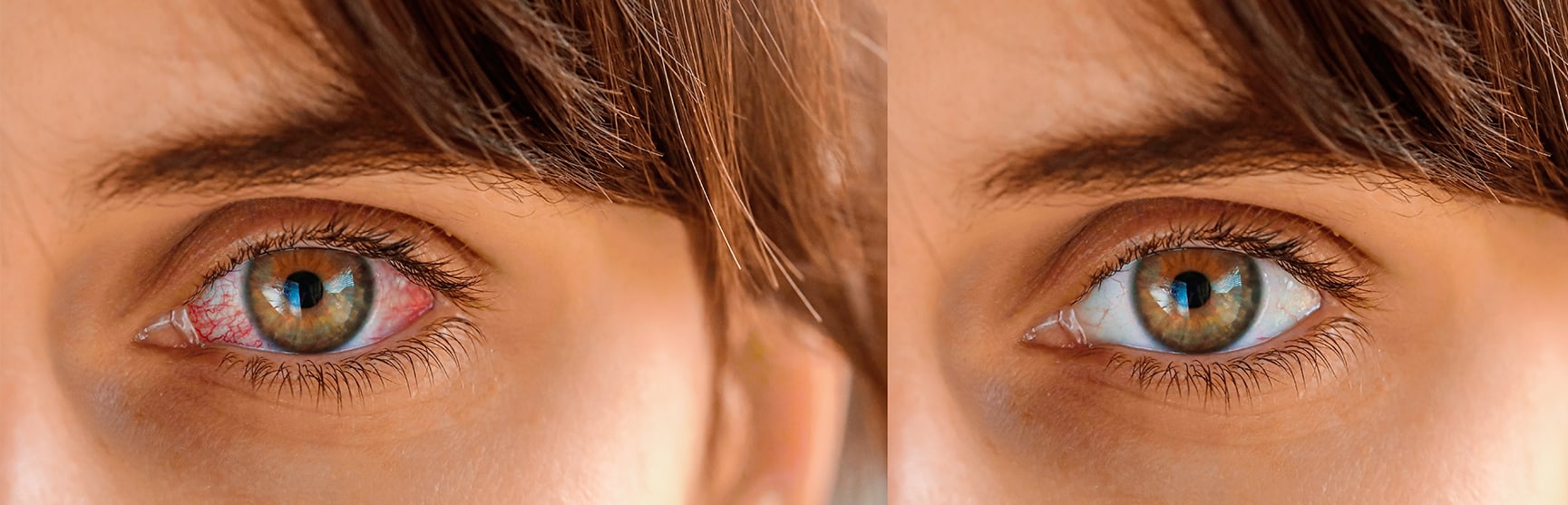
    Here are the most common causes and risk factors:

    1. Age
    Macular degeneration is the number one cause of vision loss in the senior population. The older a person gets, the higher their risk of developing AMD. According to the National Eye Institute, ”a large study found that people in middle-age have about a 2 percent risk of getting AMD, but this risk increased to nearly 30 percent in those over age 75.”

    2. Gender
    Women are more likely to develop age related macular degeneration than men.

    3. Race
    It occurs in all races but it is more common in Caucasians.

    4. Iris Color
    The research shows that there is less pigment in blue eyes, and green eyes for that matter, than there is in brown eyes, so more light is able to penetrate blue eyes. This makes lighter eyes more sensitive to light and is what makes people with blue eyes more likely to have age-related macular degeneration. Macular degeneration is caused when the light sensitive cells in the eyes start to die, which can eventually result in blindness.

    5. Heredity
    My husband’s father has wet macular degeneration as well as did many of my husband’s aunts and uncles. The lifetime risk of developing late-stage macular degeneration is 50% for people who have a relative with macular degeneration versus 12% for people who’s relatives do not have macular degeneration (4x the risk).

    6. Smoking
    Smoking is the single most controllable risk factor that contributes to the development of macular degeneration. Every cigarette that is smoked causes damage to one’s vision. Smoking causes vasoconstriction or narrowing of the blood vessels which reduces the blood supply to the eyes. Smoking also creates free radicals which causes cellular damage while decreasing ones levels of antioxidants.

    7. Diet Low in Dark Leafy Greens
    A Harvard study done by Dr. Johanna Seddon in 1994 concluded that those who ate at least 5 servings of dark leafy greens per week had a 43% lower risk of developing AMD than those who ate small amounts or none at all. These lutein rich greens include kale, collard greens, spinach, Swiss chard, parsley, mustard greens, romaine lettuce and beet greens.

    8. Diet Low in Omega 3 Fatty Acids
    Thanks again to Dr. Seddon and her researchers at Harvard University, they discovered that people with diets of a 3:1 ratio of omega-6 to omega-3 fatty acids have less macular degeneration. Almost all of our processed foods and vegetable oils contain omega-6 which means that the average American gets way to much omega-6. Because omega 6 competes with omega 3, if we have too much omega-6 then the omega-3 we do take isn’t utilized properly.

    9. High Blood Pressure
    Dr. Michael A. Samuel the author of Macular Degeneration: A Complete Guide for Patients and Their Families writes “If you have high blood pressure that is fairly well controlled, you have double the risk of wet AMD compared to someone who does not have hypertension at all. If you have uncontrolled high blood pressure that goes above 160/90 you are three times more likely to develop wet AMD.” (Ophthalmology 2003;110: 636-643).

    10. Exercise
    Those with an active lifestyle were 70% less likely to develop macular degeneration than those who did little or no exercise according to researchers from the University of Wisconsin. The Wisconsin study that began in 1988, tracked almost 4,000 men and women (between the ages of 43-86) over a 15 year period by conducting regular eye tests and recording levels of exercise.

    After accounting for other risk factors such as age, sex, history of arthritis, systolic blood pressure, body mass index, smoking, and education, those with a baseline active lifestyle of walking three times or more a week, were 70% less likely to develop macular degeneration than those who did little exercise.

    Reference:

    • blurtit.com
    • pmc.ncbi.nlm.nih.gov/articles/PMC3866712
    • enhancedvision.com/low-vision-info/eye-conditions/causes-of-macular-degeneration.html

    The problem with baby’s eyes is very hard to diagnosis due to its acuteness and severity. Mainly small kids are too young to actually tell about the difference between clear vision and the blur vision. Normally when kids reach an age of 4 years or so, their parents must be constantly looking out for such signs.

    Normally there are some telltale signs which tell much about the presence of baby eyes problem but they are not that evident in toddlers and babies eyes.

    According to a recent research, as many as 20 percent of kids at an age of 3 to 5 years are more prone to eye problems and parents need to pay attention towards this element. If this is ignored at that age, it can lead to more problems later on. It is therefore necessary to get a vision screening of kids every six months. Apart from this screening, there are some other things which parents can do to solve baby eyes issue.

    Baby eyes problem are either due to congenital issues or acquired. Congenital occurs due to developmental issues. When damage is due to the intrauterine development after an intake of any drugs by mother or some infections. Mothers with congenital problems have no symptom common. On the other hand in the case of acquired baby eyes problem starts in the initial 28 days of baby’s life. This is normally infective since origin.

    It is always pretty easy for parents to find out the presence of baby eyes problem.

    • Farsightedness: in this case the distinct objects appear clear to babies while they feel problem when focusing on close things. Kids mainly rub their eyes more often and try to sit closer to television. At times they even squint to make their vision clear. When doing some work, like drawing or reading, kid may not be able to stay focused for a longer period of time. The extreme farsightedness causes inability to view even the far end things. Naturally new babies are born with some signs of farsightedness but with the passage of time, their eyes are adjusted to normal view. If fail to treat, it can have drastic effects on the baby eyes.

    • The nearsightedness is associated with an ability to see near objects while difficulty in focusing the far ends things. Such kids squint when view near objects and also complain of an inability to see chalkboard in school from far distance. Either a child is given spectacles or making him sit in the front row can also resolve problem. If the both eyes of a baby are even, kids of age 4 years or so are not given spectacles, however in case of strain problem has to be identified to avoid further problems.

    • The last problem that baby eyes may get is astigmatism, but this can only be diagnosed by professional as layman can’t figure out

    How Can I Tell if My Child Has Myopia

    It is always better to take babies to pediatric if you feel any problem with their eyes.

    Amblyopia (Lazy Eye)

    Amblyopia, also known as lazy eye, is a condition in which one or both eyes do not develop normal vision due to various factors that cause the visual part of the brain to function abnormally. This weakens the eye and can lead to long-term vision problems. If recognized early, amblyopia generally responds well to treatment. Amblyopia therapy can include glasses, patching, eye drops, and sometimes surgery. Click here to learn more about amblyopia.

    Astigmatism-Farsightedness-Nearsightedness

    These three conditions, or refractive errors, are the most common eye problems in children and adults and are most often caused by abnormalities in the surface of the eye that prevent light from properly being focused on the retina. Click here to learn more about these refractive errors.

    Childhood Tearing/Epiphora

    Epiphora is the term for excessive tearing. Childhood epiphora is often noted soon after birth, but can be acquired later. When noted during infancy, it is usually due to blockage of the tear drainage system. This type of tearing often improves spontaneously by 6 to 12 months of age. Medical treatment includes tear sac massage and eye drops, but if tearing persists, surgical probing of the drainage system may be required. Other rare causes of childhood tearing include pediatric glaucoma and ocular surface diseases.

    Cortical Visual Impairment

    Cortical visual impairment (CVI) is vision loss due to any abnormality of the visual center in the brain. The eyes are normal, but the visual interpretation center in the brain does not function properly and prevents normal vision.

    Developmental Abnormalities

    During development of the fetus, abnormalities in the visual system can occur. Some developmental abnormalities include coloboma, microphthalmia (small eye), and optic nerve hypoplasia. These abnormalities often result in vision loss.

    Double Vision

    Double vision (diplopia) is typically caused by misalignment of the eyes (strabismus), which causes one to see an object in two different places at the same time. The object can be displaced in a horizontal, vertical, or diagonal fashion. Double vision can result from many conditions and should be evaluated at the time of onset. Treatment for double vision can include prism glasses, strabismus surgery, or Botox injections.

    Genetic Eye Disease

    Many eye diseases have a known genetic abnormality. These diseases are often inherited and frequently there are other family members who have had the disease. In cases of known inherited eye disease in the family, early evaluation is important. The Dean McGee Eye Institute has an ophthalmic genetic counselor who routinely sees patients in the clinic.

    Nystagmus

    Nystagmus is an involuntary, rhythmic oscillation of the eyes. The eye movements can be side-to-side, up and down, or rotary. Nystagmus may be present at birth or acquired later in life. It may result from abnormal binocular fixation early in life, and may also accompany a number of eye disorders and neurological diseases.

    Pediatric Cataract

    A cataract is a cloudiness or opacification of the normally clear lens of the eye. Depending on the size and location, the cataract can interfere with light passing to the retina and cause blurred vision. Cataracts are typically associated with older adults, but cataracts can occur at birth or during childhood. Early detection and treatment of cataracts are critical in infants and young children in order to restore normal visual development. A white area in the pupil and misalignment of the eye can be a sign of cataract.

    Pediatric cataracts that significantly obstruct vision require surgery. Patients subsequently require treatment with eyeglasses, bifocals, or contact lenses, and eye-patching. Often, pediatric cataracts result in some degree of lazy eye (amblyopia) and strabismus as well.

    Pediatric Glaucoma

    Glaucoma is a condition that is associated with high pressure within the eye. This pressure can damage the optic nerve, which is critical for vision, resulting in permanent vision loss. Pediatric glaucoma is a rare condition that can present in the newborn or during childhood. Signs and symptoms of pediatric glaucoma include cloudy corneas, tearing, frequent blinking, light sensitivity, and redness of the eye.

    Pediatric Ptosis (Drooping Eyelid)

    Ptosis, or drooping of the upper eyelid, occurs in both children and adults. Children can be born with ptosis (congenital) or acquire it during childhood. Neurological diseases can also trigger it.

    Ptosis is caused by weakness in the muscle that elevates the eyelid. A droopy eyelid can block light passing to the retina in the back of the eye and/or create significant astigmatism that produces a blurry image in the affected eye. These situations cause lazy eye (amblyopia) and, if untreated, can result in permanent loss of vision. In addition, children may develop a chin-up head position due to the droopy eyelid. If the ptosis is significant, surgical correction may be necessary.

    Retinopathy of Prematurity (ROP)

    Retinopathy of prematurity (ROP) is an eye disease that occurs in some premature infants. It results from abnormal development of the blood vessels in the retina. ROP is progressive, starting with mild changes and sometimes progressing to severe, sight-threatening changes. Most infants with ROP improve spontaneously, but some develop severe changes that require laser treatment or injections of medicine into the eye.

    Complications of ROP can include strabismus (eye misalignment), myopia (nearsightedness), cataract, and, in severe cases, blindness from retinal detachment. Premature infants at risk of ROP are identified in the hospital and enrolled in a routine screening protocol.

    Strabismus (Crossed Eyes)

    Strabismus is the term for misalignment of the eyes in which an eye may be turned inward, outward, upward, or downward. Strabismus in children can result in lazy eye (amblyopia) and cause permanent loss of vision if treatment is delayed. Adults and older children often experience double vision (diplopia). Depending on the type and cause of the strabismus, treatment may include eyeglasses, prisms, surgery, Botox injection, or eye-patching therapy.

    Reference:

    • bhszone.com
    • dmei.org/services-specialties/pediatric-ophthalmology-strabismus/common-pediatric-eye-problems/

    Related To: Finding out the Infant’s Eye Color

    پلک زدن یک عمل سریع به هم خوردن چشمان است. عمل پلک زدن از این جهت برای چشم ضروری است که باعث پخش شدن اشک در چشم و عبور آن از روی قرنیه و از بین بردن عفونتهای احتمالی می‌شود. به صورت متوسط عمل پلک زدن حدود 300 تا 400 میلی ثانیه به طول می انجامد. این سرعت تحت عواملی چون وارد شدن یک شی ریز خارجی مثل ریزه چوب، ضربات وارد شده به چشم، مصرف دارو و عفونت تغییر می‌کند.

    هر فرد تقریباً در هر 2 تا 10 ثانیه یک بار پلک می‌زند.

    پلک زدن مانند نفس کشیدن است، شما پلک می‌زنید بدون اینکه متوجه باشید. چندین بار در دقیقه پلک‌ها گرد و غبار را از چشم‌ها دور کرده و آنها را مرطوب می‌کنند.

    اگر چشم به هر دلیلی تحریک شود، مثل خوردن چیزی به آن یا نور شدید، این حرکت آرام پلک‌ها به چشمک‌های سریع و پیاپی تبدیل می‌گردد. پلک زدن زیاد هم می‌تواند علامت اخطاری باشد که چشم خشک است یا چیزی در آن افتاده است که با پلک زدن عادی خارج نمی‌شود.
    ترکیب تحریک‌کنندهٔ خشکی چشم و استفاده از لنز می‌تواند چشم را مثل یک چراغ چشمک‌زن کند. پلک زدن زیاد ممکن است واکنشی به‌وجود چیزی زائد بر روی لنز باشد.
    پلک افراد سالمند با سابقهٔ سکته یا بیماری پارکینسون (لقوه) گرفتگی نشان داده و باعث چشمک‌زدن زیاد و قوی می‌شود. این نوع گرفتگی با اضطراب تشدید شده و گاهی با انقباض ماهیچه‌های صورت همراه است.

    Blinking is a normal protective reflex of the eye. It helps lubricate the front of the eyeballs, as well as shields the eyes from bright light, dust and other debris.

    Types of blink: There are three types of blink.

    • Spontaneous blink Spontaneous blinking which is done without external stimuli and internal effort. This type of blinking is conducted in the pre-motor brain stem and happens without conscious effort, like breathing and digestion.
    • Reflex blink A reflex blink occurs in response to an external stimulus, such as contact with the cornea or objects that appear rapidly in front of the eye. A reflex blink is not necessarily a conscious blink either; however it does happen faster than a spontaneous blink. Reflex blink may occur in response to tactile stimuli (e.g. corneal, eyelash, skin of eyelid, contact with eyebrow), optical stimuli (e.g. dazzle reflex, or menace reflex) or auditory stimuli (e.g., menace reflex)
    • Voluntary blink Voluntary blink is larger amplitud: The normal blinking rate in a newborn is as low as twice a minute and it increases to 14-17 times per minute in teenagers. This rate will normally increase if the eyes are exposed to bright light, temperature variation and humidity. Eyestrain caused by reading in poor light, lack of sleep or too much screen time – especially in children – can lead to excessive blinking.

    Other causes of excessive blinking include inflammation of the eyelids, an irregular front surface of the eye, ocular allergy, habitual tics, refractive errors (the need to wear glasses), divergent squint or turning out of the eye. A rare cause of excessive blinking can be an undiagnosed neurological disorder.[3]

    • Near-sightedness – One of the most common reasons for your child’s excessive blinking is near-sightedness. Visit your closest specialist and have your child’s eye checked. They may require glasses.
    • Allergies – Like in adults, if your child is blinking too much and has watery eyes or excessive discharge around the eyes, this could be due to an allergy.
    • Dryness – Another common reason for excessive blinking is extreme dryness of the eyes, this condition can cause a child not only to blink but may cause burning sensations or an itch in the eye. Make sure your child doesn’t rub their eyes. Doctors will usually prescribe teardrops or hydrating eye drops to help ease the irritation.
    • Strain – A lot of stress on the eyes can cause excessive blinking, this can be due to reading with bad lighting, too much time on screens or even lack of sleep.
    • OCD – Obsessive Compulsive Disorder (OCD) is a psychological condition that a lot of children have that often goes undiagnosed. If the OCD is bad enough, it could cause severe blinking or other facial tics.
    • Anger – Emotional and psychological instability can cause numerous facial tics. One of the most common tics associated with extreme anger is excessive blinking, this anger usually indicates the need for therapy. It is recommended that you talk to a child psychologist at the earliest if your child shows severe anger issues.
    • Blepharitis – This is an infection caused due to dandruff or bad bacteria in the eyes, it is extremely common and is known to be a reason for excessive blinking in your child.[4]

    Parents describe their concerns about excessive blinking in a variety of ways, such as an increased frequency of blinking, shutting eyes tightly, awkward rolling and widening of the eyes, and too much rubbing of the eyes.

    The management of excessive blinking includes treating the underlying cause. A paediatric ophthalmologist should carry out a thorough eye examination to rule out the major concern: visual problems. The treatment could be as simple as getting glasses for any significant refractive error to reduce the visual stress or using topical lubricant eye drops regularly for dry eyes and ocular allergy.

    In rare cases, eye twitching can be a sign of certain brain and nervous system disorders, especially if it is accompanied by other signs and symptoms like multiple tics or auditory (vocal) tics. In such cases, an appointment with a paediatric neurologist is recommended.

    ؟ Will Blinking More Help Dry Eyes

    Healthy visual habits can prevent excessive blinking in children:

    - Wear sunglasses or a wide brim hat outdoors.

    - Avoid reading in poor light.

    - Take frequent breaks from digital screens.

    - Wash your hands before you touch your eyes.

    - Eat a balanced diet and stay hydrated.[3]

    The blink reflex is a reflex which is designed to naturally protect the eyes. Most animals with eyes have some form of this reflex, and the reflex is present from the time that an animal first opens its eyes. Abnormalities in this reflex can indicate that there is a neurological problem and may increase the risk of incurring eye damage because the eyes are no longer able to protect themselves reflexively.

    Several different stimuli can trigger this reflex. Anything which touches the cornea will cause someone to blink, and people usually blink when objects appear to be on a collision course with the eye, as for example when something is thrown in the direction of someone's head. Very bright light also stimulates the blink reflex, as do loud noises.

    Multiple cranial nerves are involved in the process of blinking, as is the brainstem. In a healthy person, the blink reflex appears in both eyes. If both eyes fail to blink, it can mean that there is a problem with one of the cranial nerves or the brainstem which is inhibiting the normal processing of the reflex. If someone fails to respond at all to a stimulus which should elicit a blink, or responds slowly, it is also a sign of a neurological issue. eResearch by Navid Ajamin -- summer 2012

    Neurological exams can include a quick assessment of the blink reflex to see how a patient responds to stimuli. This can be part of the process of checking on a patient who is believed to be nonresponsive or in a coma as well, because the reflex should kick in unless a patient has severe damage to the brainstem. It is also possible to assess the blink reflex in a test which involves placing electrodes around the eyes and providing very mild electrical stimulation while recording the responses.[5]

    Some of the best ways to stop children from blinking excessively would be by taking preventive measures and completing the course of medication prescribed to you. In addition, some measures you may consider are:

    • Protecting eyes: Using protective eyewear guards the eyes from dust, sunlight and other irritants
    • Preventing dry eyes: Omega 3 fatty acids help prevent dryness in the eyes by stimulating the tear glands and reducing inflammation
    • Helping manage stress: Help your child manage stress with activities like meditation, relaxation techniques and yoga to stop excessive blinking
    • Ensuring a healthy diet and lifestyle: Eating nutrition-rich food, drinking plenty of water, and adopting a healthy lifestyle helps maintain healthy eyes and reduce excessive blinking
    • Cleaning and nourishing eyes: Washing eyes frequently with clean water and consuming vitamins and minerals like Zinc and Vitamin A, either through supplements or through food sources like bananas and strawberries is sure to help reduce excessive blinking.

    Related image

    There are numerous ways to prevent constant blinking in children, these include:

    • Ensuring your child always reads and watches TV or works on a computer in a room with plenty of light.
    • Ensuring your child sleeps the required 8-10 hours
    • Monitoring their screen time and limiting it
    • Making sure they get some exercise
    • If required, giving them vitamin supplements [4]

    درمان چشمک‌زدن
    در بیشتر موارد، درمان پلک زدن زیاد مرطوب نگاه داشتن چشم و جلوگیری از تحریک آن است.
    -
    از اشک مصنوعی استفاده کنید. اشک مصنوعی که شبیه به اشک طبیعی است چشم خشک‌شده در اثر دود یا عوامل دیگر را مرطوب می‌کند.
    - لنزهایتان را خیس نگاه دارید. کسانی که از لنز چشمی استفاده می‌کنند باید در طول روز چشم را با مواد مرطوب کننده بشویند. به‌دنبال مرطوب‌کننده‌های بدون مواد اضافی باشید، چون این مواد باعث تحریک چشم می‌شوند.
    - کمی دیر لنز را در چشم بگذارید. صبح پیش از گذاشتن لنزها در چشم، بگذارید چشم نفسی بکشدو پس از یک ساعت لنز را در آن قرار دهید. این کار به چشم امکان تطبیق با محیط داده و ناراحتی آن را کاهش می‌دهد.
    از لنزهای تراوا استفاده کنید. لنزهای تراوا رسیدن گاز اکسیژن بیشتر به چشم را امکان‌پذیر می‌کنند و کمتر باعث خفگی و تحریک‌ آن می‌شوند. به‌علاوه، مواد و ذرات تحریک‌کننده بر سطح لنز را کمتر به خود می‌گیرند.
    از لنزهای یک‌بار مصرف استفاده کنید. لنزهای یک‌بار مصرف را یک هفته در چشم بگذارید و سپس آن را دور بیندازید و لنزهای تازه‌ای به‌جای آنها بگذارید. البته در فاصلهٔ آن یک شب را بدون لنز بخوابید. این کار به چشم فرصت استراحت می‌‌دهد. این لنزهای نازک را نشوئید و از مصرف دوبارهٔ آنها خودداری کنید.
    -
    قطرهٔ چشمی مصرف کنید. وقتی شب بدون لنز می‌خوابید، قبل از خواب قطره، ضداحتقان ضدحساسیت را که پزشک تجویز می‌کند در چشم بریزید. این قطره‌ها خارش و ورم را کم کرده و حساسیت به لنز را کاهش .می‌‌دهند. اگر باز هم مشکلی دارید لنزهایتان را بردارید و هرچه زودتر به پزشک مراجعه کنید.

    - ماهیچه‌های چشم را معالجه کنید. اگر چشمک زدن شما ناشی از گرفتگی‌های غیر ارادی ماهیچه‌ها در اثر اختلال دستگاه عصبی است، پزشک داروئی برای کنترل این گرفتاری‌های ماهیچه‌ای تجویز خواهد کرد.

    پرش پلک چشم یا تیک عصبی چشم چیست؟

    پرش پلک چشم یا تیک عصبی چشم چیست؟

    پرش یا تیک عصبی چشم (eye twitching) – یا اگر دقیق‌تر بگوییم پرش پلک چشم – یک اسپاسم و گرفتگی غیر قابل کنترل ماهیچه‌های پلک چشم است. در بیش‌تر موارد، این عارضه تنها چند دقیقه طول خواهد کشید، اما برخی اوقات ممکن است تا روزها و یا حتی بیش‌تر ادامه داشته باشد.

    زمانی که فرد دچار پرش چشم می‌شود، تصور می‌کند که همه‌ی اطرافیان متوجه‌ی مشکل او هستند. اما در بیش‌تر موارد این مشکل به راحتی قابل مشاهده توسط دیگران نیست. در پزشکی به مشکل پرش پلک چشم مایوکیمیا (myokymia) گفته می‌شود.

    شایع‌ترین علل پرش چشم چیست؟

    دلایل بروز این عارضه عبارتند از:

    • استرس
    • خستگی
    • کافئین
    • خستگی چشم
    • الکل
    • خشکی چشم
    • مشکلات تغذیه‌ای
    • حساسیت‌ها
    • چگونه می‌توان پرش چشم را متوقف کرد؟

      برای متوقف کردن این مشکل، ابتدا باید علت بروز آن را شناخت. برخی اوقات، تغییر اندکی در رژیم غذایی و سبک زندگی تا حد قابل قبول و زیادی خطر ابتلا به این مشکل را کم کرده و حتی به رفع آن به صورت کامل کمک خواهد کرد.

    • روش رفع و درمان پرش چشم

      علت بروز پرش چشم و راهکارهای رفع آن را در ادامه بررسی خواهیم کرد:

      استرس

      استرس یکی از رایج‌ترین دلایل پرش چشم است. یوگا، تمرینات هوازی، وقت گذرانی با دوستان و خانواده و سبک کردن جدول برنامه‌ی روزانه و اختصاص وقت بیش‌تر برای استراحت، از جمله راهکارهای پیشنهادی برای کاهش استرس بوده و احتمالاً به از بین رفتن پرش پلک چشم کمک خواهد کرد.

    • خستگی

      کمبود خواب که ممکن است به دلیل استرس ایجاد شده و یا علت دیگری داشته باشد، می‌تواند بروز پرش چشم را تحریک کند.

      خستگی چشم

      خستگی چشم ناشی از کار کردن زیاد با کامپیوتر، تبلت یا گوشی‌های تلفن هوشمند – یکی از علت‌های رایج پرش پلک چشم است.

      در هنگام کار با ابزار دیجیتال قانون 20 – 20 – 20 را اجرا کنید. هر 20 دقیقه یکبار از رایانه‌ی خود چشم برداشته و به مدت 20 ثانیه به نقطه‌ای در دور دست (حداقل فاصله‌ی 20 فوتی یا 6 متری) خیره شوید. این کار خستگی چشم که عامل تحریک کننده‌ی بروز پرش چشم است را کاهش خواهد داد.

    • کافئین

      مصرف فراورده‌های حاوی کافئین هم عامل تحریک کنننده‌ی این مشکل چشمی به شمار می‌رود. به مدت یک و یا دو هفته مصرف چای، قهوه و نوشیدنی‌های گاز دار را متوقف کرده و یا انواع بدون کافئین آن را مصرف کنید.

      الکل

      اگر بعد از مصرف نوشیدنی‌های حاوی الکل دچار پرش چشم شدید، باید مصرف آن را متوقف کنید. چرا که مصرف این ماده باعث بروز آن شده است.

      خشکی چشم

      بسیاری از افراد بزرگسال به خصوص بعد از سن 50 سال به خشکی چشم دچار می‌شوند. این عارضه خصوصاً در میان افرادی که با رایانه کار می‌کنند، داروهای خاصی – برخی آنتی هیستامین‌ها و داروهای ضد افسردگی – مصرف می‌کنند، از لنزهای تماسی چشمی استفاده می‌کنند و یا به مقدار زیاد الکل و کافئین مصرف می‌کنند، بسیار شایع‌تر از دیگر افراد است.

      اگر به پرش چشم مبتلا هستید و در عین حال در چشم‌های خود احساس خشکی بیش‌ از حد دارید، حتماً با پزشک خود مشورت کنید. پزشک در چنین مواقعی قطره‌های اشک مصنوعی تجویز می‌کند، این قطره‌ها رطوبت چشم‌ها را تامین خواهد کرد و منجر به توقف پرش چشم خواهد شد و از طرفی احتمال بروز آن را در آینده بسیار کاهش خواهد داد.

      مشکلات تغذیه‌ای

      برخی گزارش‌ها حاکی از این است که کمبود برخی از مواد همچون منیزیم می‌تواند به اسپاسم پلک چشم منجر شود. این گزارش‌ها را نمی‌توان با قاطعیت تایید کرد، ولی می‌توان از آن به عنوان یکی از دلایل احتمالی بروز این مشکل یاد کرد.

      اگر نگران هستید که رژیم غذایی شما، همه‌ی نیازهای بدن برای داشتن دید سالم‌تر را پوشش نمی‌دهد، قبل از خرید مکمل‌های غذایی و ویتامین‌ها حتما با پزشک خود مشورت کنید.

      حساسیت‌ها

      افراد مبتلا به آلرژی‌ها یا حساسیت چشمی، دچار خارش، تورم و اشک چشم زیاد می‌شوند. فرد به دلیل داشتن حساسیت چشم خود را زیاد می مالد و همین باعث آزاد شدن هیستامین از پلک شده و در نتیجه به پرش پلک چشم می انجامد.

      در برخی از مواقع قطره‌های چشمی که برای کاهش علایم حساسیت ساخته شده‌اند، کمک کننده خواهند بود. اما آنتی هیستامین موجود در این قطره‌ها منجر به خشکی چشم خواهد شد. پیش از مصرف هرگونه ماده‌ای اگر علائم حساسیت و یا پرش چشم دارید با پزشک خود مشورت کنید.

    Reference:

    1. fa.wikipedia.org
    2. pezeshk.us
    3. moorfields.ae
    4. parenting.firstcry.com
    5. wisegeek.com/what-is-the-blink-reflex.htm
    6. iovs.arvojournals.org/article.aspx?articleid=2711429

    See also:

    • What Makes a Child Tic?
    • Excessive blinking in children

    ملتحمه (conjunctiva) غشاء نازك و شفافي است كه روي سفيدي چشم (صلبيه) و سطح داخلي پلك ها را پوشانده است. كنژنكتيويت التهاب اين غشاء است كه بدلايل عفوني و يا غير عفوني رخ مي دهد.

    انواع كنژنكتيويت:

    از ميان انواع غير عفوني، شايعترين نوع كنژنكتيويت نوع حساسيتي يا آلرژيك است. اين مشكل در افرادي كه حساسيت فصلي دارند نيز ديده مي شود و ممكن است بر اثر حساسيت به موادي از قبيل مواد آرايشي، عطر و يا داروها نيز رخ دهد. اين نوع از كنژنكتيويت مسري نيست.

    نوع عفوني كنژنكتيويت بر دو نوع است. نوع ويروسي كه عامل بيماري ويروس بوده و معمولا با يك سرما سرماخوردگي و گلو درد همراه است. نوع دوم يا كنژنكتيويت ناشي از باكتري (باكتريال) معمولا بر اثر آلودگي با باكتري هايي نظير استافيلوكك و استرپتوكك ايجاد مي شود و شدت آن به نوع و گونه باكتري بستگي دارد.

    Types of pink eye: Bacterial, viral and allergic conjunctivitis

    Viral conjunctivitis

    This is the most common type of pink eye. Since it is caused by a virus, usually an adenovirus, it is extremely contagious. It can spread through respiratory droplets (from coughs and sneezes) or by touching your eye before washing your hands. Swimming pools can harbor the viruses that cause pink eye, and so can personal items like towels and pillowcases. It can also develop along with a cold, flu or other respiratory infection caused by a virus.

    Adenoviruses are extremely common, but less-common viruses can also cause viral pink eye. For example, herpes viruses can cause a more serious form of pink eye. Molluscum contagiosum (a poxvirus infection) can cause chronic pink eye if it enters the eyes. And, in rare cases, the coronavirus responsible for COVID-19 can also cause viral pink eye.

    The viral form usually starts in one eye (unilateral) and then spreads to both (bilateral). Common signs and symptoms include burning or itchy, red eyes with clear, watery discharge.

    Since antibiotics aren't effective against viruses, there is no cure for viral pink eye. However, it usually goes away by itself after a short time. Your eye doctor may prescribe other medicated eye drops to help you feel more comfortable and decrease the risk of spreading the virus. You can also ask your eye doctor about safe home treatments for pink eye symptoms.

    Some people with conjunctivitis (“pink eye”) may experience mucopurulent discharge — a mix of mucus and pus. This usually suggests a bacterial infection, but not always. Too much discharge might also mean a severe infection.

    Conjunctivitis — more commonly known as “pink eye” — is an infection of the mucus membranes that coat your eyes and the insides of your eyelids. This membrane is called your conjunctiva.

    Mucopurulent refers to a type of liquid discharge. “Muco” relates to mucus, and “purulent” relates to pus. So mucopurulent discharge contains both mucus and pus.

    There are many causes of conjunctivitis. It could be due to a virus or the result of allergies, among other causes. Regardless of the cause, mucopurulent discharge from your eyes may be a symptom of pink eye.

    علائم و نشانه هاي كنژنكتيويت:

    بارزترين علامت كنژنكتيويت قرمزي چشم بر اثر التهاب است. علائم انواع كنژنكتيويت را مي توان بصورت زير خلاصه كرد:

    كنژنكتيويت آلرژيك: هر دو چشم را درگير كرده و سبب خارش و قرمزي همراه با اشكريزش و تورم پلك ها مي شود. بيمار ممكن است بدليل حساسيت آبريزش بيني هم داشته باشد.

    كنژنكتيويت ويروسي: معمولا با درگيري يك چشم شروع شده و ممكن است در ادامه چشم ديگر را نيز درگير كند. علائم ديگر شامل اشكريزش شديد، ترشح آبكي و قرمزي چشم هستند.

    كنژنكتيويت باكتريال (ميكروبي): هر دو چشم را درگير كرده و سبب ترشح غليظ مي شود كه ممكن است بخصوص هنگام بيدار شدن از خواب سبب چسبيدن پلك ها به هم شود. علائم ديگر عبارتند از: تورم ملتحمه، قرمزي و اشكريزش. اين نوع از كنژنكتيويت معمولاً ابتدا يك چشم را درگير مي كند ولي به سادگي به چشم ديگر منتقل مي شود.

    تشخيص:

    كنژنكتيويت معمولا در يك معاينه ساده چشم پزشكي با استفاده از Slit Lamp تشخيص داده مي شود. در بعضي از موارد چشم پزشك ممكن است براي تشخيص نوع باكتري عامل كنژنكتيويت ترشحات چشم را براي كشت به آزمايشگاه بفرستد.

    پيشگيرى :

    از آنجا كه انواع ويروسي و باكتريال كنژنكتيويت مسري بوده و براحتي منتقل مي شوند، پيشگيري نقش بسيار مهمي در جلوگيري از انتقال آلودگي به چشم ديگر بيمار و نيز افراد ديگر دارد. براي پيشگيري مؤثر توجه به نكات زير لازم است:

    • شستشوي مرتب دستها و خودداري از ماليدن چشم. آلودگي در بسياري از موارد بر اثر خاراندن چشم درگير و انتقال از طريق دست آلوده اتفاق مي افتد.
    • استفاده نكردن از حوله مشترك
    • استفاده نكردن از لوازم آرايشي مشترك نظير خط چشم، سايه چشم و ...
    • استفاده نكردن از قطره هاي چشمي مشترك
    • شنا نكردن (بعضي از باكتري ها مي توانند از طريق آب منتقل شوند)
    • ضد عفوني كردن ميز و پيشخوان محل كار يا آشپزخانه منزل
    • خودداري از دست دادن با ديگران
    • عدم استفاده مجدد از دستمال جهت پاك كردن چشم. بهتر است از دستمال كاغذي و بصورت يك بار مصرف استفاده شود.

    درمان :

    كنژنكتيويت آلرژيك: كمپرس سرد و استفاده از اشك مصنوعي در موارد خفيف كمك كننده است. در موارد شديدتر از داروهاي ضد التهابي غير استروئيدي و آنتي هيستامين ممكن است استفاده شود. در بعضي از بيماراني كه دائما كنژنكتيويت آلرژيك دارند ممكن است از قطره هاي استروئيدي نيز استفاده شود.

    كنژنكتيويت ويروسي: نوع ويروسي بيماري مانند سرماخوردگي درمان خاصي ندارد و معمولا براي آن داروي خاصي تجويز نمي شود ولي علائم بيماري را مي توان با استفاده از كمپرس سرد و اشك مصنوعي تخفيف داد. در موارد خيلي شديد نيز مي توان از قطره هاي استروئيدي براي كاهش التهاب استفاده كرد. كنژنكتيويت ويروسي معمولا ظرف سه هفته بهبود مي يابد.

    كنژنكتيويت باكتريال: معمولا با استفاده از قطره ها يا پماد هاي آنتي بيوتيك درمان مي شود.

    نكته آخر:

    هر چند كنژنكتيويت معمولاً عفونتي خفيف بوده و مشكل چنداني ايجاد نمي كند ولي گاهي ممكن است شدت يافته و منجر به مشكلات جدي تري شود بنابراين توصيه مي شود در صورت بروز علائم حتماً به چشم پزشك مراجعه كنيد.

    Reference:

    • healthline.com/health/eye-health/mucopurulent-conjunctivitis
    • allaboutvision.com/conditions/conjunctivitis-types.htm

    See also: What is Mucopurulent Conjunctivitis?

    شايد بتوان گفت بينائى مهم‌ترين حس انسان است. نه تنها به اين علت که در ارتباط با محيط خارج از بدن، نقش مهمى دارد، بلکه از اين‌رو که اختلال ديد، سبب کاهش يادگيرى نيز مى‌شود. شخص با حس بينائى مى‌تواند از خود محافظت کند، تعادل خود را نگه‌دارد، خلاقيت داشته باشد و بالاخره از ديدن محيط اطراف لذت ببرد. لذا از چشم‌ها بايد نهايت مواظبت به‌عمل آمده و براى بهداشت آن به نکات زير توجه گردد:

    • هرگز نبايد دست آلوده را به چشم ماليد. دست و صورت خصوصاً دست‌ها بايد مرتب با آب و صابون شسته شود. از شستن دست و صورت در آب‌هاى آلوده و کثيف، جداً بايد خوددارى گردد.
    • بايد از حوله و دستمال شخصى و تميز استفاده کرد و هرگز حوله و دستمال ديگران را مورد استفاده قرار نداد.
    • مطالعه بايستى در نور کافى انجام گيرد و از خواندن نوشته‌هاى خيلى‌ريز خوددارى شود.
    • به‌هنگام مسافرت با اتومبيل و ساير وسايط ‌نقليه، حتى‌المقدور از مطالعه خوددارى شود.
    • فاصله چشم از کتاب بايد حدود ۳۰ سانتى‌متر و زاويه کتاب نسبت به سطح مورد مطالعه ۴۵ تا ۷۰ درجه باشد.
    • از خيره‌شدن به نور شديد، مانند خورشيد و لامپ‌هاى پرنور خوددارى شود. هم‌چنين در مقابل آفتاب، باد، گرد و خاک، از چشم‌ها محافظت شود.
    • براى خواب و استراحت، اتاق بايد تاريک باشد تا اعصاب چشم به‌خوبى استراحت کند.
    • مصرف غذاهاى داراى ويتامين A و پروتئين در حفظ سلامت چشم اهميت به‌سزائى دارد.

    اختلالات بينائى در سلامت دانش‌آموز و پيشرفت تحصيلى او اثر زيادى دارد و بايد مورد توجه مسئولان آموزشگاه قرار گيرد. آموزگار، مدير، پزشک، مربى بهداشت در تأمين سلامت بينائى شاگردان مسئوليت مشترک دارند. آموزگار به‌علت تماس دائمى با شاگردان، بهتر از هر شخص ديگر با توجه دقيق، به خواندن و نوشتن و کار کردن آنها مى‌تواند افرادى را که داراى اختلال يا ضعفى در قوه بينائى هستند، بشناسد و پس از آزمايش، آنها را نزد پزشک هدايت نمايد.

    به‌طور کلى علائمى که شخص با ديدن آن علائم، بايد متوجه اختلالاتى در دستگاه بينائى خود گردد، عبارتند از:

    • - اگر اشياء و نوشته‌ها را در فاصله معمولى به‌خوبى نتوان مشاهده نمود و يا هنگام نگاه‌کردن به آنها احساس ناراحتى کرد.
    • - اگر در هنگام غروب آفتاب نتوان به‌خوبى ديد.
    • - اگر احساس شود که چشم زود خسته مى‌شود.
    • - اگر گاهى سردرد، به‌خصوص هنگام غروب عارض شود.
    • - اگر چشم، خارش و سوزش داشته باشد و يا درد بگيرد.
    • - اگر صبح‌ها، هنگام بيدار شدن، در گوشه چشم ترشحاتى جمع شود.


    با مشاهده علائم بالا، لازم است به چشم پزشک مراجعه نموده و دستورات وى را به دقت به‌کار برید.


    اختلالات بينائى

    دوربينى، نزديک‌بينى، و آستيگماتيسم، از اختلالات عمده بينائى در دانش‌آموزان به شمار مى‌رود.
    دوربينى

    در افراد دوربين، اشعه‌هاى نورانى که به‌طور موازى به چشم مى‌تابد، در عقب شبکيه، تشکيل تصوير مى‌دهد که اين اختلال گاهى علل ارثى دارد. دانش‌آموزانى که به اين عارضه دچار هستند، چشمان خود را زياد مى‌مالند. در موقع کارهاى ظريف مضطرب و بى‌‌قرار هستند. در موقع خواندن يا نوشتن، سر را دورتر از حد معمول نگاه مى‌دارند. به خواندن يا ديدن کتاب‌هاى مصور تمايلى نشان نمى‌دهند. درد چشم، سردرد، التهاب پلک، تيک صورت و گاهى سرگيجه دارند. براى درمان دوربيني، از عينک‌هاى با عدسى محدب استفاده مى‌گردد.

    نزديک‌بينى

    در افراد نزديک‌بين، اشعه نورانى که به چشم مى‌تابد جلوتر از شبکيه، تشکيل تصوير مى‌دهد. نزديک‌بينى به حالت و عادت کسانى که چشم خود را تنگ مى‌کنند تا بهتر ببينند، اطلاق مى‌شود. فاکتورهائى نظير کار نزديک، کمى‌ نور، کمبود ويتامين‌ها و غيره در ايجاد نزديک‌بينى مؤثر هستند. دانش‌آموزانى که به اين عارضه دچارند، اغلب موقع نگاه‌کردن به دور، گردن مى‌کشند. در موقع نوشتن يا خواندن، سر را از حد معمول نزديک‌تر مى‌برند. گاهى يک چشم را مى‌بندند و با تنگ نمودن چشم ديگر، سعى در تطابق اجسام دارند. سردرد و آب‌ريزى از چشم نيز مشاهده مى‌گردد. براى درمان نزديک‌بيني، از عينک‌هاى با عدسى مقعر مناسب استفاده مى‌گردد.

    آستيگماتيسم

    اين عارضه از عيوب انکسارى چشم است. در اين عارضه، تصوير يک نقطه همان يک نقطه درک نمى‌شود، بلکه از يک نقطه از هر جسم خارجي، دو خط افقى ديده مى‌شود. در اين حالت بعضى از اشعه‌هاى نوراني، يکديگر را روى شبکيه قطع مى‌کنند، در حالى‌که محل تلاقى پاره‌اى ديگر، در جلو يا عقب شبکيه است و تصويرى که ايجاد مى‌کند، زياد روشن و واضح به‌نظر نمى‌رسند. دانش‌آموزان مبتلا به آستيگماتيسم، اغلب سر را به کتاب يا تصوير نزديک مى‌کنند، سردرد دارند و چشم را هنگام مشاهده تصاوير ظريف مى‌مالند. تصحيح چشم آستيگمات اغلب ساده بوده و با استفاده از عدسى استوانه‌ائى که در جهت مناسبى از چشم قرار داده مى‌شود، انجام مى‌گيرد. مگر در بعضى حالات نادر که تصحيح آن بسيار مشکل بوده و گاهى حتى غيرممکن است.

    Reference: www.pezeshki.net

    As cold winter months arrive, many people will live in dry, indoor conditions with the heat on all day, which contributes to the season’s being the most common time people complain about dry, itchy and watery eyes.

    Dry eye occurs when there is low tear production, or when the tear quality is poor and the tears evaporate too quickly. When people blink, tears spread evenly over the eye, keeping them smooth, healthy and clear. This is an important step for healthy, comfortable vision. However, it is estimated that 4.88 million Americans age 50 and older have dry eye, and suffer from irritated, burning and scratchy eyes.

    In most cases, dry eye can be managed successfully; but colder weather is a common dry eye irritant of which people may be unaware. Dry eyes are often a common complaint for contact lens wearers during the colder, winter months.

    Winter brings some of the most popular holidays of the year, but the season also comes with extreme temperatures and precipitation. Whether you love the season for its festivities or hate the season for its weather, you have to take steps to protect yourself from the potential hazards of winter conditions.

    While you probably remember to drive more carefully and bundle up before going outside, you may overlook the seasonal threats to your eye health. These hazards often seem less obvious than an icy road or freezing morning, but eye health issues can be just as dangerous.

    1. DRYNESS Cold outdoor air and heated indoor air often have less moisture in them than other environments. In the winter, you may experience dry skin, chapped lips, and dry eyes due to this low humidity. Cold winter winds may also dry your eyes out.To minimize the drying ability of winter air, keep yourself hydrated and increase your intake of omega-3s. You may also want to run a humidifier in your home to improve the quality of your indoor air.
    2. EXCESS TEARING While some people experience a lack of tears in the winter, others have the opposite problem. Excess tearing and runny eyes can occur due to cold air, biting winds, or seasonal allergies. Pay attention to when your eyes tear up to determine the cause.If your eyes start to water when you step outside or when the wind blows your way, wear sunglasses or goggles to protect your eyes. If you experience excess tearing while indoors, try an allergy medication and appropriate eye drops to reduce the effect of seasonal allergies. If you cannot determine the cause of your watery eyes or if over-the-counter treatments have no effect, especially if the wateriness alters your vision, see an optometrist.
    3. LIGHT SENSITIVITY Winter skies can seem dark and gloomy, but snowfall and ice create many reflective surfaces that can dramatically increase the amount of light. If you have sensitive eyes, you may experience even more blinking, discomfort, and other symptoms in bright winter light.Some individuals develop new light sensitivity during winter due to a condition known as “snow blindness,” which we’ll discuss in more detail in section five. Always protect your eyes when going outdoors for long periods of time, including when walking, shoveling snow, or performing other routine activities.
    4. REDNESS Harsh winter conditions can cause redness, tenderness, and inflammation in the eye area. You may have swollen eyelids or discoloration of your eye itself. You may also notice eyelid spasms or involuntary tics if your eyes become particularly irritated.This redness could result from dry eye, seasonal allergies, or snow blindness. To reduce the discomfort of inflamed eyes, apply a cool compress like a damp washcloth and take an over-the-counter pain killer. If your symptoms persist, see an eye doctor to determine the cause of the irritation.
    5. SUNBURN When you picture a sunburn, you likely imagine redness and blisters on the skin, but long periods of light exposure can damage your eyes too. Eye sunburn and snow blindness often occur simultaneously. If you notice an increase in your light sensitivity, your eyes may have sustained UV damage, especially if you also experience itchiness or pain.You are most vulnerable to UV damage when participating in outdoor activities at high elevations. If you enjoy winter sports like skiing and sledding, always wear eye protection. If you experience the symptoms of eye sunburn, see your optometrist. Treatment can decrease your acute discomfort and decrease the risk of long-term complications, including vision loss and macular degeneration.
    6. VISION CHANGES While many winter eye health problems result from increased light or decreased moisture, you can also experience eye conditions caused by the cold temperature. Extremely low temperatures cause the blood vessels in and around the eyes to constrict, and this constriction can cause immediate vision changes, such as blurriness and double vision. These changes are most likely to occur when you stay outside for long periods of time in temperatures that are well below freezing.If you notice vision changes while out in the cold, move to a warm area as soon as possible. If your normal vision doesn’t return after 30 minutes or so, seek medical attention. An optometrist may use medicated eye drops to help the blood vessels in your eye dilate back to their normal size.

    If you experience any of the seasonal problems listed above, consult with an eye doctor. While some eye health issues will clear up as the temperatures rise, others may become more uncomfortable and potentially dangerous without medical attention.

    A common trigger for dry eye can be the humidity in the air. University of Alabama at Birmingham Department of Ophthalmology instructor Marissa K. Locy, O.D., says there are a variety of tools people can use to prevent dry eye in colder weather.

    “On average, the humidity drops in the winter with the colder weather,” Locy said. “In addition, most people turn on the heat in their homes or offices to combat the cold. So, what you end up having is lower humidity outside, and even lower humidity inside, making for warm, dry conditions where moisture can evaporate from the eye faster than normal.”

    This can leave the eyes feeling gritty, dry, stuck and irritated. For those susceptible to dry eye or think they might be suffering from dry eye, here are some protective steps to take:

    Use a humidifier: If you spend time in heated environments, use a humidifier to add some moisture back into the air. This can help restore humidity, and moisture to the eyes.

    Clean your contacts: With cold weather, your eyes could dry even more with contacts, so make sure you are always wearing clean contacts to reduce risk of infection and itching.

    Drink lots of fluids: Keeping your body hydrated will help maintain moisture in your eyes.

    Protect your eyes: If you know you will be outdoors in harsh weather conditions — extreme cold or wind — make sure to wear eye protection or a hat with a visor to keep the wind and particles from getting in your eyes.

    Divert heat from your face: You might not feel it at the time, but blowing heat directly onto your face is drying up moisture in your eyes. Turn the vents in your car down toward your lower body to prevent this direct contact.

    See your doctor: Occasional bouts of dry eyes can eventually progress into dry eye disease.

    People with dry eyes may experience itchy, aching, fatigued, burning or red eyes. Blurred vision and sensitivity to light may also occur. Although dry eye cannot be cured, it is possible to relieve symptoms with eye drops and ointments, medication, or plugging some of the tear ducts to slow the drainage of tears once they collect on the eye’s surface. Certain new treatments can provide significant relief. eResearch by Navid Ajamin -- spring 2010

    Reference:

    • uab.edu/news/youcanuse/item/8951-cold-weather-woes-and-dry-eyes
    • allabouteyes.com/cold-weather-woes-6-ways-winter-can-affect-eyes

    The risk for a patient of suffering from disturbing visual side effects such as halos, double vision (ghosting), loss of contrast sensitivity (foggy vision) and glare after LASIK depends on the degree of ametropia before the laser eye surgery and other risk factors. For this reason, it is important to take into account the individual risk potential of a patient and not just the average probability for all patients. The following are some of the more frequently reported complications of LASIK .

    LASIK or Lasik (laser-assisted in situ keratomileusis) is a type of refractive surgery .PRK, (also called ASA, Advanced Surface Ablation).

    What are different types or techniques of laser eye surgery?

    PRK (photorefractive keratectomy) is a type of refractive surgery ...


    PRK, LASIK and LASEK are acronyms for three different laser eye surgery techniques or types:

    PRK - Photo-refractive keratotomy

    LASIK - Laser in-situ keratomileusis

    LASEK - Laser in-situ epithelial keratomileusis

    IntraLASIK, also known as Femto-LASIK or All-Laser LASIK, is a form of refractive eye surgery similar to LASIK that creates a corneal flap with a femtosecond laser microkeratome rather than with a mechanical microkeratome, which uses a steel blade. The only difference between LASIK and IntraLASIK is the method by which the LASIK flap is created.[3]

    Complications due to LASIK have been classified as those that occur due to preoperative, intraoperative, early postoperative, or late postoperative sources.[2]

    Lasik and other forms of laser refractive surgery (i.e. PRK, LASEK and Epi-LASEK) change the dynamics of the cornea. These changes make it difficult for your optometrist and ophthalmologist to accurately measure your intraocular pressure, essential in glaucoma screening and treatment. The changes also affect the calculations used to select the correct intraocular lens implant when you have cataract surgery. This is known to ophthalmologists as "refractive surprise." The correct intraocular pressure and intraocular lens power can be calculated if you can provide your eye care professional with your preoperative, operative and postoperative eye measurements.[2]

    Image result for lasik side effects long term

    FDA clinical trials demonstrate alarming numbers of patients experience complications such as dry eyes and night vision problems after LASIK. A published review of data from twelve FDA clinical trials for LASIK, including newer technology, reveals that six months after LASIK, 17.5% of patients report halos, 19.7% report glare (starbursts), 19.3% have night-driving problems and 21% complain of eye dryness.

    Source: Bailey MD, Zadnik K. Outcomes of LASIK for myopia with FDA-approved lasers. Cornea 2007 Apr;26(3):246-54.

     Before the surgery, you may get tests that include:

    After that, your surgeon will answer any questions you have. Then, you’ll schedule your surgery.

    • Corneal thickness measure
    • Refraction
    • Corneal mapping
    • Eye pressure check

    Many PRK patients experience some discomfort in the first few days after surgery, and almost all experience sensitivity to light. Other rare but potential side effects may include:

    • loss of vision that can't be corrected with eyeglasses or contact lenses.
    • permanent changes to night vision that include seeing glare and halos.
    • double vision.
    • severe or permanent dry eye.
    • diminished results over time, especially in older and farsighted people.[5]
    • Loss of best vision achievable with glasses.
    • Glare symptoms related to light scattering or halos around images.
    • Corneal haze related to abnormal wound healing after PRK. This is rarely a source of vision problems and usually improves with time. A medicine called mitomycin C is sometimes used during surgery to minimize the risk of haze after PRK.
    • Corrections for larger amounts of farsightedness and astigmatism may be more prone to regression (partial loss of the treatment effect) after PRK than after LASIK. This may depend on the laser system used, and adjustments are often made in the targeted correction to account for historical trends with that particular laser system. Your surgeon will be able to provide guidance based on your particular situation.

    Other potential side effects include delayed surface healing or mild corneal irregularity that could affect the best vision achieved with glasses. Usually, the quality of vision long-term is as good as that achieved with LASIK.[4]

    Patients place their trust in LASIK surgeons to disclose risks and potential problems of the surgery. LASIK surgeons behave more like used car salesmen than physicians, talking up the supposed benefits of LASIK while downplaying or concealing complications.

    If you suffer from dry eyes, night vision problems, or other complications of LASIK, it is very important that you file a MedWatch report with the FDA so that your complications will be counted.[1]    eResearch by Navid Ajamin -- spring 2010

    Related to:

    • ABC Good Morning America 2/25/2010 Former FDA Regulator Says LASIK Side Effects Weren't Taken Seriously Enough

    • LasikTube - Videos of bad LASIK outcomes
    • FDA Advisory Panel Chairman Rejects LASIK

    Reference:

    1. lasikcomplications.com
    2. en.wikipedia.org/wiki/LASIK
    3. allaboutvision.com/visionsurgery/lasik_complication
    4. my.clevelandclinic.org/health/treatments/8596-photorefractive-keratectomy-prk-eye-surgery
    5. healthline.com/health/prk#side-effects
    • ميزان واقعي خطر نامعلوم است و كمتر از مقدار واقعي آن اعلام مي گردد قبل از جراحي معمولا به بيماران گفته مي شود كه خطر بروز عوارض جانبي ناشي از ليزيك 1% و يا حتي كمتر از آن در صورتي كه توسط يك جراح با تجربه انجام پذيرد مي باشد . بايد توجه نمود كه اين خطر مربوط به يك چشم است و خطر كلي بروز صدمه دائمي به حداقل يك چشم با اين ارقام واقعا 2 درصد است آيا واقعا عالي نيست ؟ درنشست فوق العاده محققين بينايي و چشم پزشكي در فلوريدا محققين گزارش دادند كه تا 21% بيماراني كه اصلاح انكساري با روش ليزري انجام داده اند از مشكلات ديد در شب كه ناشي از كاهش حساسيت CONTRAST و همچنين GLARE و هاله مي باشد رنج مي برند. بر طبق يك مطالعه ديگر 12% از بيماران بعد از عمل ليزيك از اختلالات بينايي در شب رنج ميبرده اند در حالي كه ارقام براي بيماران PRK رقم 6% را نشان مي دهد در يك مطالعه ديگر 22.8% از بيماران ليزيك شده بعد از عمل دچار حساسيت به نور شده اند و 7.1% از سردرد رنج مي برده اند و 31.9% از GLARE شكايت داشته اند و 32% هم در رانندگي در شب مشكل داشته اند.

     در يك مطالعه كه در مجله REVIEW OF OPTOMETRY به چاپ رسيد عوارض جراحي ليزيك به شرح زير آمده است:

    - عوارض ضمن عمل فلاپ 2.7 %

    - عوارض بعد از عمل فلاپ 4%رشد اپي تليال به درون چشم

    -  14.7%نقايص اپي تليال

    - پنج درصدچين و چروك فلپ

    - 5.9%اين عوارض اغلب توليد آستیگما تیسم غير منظم مي كند همانطور كه مي دانيد آستيگماتيسم منظم با عينك قابل اصلاح است در حاليكه آستيگماتيسم نامنظم با عينك قابل اصلاح نمي باشد و در اين موارد چاره اي جز استفاده از عدسيهاي تماسي سخت (RGP) نمي باشد ولي بدليل مسطح شدن قرنيه بعد از ليزيك يافتن لنزي كه بخوبي بر روي قرنيه در مركز قرار گيرد تقريبا غير ممكن مي باشد نتيجه اينكه لنزهاي RGP در وضعيت بعد از ليزيك براي اين افراد بسيار ناراحت كننده خواهد بود و بنابر اين نمي توان از آن براي رفع آستيگماتيسم نامنظم ايجاد شده بهره برد .

    بسياري از چشم پزشكان از خطرات واقعي ليزيك بي اطلاع هستند و اغلب ترجيح مي دهند از پذيرش بيماران دچار عوارض بعد از ليزيك خودداري كنند زيرا درمان چنين بيماراني مطمئنا سود كمتري نسبت به عمل بر روي يك بيمار جديد دارد بنابر اين اين پزشكان هرگز آگاهي درستي از ميزان ضايعه اي كه توسط ليزيك ايجاد شده پيدا نمي كنند .

    • تكنولوژي ليزيك هنوز در ابتداي راه است : جامعه جراحي انكساري از نظر اقتصادي به خواست هاي زود گذر افراد متكي است . اين تمايل طبيعي انسان براي جراحان انكساري و تجهيزات پزشكي آنها تعداد زيادي بيمار فراهم مي كند كه بدين وسيله جراحان تكنيك ها و ابزار كارشان را توسعه مي دهند .براي مثال در حال حاضر هيچ اتفاق نظري در مورد اينكه چه نوع ليزر اگزايمري بهترين نتيجه را مي دهد وجود ندارد روش ablat قرينه توسط ليزر Nidek با ليزر visx متفاوت است با اين وجود هر دو خود را بهترين مي دانند . خيلي از جراحان فلپ ليزيك را از يك طرف تا طرف ديگر با ميكروتوم chiron بر مي دارند در حاليكه ساير جراحاني كه در كار با ميكروتوم Hansatom تبحر دارند يك فلپ فوقاني از بالا به پايين را بر مي دارند .جراحي انكساري هنوز در ابتداي راهست بطوريكه بسياري از جراحان ابزارهاي خاص خودشان را براي كارهايي مانند برش ، برداشتن و شستن فلپ ابداع و روانه بازار مي نمايد بسياري از تلاشهايي كه براي توسعه ليزيك صورت مي گيرد با هدف سود آوري سريع با ترويج فروش ابزار جراحي و پذيرش تكنيكهاي جديد جراحي و پذيرش تكنيك هاي جديد جراحي انجام مي گيرد . بازاريابان نوعي از مشتريان را كه با عنوان پذيرندگان اوليه مشهورند مي شناسند اينها مايل به خريد جديدترين محصولات و تكنولوژي مي باشند . جراحان انكساري نيز از اين موصوع با اطلاع هستند وبيشتر كتابهاي جراحي انكساري داراي حداقل يك يا دو فصل در مورد چگونگي كسب سود با هدف قرار دادن اين افراد مي باشد .بحث استراتژي هاي بازار يابي در كتابهاي پزشكي بسيار قابل توجه مي باشد و بخشهاي متعددي از اين كتابها درباره طبيعت تجاري اين زمينه فعاليت از پزشكي مي باشد در مورد بازيهاي كامپيوتري يا laptop اگر ما همان ابتداي كار آنها را قبول كنيم مشكلي پيشم نمي آيد ولي در مورد يك روش جراحي با آثاري غير قابل برگشت بر روي چشم چه پيش مي آيد يكي از پزشكان در اين باره مي گويد :‌ ما صدها و هزاران چشم پزشك را براي استفاده از تكنيك آموزش مي دهيم كه همزمان شامل نحوه انجام كار و شناسايي عوارض پيامد هاي احتمالي آن مي باشد . آيا هم براي بيماران و هم براي حرفه ما بهتر نبود تا به شيوه اي تدريجي عمل مي نموديم تا تكنيك را بر روي تعداد كمتري از بيماران ( يا حتي در آزمايشگاه ) امتحان مي نموديم و آموزش عمومي پزشكان را براي زماني نگه مي داشتيم كه تكنيك ها را آزمايش نموده بوديم و نتايج قابل قبولي در باره صحت و كارآيي تكنيك بدست آورده بوديم ؟يكي ديگر از جراحان برجسته انكساري معتقد است كه روش ليزيك هنوز كامل نشده است يك گروه ديگر اظهار مي دارند كه هر چه كه تكنولوژي و تكنيها توسعه مي يابند ما بايد درك بهتري از اهميت تداخل عمل ليزر و بافت ، ترميم زخم قرنيه و نقش عوامل دارويي در تعديل نتايج انكساري بدست آوريم . اين پيشرفت ها بايد در جهت قابل پيش بيني نمودن PRK و ليزيك همراه با عوارض جانبي كمتر بوده باشد . آيا شما مي خواهيد كه چشمهاي شما همان چشماني باشد كه به اين پزشكان ياد مي دهد چگونه عمل ليزيك را ايمن تر و موفقيت آميز تر نمايد .
    • ليزيك ايجاد انحرافات اپتيكي مي كند كه چشم پزشكان اطلاعات كاملي درباره آن ندارند .جراحي انكساري تلاش مي كند خطاهاي انكساري اسفريك و سيلندريك را حذف نمايد اما در اين روش به ساير انحرافات اپتيكي چشم توجهي نمي گردد بر طبق نظر دكتر ريموند از دپارتمان چشم پزشكي دانشگاه تگزاس اين انحرافات طبيعي توام با انحرافاتي كه در اثر جراحي انكساري ايجاد مي شود مي تواند باعث كاهش عملكرد سيستم بينايي عليرغم حذف خطاهاي انكساري اسفروسيلندريك گردد . انحرافات اپتيكي كه در اثر جراحي ايجاد مي گردند عليرغم اهميتي كه در عملكرد ايده آل سيستم بيناي دارند كمتر مورد توجه قرار گرفته اند .علاوه بر اين قرنيه طبيعي تقريبا عاري از هر گونه مشكلي است در حالي كه قرنيه بعد از عمل داراي عملكردي توام با انحرافات اپتيكي است و اغلب بي ثبات و عملكرد آن در شب و يا در بيماراني كه داراي مردمك بزرگتر از متوسط هستند بدتر مي گردد لنز هاي تماسي و عينك فيزيولوژي اپتيك چشم را به طور دائمي تغيير نمي دهند ولي جراحي انكساري اين كار را مي كند .يكي از چشم پزشكان مي گويد : ما واقعا اپتيك چشم را در موقع عمل ليزيك تخريب مي كنيم اگر مردمك كوچك باشد مشكلي پيش نمي آيد ولي وقتي مردمك بزرگ شود مانند وضعيتي كه در شب ايجاد مي شود مشكل ايجاد مي گردد .
    • ليزيك باعث كاهش حساسيت به Contrast مي گردد .بيشتر مردم چيزي درباره حساسيت به كنتراست نمي دانند مگر اينكه در اثر ليزيك آن را از دست داده باشند نمونه اي از كاعش حساسيت به كنتراست شخصي است كه به طرف شما مي آيد در حالي كه نور از پشت سرش مي تابد و خودش مانند شبحي به نظر ميرسد در وضعيت قبل از ليزيك مشخصات چهره اين فرد بخوبي قابل شناسايي است از دست رفتن حساسيت به كنتراست دائمي است و به درجات متغير افرادي را كه ليزيك مي كنند گرفتار مي سازد .اخيرا مطالعه اي در مركز مطالعات جراحي انكساري انجام شده و در آن بيماران بين دو تا هفت سال بعد از جراحي انكساري تحت نظر بوده اند نشان مي دهد كه 58 درصد از آنان در تست حساسيت كنتراست براي رانندگي شب مردود شده اند .
    • ليزيك باعث كاهش ثبات ساختماني قرنيه مي شود .با افزايش سن قرنيه به طور طبيعي نازكتر مي گردد و همراه با اين برداشتن بافت قرنيه از طريق جراحي ليزيك مي تواند مشكلات جدي را به دنبال داشته باشد مطالعات اخير نشان مي دهد كه افرادي كه تحت عمل ليزيك قرار گرفته اند در معرض خطر بيشتري براي بروز كراتكتازي Iatrogenic ناشي از نازك شدن قرنيه هستند اكتازي ضعف ثبات ساختماني قرنيه است كه باعث مي شود قرنيه به طرف جلو خميده شود و منجر به تاري ديد ، بازگشت ميوپي (نزديك بيني ) ، ديد متغير و حتي نازك شدگي بيشتر قرنيه گرد در بدترين شكل اكتازي تنها درمان پيوند تمام عيار قرنيه مي باشد حتي بيماران داراي مقادير كم نزديك بيني نيز ممكن است دچار اكتازي ناشي از ليزيك گردند
    • ليزيك باعث ايجاد فتوفوپي يا ترس از نور مي گردد : فتوفوپي حساسيت دردناك به نور است كه اغلب ناشي از ليزيك مي باشد اگرچه كه علت اصلي آن معلوم نيست بعضي از جراحان معتقدند تفرق نور ، glare ، و التهاب خفيفي بعد از ليزيك مي تواند علت بروز اين مشكل باشد.
    • نزديك بيني بالاخره با شما باقي خواهد ماند : جراحي انكساي براي حذف نزديك بيني باعث بهبود ديد دور مي شود اما ممكن است باعث بدتر شدن ديد نزديك گردد و دير يا زود بايد به سراغ عينك هاي ديد نزديك براي مطالعه رفت .ميوپي باعث كاهش نياز به عينك هاي مطالعه مي گردد و با افزايش سن ، سختي لنز نيز افزايش مي يابد و به تدريج نزديك بيني دوباره باز مي گردد در موارد خفيف ميوپي براحتي مي توان براي مطالعه عينك را برداشت اما مشكل در مورد موارد شديدتر ميوپي مشكل بروز مي كند زيرا نياز به عينك هاي دو ديد پيدا مي شود .
    • ليزيك باعث افزايش خطر بروز كاتاراكت مي گردد .ليزيك از دو طريق باعث افزايش خطر بروز آب مرواريد مي گردد نخست داروهاي استروئيدي مانند Tobradex و Alrex و pred fort و FML كه داروهاي استاندارد براي درمان عوارض بعد از ليزيك هستند . در صورت استفاده مي توانند كاتاراكت خلفي تحت كپسولي ايجاد نمايند . اگر چه كه استروئيد بعد از ليزيك بصورت طولاني مصرف نمي شوند عوارض متعدد ديگري وجود دارند (مانند خراش قرينه،‌ عفونت و كدورت)‌كه ممكن است به درمان طولاني مدت با قطره هاي استروئيدي نياز داشته باشند .راه دوم :‌ پوشش ضد UV روي شيشه هاي عينك يك سد محكم در برابر اشعه UV خورشيد كه اثر كاتاراكت زايي دارد مي باشد عدم استفاده از عينك و حدف اين سد محكم باعث قرار گرفتن بيشتر چشم شما در معرض نور خورشيد مي شود و بتدريج بروز كاتاراكت را در سنني بالاتر افزايش مي دهد .
    • ليزيك باعث ايجاد خشكي چشم مي گردد:‌ چشم آن خشك بعد از ليزيك Post Lasik Ory Eye‌) PLDE) كاملا شايع است و درمان آن يكي از مشكلات مهم جراحان انكساري مي باشد .براي اين بيمار وضعيتي دردناك و ناراحت كننده براي مدتي نامحدود ايجاد مي گردد اگر چه كه همه بيماران دچار اين مشكل نمي شوند ولي درصد بالايي از بيماران با اين مشكل مواجه اند .دليل آن هم اين است كه ليزيك باعث اختلال در مكانيسم هاي طبيعي توليد اشك مي گردد . نخست گذاشتن اسپكولوم براي آماده كردن بيمار براي جراحي باعث اختلال عمل پلك مي گردد و وضعيتي را بوجود مي آورد كه به آن لاگوفتالموس مي گويند . اسپكولوم همچنين باعث صدمه به مجموعه بالا برنده پلك مي گردد و مي تواند كاهش ميزان پلك زدن و يا نيمه بازماندن آن در موقع خواب گردد و در نتيجه باعث افزايش تبخير اشك گردد.ثانيا گذاشتن حلقه مكش در لمب براي ثابت ساختن چشم براي ايجاد فلپ توليد اشك توسط Gobletcellsرا مختل مي نمايد اين سلولهاي مسئول ترشح اشك پايه در چشم هستند .ثالثا پروسه برش فلپ بسياري از اعصابي را كه غدد اشكي را وادار به ترشح مي نمايند قطع مينمايد تقريبا هفتاد درصد اعصاب قرينه در ضمن ايجاد فلپ آسيب مي بيند .قطع اين اعصاب باعث بي حسي سطح قرنيه مي گردد .رابعا ablate‌ قرنيه با ليزر باعث تخريب بيشتر اعصاب مي گردد .خامسا ليزر انحناي قرنيه را تغيير مي دهد و ايجاد و نگهداري مداوم لايه اشكي را مشكل مي سازد .بيماران ليزيك تا حدقال دو ماه بعد از عمل دچار خشكي چشم خواهند بود قطره هاي چشمي بايد بطور دائم بكار برده شوند و ممكن است شخص نيمه شب با چشم هاي خشك و قرمز بيدار شود و نياز به قطره هاي اشك پيدا نمايند . خشكي چشم همچنين مي تواند باعث كاهش حدت بينايي گردد .
    • خطرات طولاني مدت ليزيك ناشناخته هستند :‌چشم پزشكان خودشان هم موافقند كه خطرات ليزيك شناخته شده نيستند . يك جنبه قضيه به برداشتن بافت قرنيه ضمن ليزيك مربوط مي شود بدين ترتيب كه براي دفعات بعدي نياز به ablate قرنيه كه ممكن است بعدها در طول عمر نياز به آن باشد درمان هاي ليزري مانند PTK ممكن است به خاطر برداشته شدن بيش از حد بافت توسط ليزيك غير ممكن باشند .يك جنبه ديگر قضيه تغيير شكل قرنيه در ليزيك ارزيابي وجود گلوكوم را براي چشم پزشكان مشكل مي سازدتست هاي تونومتري بر روي قرنيه بعد از ليزيك بدليل تغيير استحكام قرنيه از دقت كافي برخوردار نخواهد بود و بنابراين ممكن است بيماران گلوكومي بسياري كشف نشده باقي بمانند .
    • در جراحي ليزيك پس از برداشتن فلپ (ورقه نازكي) از قرنيه ليزر انجام مي شودو بعد از انجام ليزر ، فلپ قرنيه به جاي خود برگردانده مي شود.
      در جراحي لازك ، اپي تليوم سطح قرنيه به كمك الكل برداشته مي شود و سپس از ليزر اگزايمر استفاده مي شودو مجددا فلپ اپي تليال سرجايش برگردانده شده و بعد از عمل از لنز تماسي پانسماني به مدت 3 تا 4 روز تا زمان ترميم مجدد اپيتليوم استفاده مي شود.
      در جراحي PRK  : اپيتليوم سطح قرنيه به روش مكانيكي برداشته شده و پس از انجام ليزر اگزايمر از لنز پانسماني روي سطح قرنيه تا بهبودي اپيتليوم استفاده مي شود.
    •  (در هر مرحله آسیبی که به چشم وارد میشود  غیر قابل جبران است که تا آخرعمر  بیمار  بایستی عوارض این جراحی ها را تحمل کند)

    نتيجه :

    ‌هر كدام از اين ده نكته را بايد هنگام تصميم به انجام عمل ليزيك در نظر گرفت بسياري از چشم پزشكان به اين نكات توجه دارند اما بيشتر جراحان انكساري و صنعت ليزر اگزايمر كه مايل به كسب حداكثر سود از راه ليزيك هستند به اين نكات توجه ندارند . قرنهاست كه عينك بهترين ديد را براي مردم فراهم نموده است .

    منبع: اپتومتری ایران

    مطالب مرتبط: هشدار انجمن چشم پزشكان در مورد خطرات جراحى ليزيك چشم

    ناخنک یا پتریژیوم pterygium ، یک ضایعه گوشتی مثلثی شکل است که از سفیدی چشم (ملتحمه) روی سیاهی چشم (قرنیه) کشیده می‌شود. معمولا در طرف بینی چشم است. این ضایعه ناشی از رشد خوش‌خیم بافت پیوندی و رگهای ملتحمه است. ناخنک معمولاً ابتدا به صورت یک سفیدی روی ملتحمه ایجاد می‌شود که به تدریج به سمت قرنیه رشد می‌کند. به علت وجود رگهای زیاد معمولاً رنگ ناخنک صورتی یا قرمز است. ناخنک گاهی ملتهب می‌شود و باعث سوزش و خارش و اشکریزش می‌شود. گاهی اوقات ناخنک رشد زیادی می‌کند و تا وسط قرنیه می‌رسد و جلوی دید را می‌گیرد. به علاوه حتی ناخنک‌های کوچکتر هم می‌توانند با تغییر شکل قرنیه باعث ایجاد آستیگماتیسم شوند و دید افراد را تار کنند.

    در ناخنک‌های خیلی بزرگ که وسط قرنیه را هم درگیر کرده‌اند ممکن است لازم باشد علاوه بر اقدامات فوق از پیوند قرنیه لایه‌ای برای اصلاح شکل قرنیه استفاده شود.

    پينگوکولا (Pinguecula)

    پينگوکولا که در بالغين فوق‌العاده شايع است. ضايعه به‌صورت ندول‌هاى زردرنگى در هر طرف قرنيه (به‌طور شايع‌تر در طرف داخل) در سطح بين پلک‌ها ديده مى‌شود. از بافت الاستيک و هيالن تشکيل مى‌شود و به‌ندرت اندازه آنها افزايش مى‌يابد. التهاب آنها شايع است. درمانى لازم نيست؛ ولى در مورد التهاب آن (پينگوکوليت)، ممکن است استروئيد ضعيف موضعى يا داروهاى ضدالتهاب غيراستروئيدى موضعى تجويز شود.

    لازم به ذکر است که برخی از تومورهای چشمی ممکن است در ابتدا با ناخنک اشتباه شوند. پس در مواردی که ناخنک رشد خیلی سریعی دارد و یا مکرراً عود می‌کند.حتماً باید با چشم پزشک مشورت کنیم.

    علت ناخنک و پیشگیری

    اشعه ماوراء بنفش خورشید (UV) در ایجاد ناخنک مؤثر است. همچنین باد شدید و گرد و خاک با تحریک چشم در ایجاد ناخنک مؤثر می‌باشند.

    بنابراین توصیه می‌شود افرادی که در مناطق آفتابی زندگی می‌کنند و مدت زیادی در معرض تابش آفتاب هستند از کلاه لبه‌دار و عینک آفتابی استفاده کنند.در افرادی که ناخنک دارند معمولاً ورود آب به چشمها (در موقع شستن دست و صورت یا حمام کردن) باعث تحریک چشم و ایجاد سوزش و قرمز شدن چشمها می‌شود. بنابراین این افراد باید دقت کنند که حتی الامکان آب وارد چشمشان نشود.

    درمان

    اگر ناخنک کوچک باشد و ظاهر ناخوشایندی نداشته باشد و باعث قرمزی و سوزش چشمها نشود نیاز به درمان خاصی ندارد.
    اگر ناخنک گهگاه ملتهب شود و سوزش و قرمزی چشم ایجاد کند مصرف قطره‌های اشک مصنوعی به صورت مکرر ممکن است باعث تسکین علائم شود. در مواردی که ناخنک التهاب شدیدی دارد می‌توان با نظر چشم پزشک یک دوره درمان با قطره‌های استروئیدی یا سایر قطره‌های ضد التهاب انجام داد.

    در مورد ناخنک‌های بزرگتر که از لحاظ ظاهری و زیبایی اهمیت دارند و یا با ایجاد آستیگماتیسم باعث تاری دید شده‌اند می‌توان با جراحی ناخنک را برداشت. البته اگر ناخنک به صورت ساده برداشته شود احتمال عود آن زیاد است و به خصوص در افراد جوان و افرادی که ناخنک ملتهب دارند در نیمی از موارد، عود دیده می‌شود. در این افراد بهتر است علاوه بر برداشتن ناخنک از روشهای تکمیلی مثل پیوند ملتحمه یا استفاده از داروهای خاص (مثلا میتومایسین) در حین جراحی استفاده شود.


    Pterygium
    A pterygium is a growth of pink fleshy tissue. It will usually start close to your nose and grow slowly towards the centre. Often there are no other symptoms, but a pterygium can irritate your eye and cause redness.If a pterygium grows onto your cornea (which is the layer in front of your iris and pupil), it can cause astigmatism and blurry vision

    Symptoms may include: eResearch by Navid Ajamin -- spring 2010

    • Blurry vision
    • Itchy eyes
    • Feeling of something being in the eye
    • Gritty sensation
    • Burning, redness, and irritation

    You can treat the irritation caused by a pterygium by using lubricating eye drops, and sometimes anti-inflammatory eye drops, which you can get from your optometrist or GP. If it's affecting your vision, or if the appearance worries you, you can have surgery to remove it.

    Slit-lamp photos of different grades of pterygium and results of lymphatic vessel endothelial hyaluronan receptor 1 immunohistochemistry for the three grades of pterygia: A and D: Grade 1; B and E: Grade 2; C and E: Grade 3. Microvessels were dramatically increased in Grades 2 and 3 pterygia. Lymphatic vessel endothelial hyaluronan receptor 1 (LYVE-1) was positive for lymphatic vessels but negative for blood vessels. Red arrows point to lymphatic vessels and blue arrows, blood vessels (original magnification=100X).

    Usually you'll have to get this done privately, but if it's severe it may be publicly funded. Talk to your GP or optometrist about your options.

    Pinguecula
    A pinguecula is a white or creamy colour, and doesn't grow across your cornea. It can cause irritation, redness, discomfort, and dryness.

    Related image
    You can also treat a pinguecula with lubricating eye drops or anti-inflammatory drops, which you can get from your optometrist or GP.

    It's very rare to have a pinguecula surgically removed, but this can happen if it's very uncomfortable or you're worried about how it looks.

    Usually you'll have to get this done privately, but if it's severe it may be publicly funded. Talk to your GP or optometrist about your options.[2]

    A Pterygium is a usually harmless condition affecting the conjunctiva of the eye – often on the side closest to the nose. If it grows across the cornea, it can cause scarring and sometimes loss of vision. The growth may also distort the shape of the cornea, causing vision problems.

    Pterygium images

    One or both eyes can be affected and it’s more common in people between 20 and 40 years.

    The exact cause of pterygium isn’t known. One explanation is that too much exposure to ultraviolet (UV) light can lead to these growths. It occurs more often in people who live in warm climates and spend a lot of time outdoors in sunny or windy environments. People whose eyes are exposed to certain elements on a regular basis have a higher risk of developing this condition. These elements include: pollen, sand, smoke and wind.

    Diagnosing a pterygium is straightforward. Your eye doctor may diagnose this condition based on a physical examination using a slit lamp. This lamp allows your doctor to see your eye with the help of magnification and bright lighting. If your doctor needs to do additional tests, they may include:

    • Visual acuity test. This test involves reading letters on an eye chart.
    • Corneal topography. This medical mapping technique is used to measure curvature changes in your cornea.
    • Photo documentation. This procedure involves taking pictures to track the growth rate of the pterygium.

    What treatments can help with pterygia?

    Artificial teardrops, which are available without prescription from pharmacies, lubricate the surface of the eye to improve comfort and redness. They can be used safely on a regular basis. If a pterygium becomes particularly inflamed non-steriodal or steriod anti-inflammatory drops may be prescribed for a short period, under supervision of an eye specialist. Decongestant drops, such as ‘Clear Eyes’ are not recommended as they can cause rebound redness, making the eyes worse. Protecting the eyes from ongoing ultraviolet light exposure may reduce the chance of a pterygium enlarging, so sunglasses should be worn when outside. When symptoms persist despite medical treatment, surgical removal of the pterygium may be required.

    Image result for pterygium yu[road

    What does pterygium surgery involve?

    Surgery may be recommended if a pterygium is causing persistent discomfort, redness or cosmetic concerns, and if it is growing progressively larger or interfering with vision or contact lens wear. Surgery can be performed on one eye or both eyes at the same time.

    Pterygium surgery is generally done under local anaesthetic as a day-stay procedure. It takes about half an hour for one eye and is not particularly uncomfortable. The pterygium is excised from the surface of the eye, and then a small piece of healthy conjunctival tissue, from under the upper lid, is transplanted onto the exposed white of the eye. Although traditionally held in place with stitches, Eye Doctors use fibrin glue in the majority of cases, to make the surgery quicker and reduce the irritation on the eye afterwards. The transplant helps the eye to heal with an improved cosmetic appearance and reduces the risk of pterygium recurrence, to about one in fifty.

    What happens after surgery?

    Following surgery a patch is placed on the eye overnight and drops are prescribed, usually about four times daily for four-six weeks. Painkillers are also recommended in the first one-two days, because the eye may be quite uncomfortable. The eye may look red for up to a month after surgery and there may be mild swelling or discomfort during this time, as the eye heals.

    Eye drops may be used to treat symptoms, such as inflammation, mild pain, itching or a feeling of having grit in the eye. Treatment for a minor irritation includes eye drops or ointments that help to lubricate and soothe the cornea. In more severe cases, you may be prescribed a short course of steroid eye drops.

    When outdoors, wear good-quality, wrap-around sunglasses as recommended by an optometrist. Choose sunglasses that meet the Australian Standard and have an EPF UV rating of either 9 or 10.

    A wide-brimmed hat will also protect your eyes from sun and wind exposure.

    A pterygium reduces the vision in several ways:

    1. Distortion of the corneal optics. This begins usually when the pterygium is greater than 2mm from the corneal edge (limbus).
    2. Disruption of the tear film. The tear film is the first lens in the eye. Pterygia are associated with eyelid inflammation, called Blepharitis.
    3. Growth over the corneal centre, which leads to dramatic reduction of vision.
    4. Induced anterior corneal scarring, which often remains after surgical removal.

    Key points about pterygium

    • Men are usually more likely to develop a pterygium than women.
    • A pterygium is a type of noncancer growth on your eye. It is often only a minor problem.
    • Many people don’t have any symptoms. But you might have symptoms such as eye irritation, itching, burning or redness.
    • You might have blurred vision if your growth covers a large part of your cornea.
    • You might only need treatments such as eye drops to reduce discomfort.
    • Some people need surgery to remove their growth, if it is causing major symptoms. But it often grows back after surgery.

    Reference:

    1. daneshju.ir
    2. healthinfo.org.nz
    3. youngeyes.com.au/blog/pterygium_126s79
    4. en.wikipedia.org/wiki/Pterygium
    5. researchgate.net
    6. healthline.com/health/pterygium#diagnosis
    7. oceyeinstitute.com/articles/92630-eye-doctor
    8. cedars-sinai.org/health-library/diseases-and-conditions/p/pterygium.html
    9. eyedoctors.co.nz/services/pterygium

    راه های درمان و بهبود آلرژی های چشمی شامل موارد زیر است:

    1- پرهیز از عامل حساسیت زا: در مواردی که بتوان ماده مسئول ایجاد آلرژی را شناسایی کرد، بهترین و مؤثرترین درمان پرهیز از تماس با این ماده است.

    2- پاک سازی و بهبود محیط زندگی: تمیز نگه داشتن خانه به منظور به حداقل رساندن گرد و غبار، پرهیز از نگه داری حیوانات خانگی، حفظ رطوبت مناسب در هوا، خنک کردن هوای منزل و استفاده از صافی های جذب کننده غبار می تواند به کنترل علائم آلرژی کمک کند.

    3- پرهیز از خاراندن چشم ها

    4- استفاده از کمپرس سرد: کمپرس سرد باعث کم شدن تحریک چشم ها و احساس خارش می شود و معمولا اثر تسکین دهنده دارد.

    5- آلرژی درمانی: در مواردی خاص، ممکن است بتوان با روش هایی مثل تلقیح ماده آلرژی زا و افزایش تدریجی مقدار آن، واکنش دستگاه ایمنی بدن به یک ماده خاص را تغییر داد و مانع بروز علائم آلرژی در فرد مبتلا گردید. البته این روش پرهزینه و وقت گیر است و در بسیاری موارد امکان پذیر نمی باشد.

    6- استفاده از داروهای مناسب:

    الف-قطره اشک مصنوعی

    قطره اشک مصنوعی گرچه اثر مستقیمی در درمان آلرژی ندارد، اما استفاده از آن باعث رقیق شدن مواد آلرژی زا تجمع یافته در چشم می شود. به علاوه تا حدی از چسبیدن مواد آلرژی زا به سطح ملتحمه جلوگیری می کند و عملکرد دفاعی لایه اشک را بهبود می بخشد. استفاده از این قطره ها به عنوان درمان کمکی به خصوص در افرادی که علاوه بر آلرژی، زمینه خشکی چشم دارند، گاه اثرات درمانی شگفت آوری دارد.

    ب-قطره های آنتی هیستامین و دکونژستانت(ضد احتقان)

    به هیچ وجه از قطره های آنتی هیستامین و دکنونژستانت نظیر قطره نفازولین + آنتازولین بیش از 2-1 هفته بصورت متوالی استفاده نکنید، زیرا مصرف طولانی مدت این قطره ها خود می تواند باعث تحریک و قرمزی چشم شود و حتی ممکن است در اثر عدم مصرف قطره، چشم شدیدا قرمز شود.

    قطره های آنتی هیستامین، اثر هیستامین در ملتحمه را خنثی می کنند و باعث کاهش خارش چشم ها می شوند، اما روی تورم و قرمزی اثر چندانی ندارند. قطره های آنتی هیستامین دو مزیت عمده نسبت به اشکال خوراکی آنتی هیستامین دارند: اولا این قطره ها سریع تر از قرص و شربت اثر می کنند. ثانیا مصرف دارو به صورت قطره کمتر از مصرف خوراکی باعث ایجاد خشکی چشم می شود.

    قطره های دکونژستانت قرمزی چشم را کم می کنند و در نتیجه ظاهر فرد بهبود پیدا می کند، اما این قطره ها اثری روی خارش ندارند. این داروها باعث تنگ شدن رگ های ملتحمه می شوند و در نتیجه قرمزی چشم کم می شود. البته در صورت مصرف طولانی مدت، خود این قطره ها می توانند باعث تحریک و قرمزی چشم شوند. به علاوه پس از چند روز مصرف مداوم، چشم اصطلاحا به این قطره ها عادت می کند و قطره بی اثر می شود و حتی ممکن است در اثر عدم مصرف قطره چشم شدیدا قرمز شود. بنابراین به هیچ وجه نباید این قطره ها را بیش از یکی دو هفته به صورت متوالی استفاده کرد. مصرف قطره های دکونژستانت در افراد مبتلا به آب سیاه(گلوکوم) ممکن است خطرناک باشد. بنابراین این افراد هرگز نباید به صورت خودسرانه این قطره ها را مصرف کنند.

    در برخی از فراورده های دارویی مثل قطره نفازولین + آنتازولین ماده آنتی هیستامین و دکونژستانت با هم بکار رفته است. مصرف این قطره ها می تواند ظرف چند دقیقه قرمزی و خارش چشم را کنترل کند و معمولا این اثر تا 4-3 ساعت قابل توجه است. بنابراین در درمان علائم حاد آلرژی می توان از این قطره ها استفاده کرد و مصرف کوتاه مدت آن ها معمولا عارضه مهمی(غیر ازسوزش چشم در موقع استفاده از قطره) ندارد. اما همان طور که گفته شد مصرف دراز مدت آن ها چندان مفید نیست و ممکن است باعث بروز عوارضی شود.

    ج-قطره های پایدار کننده ماست سل ها

    همان طور که گفته شد واسطه اصلی ایجاد آلرژی ماده ای به نام "هیستامین" است که از گروهی از سلول های ایمنی به نام "ماست سل ها" آزاد می شود. داروهای پایدار کننده ماست سل ها مانع آزاد شدن هیستامین از ماست سل ها می شود و در نتیجه از بروز واکنش های آلرژیک جلوگیری می کنند. این داروها در تمام انواع حساسیت مؤثر است؛ در افراد مبتلا به حساسیت فصلی و کراتوکنژنکتیویت بهاره می توان از یکی دو هفته پیش از شروع فصل حساسیت این قطره ها را استفاده کرد، تا از بروز علائم حساسیت جلوگیری شود.

    از آنجا که این داروها نسبتا بدون عارضه هستند می توان مصرف آن ها را برای یک دوره طولانی (مثلاً چند ماه) ادامه داد بدون این که مشکل خاصی ایجاد شود. البته باید توجه داشت که شروع اثر این داروها نسبتا کند است و برای آغاز بهبودی با مصرف این داروها معمولا چند روز زمان لازم است. از داروهای این گروه می توان به کرومولین سدیم اشاره کرد. البته در حال حاضر داروهای قوی تر و مؤثرتری نیز از این دسته در دسترس است.

    د-قطره های ضد التهاب غیر استروئیدی

    این قطره ها با اثر ضد التهابی خود باعث کاهش علائم آلرژی، به ویژه خارش می شوند. اما مصرف دراز مدت آن ها می تواند سوزش و خارش چشم را بدتر کند. از این دسته دارویی می توان به قطره ولتازن(دیکلوفناک) اشاره کرد.

    ه-قطره های ضد التهاب استروئیدی

    این قطره ها مثل بتامتازون، پردنیزولون و دگزامتازون در درمان حساسیت های چشمی بسیار مؤثرند، اما به علت عوارض جلدی بالقوه مصرف آن ها محدود به آلرژی های شدیدی است که با درمان های معمولی بهبود پیدا نکند. به علت احتمال بروز عوارض جلدی مثل آب مروارید(کاتاراکت) و گلوکوم(آب سیاه) مصرف این داروها باید حتماً با تجویز چشم پزشک و برای مدت کوتاه انجام گیرد.

    و- آنتی هیستامین های خوراکی

    مصرف آنتی هیستامین خوراکی معمولا در درمان آلرژی چشمی چندان مؤثر نیست، اما در مورادی که آب ریزش از بینی یا حساسیت پوستی(بویژه درماتیت آتوپیک) به همراه حساسیت چشمی وجود دارد، مصرف آنتی هیستامین خوراکی می تواند به کنترل این علائم کمک کند.

    چند نکته مهم:

    1. در موراد آلرژی های شدید چشمی و یا آلرژی های طولانی مدت، حتما به چشم پزشک مراجعه کنید.
    2. به یاد داشته باشید که آلرژی یک زمینه مزمن است و ممکن است مکررا عود کند.
    3. آلرژی ها آزار دهنده اند، اما اغلب خطرناک نیستند.

    What Is the Treatment for Eye Allergies?
    The first and best option is to avoid contact with substances that trigger your eye allergies. If that is not enough, consider using:

    • Saline eye drops to wash away the allergens
    • Over-the-counter medicine or eye drops (short-term use)
    • Prescription treatments from your doctor
    • Allergy shots (immunotherapy) from your doctor

    Eye allergy symptoms may disappear completely when the allergen is removed or after the allergy is treated. Talk to your pharmacist and health care provider about what is best for you.

    Related image

    How Can I Prevent Eye Allergies?
    The first and best option is to avoid contact with things that trigger your eye allergies.

    Other tips are:

    • Don’t touch or rub your eye(s).
    • Wash your hands often with soap and water.
    • Wash your bed linens and pillowcases in hot water and detergent to reduce allergens.
    • Use allergen covers (encasements) for pillows, comforters, duvets, mattresses and consider using them for box springs.
    • Keep pets out of the bedroom to reduce pet dander allergen in your bedding.
    • Wear sunglasses and a wide-brimmed hat to help keep pollen from getting into your eyes.
    • Keep windows closed during high pollen and mold seasons. Use the air conditioner in your car and home.

    • درمان آلرژی های حاد با استفاده از قطره های آنتی هیستامین- دکونژستانت چشمی در یک دوره کوتاه به بهبود علائم کمک می کند.
    • در آلرژی های مزمن، بهترین دارو برای استفاده دراز مدت، قطره های پایدار کننده ماست سل ها(mast cells) است.
    • قطره های استروئیدی حتما باید در یک دوره کوتاه و تحت نظر چشم پزشک مصرف شوند.
    • از مصرف خودسرانه این داروها (بتامتازون - دگزامتازون - پردنیزولون) جدا پرهیز کنید.
    • قطره های خنک معمولا خارش چشم را بیشتر تسکین می دهند.
    • قطره های چشمی را در یخچال نگهداری کنید.

    آلرژي هاي چشمي يكي از شايعترين علت هاي مراجعه به چشم پزشك است بطور متوسط از هر 10 تا 15 نفر يك نفر در طول عمر خود دچار علائم حساسيت هاي چشمي مي شود. علائم آلرژي چشمي در افراد مختلف متفاوت است و طيف وسيعي از علائم از سوزش و خارش خفيف تا علائم شديد و آزار دهنده و گاه خطرناك را در برمي گيرد. به علاوه، حساسيت هاي چشمي اغلب با ساير علائم آلرژي مثل تب يونجه (آبريزش از بيني) و حساسيت هاي پوستي (مثل درماتيت آتوپيك) همراه است. گرده گل ها و گياهان، مو و كرك و پر حيوانات، داروها و لوازم آرايش نقش اساسي را در ايجاد آلرژي چشمي دارند. البته گاهي اوقات هم تحريك چشم ناشي از مواد محرك (مثل دود سيگار يا گرد و غبار) علائمي ايجاد مي كند كه با آلرژي چشمي اشتباه مي شود.

    چشم دريچه اي به جهان خارج است. وقتي چشم ها باز هستند ملتحمه در تماس مستقيم با مواد حساسيت زاي موجود در هواست. ملتحمه كه بافت پوششي مخاطي قسمت جلوي كره چشم و سطح داخلي پلك هاست يك سد دفاعي در برابر محيط خارج است. در ملتحمه تعداد زيادي رگ، سلول هاي ايمني و مواد دفاعي وجود دارد كه باعث حفاظت لايه هاي داخلي تر كره چشم در برابر عفونت ها و عوامل بيگانه مي شود. به علاوه اشك چشم نيز داراي سلول ها و عوامل ايمني مي باشد كه به دفاع در برابر عوامل خارجي كمك مي كند بنابراين بسياري از واكنش هاي دفاعي در جلوي كره چشم در سطح ملتحمه اتفاق مي افتد. هرگاه كنترل طبيعي اين واكنش هاي دفاعي به هم بخورد و يا واكنش هاي دفاعي به جاي عوامل بيگانه در برابر عوامل خودي اتفاق بيافتد واكنش هاي حساسيتي ايجاد مي شود. مهمترين عوامل ايجاد واكنش هاي حساسيتي ماده اي به نام "هيستامين" است كه از گروهي از سلول هاي ايمني به نام "ماست سل ها" مي شود. هيستامين باعث ايجاد خارش مي شود. به علاوه هيستامين موجب گشاد شدن رگ هاي موجود در ملتحمه مي شود و همين امر باعث مي شود كه چشم قرمز به نظر برسد. البته به جز هيستامين مواد ديگري نيز مي توانند در واكنش هاي آلرژيك دخيل باشند اما هيستامين مهمترين واسطه ايجاد علائم حساسيت است.

    اصولاً واكنش هاي حساسيتي در افراد جوان (سن زير 30 سال) شايع تر است. به علاوه احتمال بروز آلرژي در كساني كه سابقه حساسيت قبلي داشته اند و يا در خانواده آن ها بيماري هاي آلرژيك وجود دارد بيشتر است. بخصوص افرادي كه آلرژي هايي از نوع تب يونجه (آبريزش از بيني حساسيتي) يا آلرژي پوستي (درماتيت آتوپيك) دارند بسيار مستعد ابتلا به حساسيت هاي چشمي هستند.

    همانطور كه گفته شد واسطه اصلي ايجاد علائم آلرژي هيستامين است. ترشح هيستامين در ملتحمه باعث بروز خارش، سوزش و اشكريزش مي شود. همچنين هيستامين با گشاد كردن رگ ها باعث مي شود كه چشم قرمز و پرخون بنظر برسد. به علاوه اين ماده موجب مي شود كه مقداري مايع از رگ ها به داخل بافت ملتحمه و پلك نشست كند در اين حالت تورم پلك ايجاد مي شود. ممكن است پلك ها آنقدر متورم شوند كه فرد حتي نتواند چشم هايش را باز كند. تجمع مايع در ملتحمه باعث مي شود كه ملتحمه ظاهر آبي پيدا كند، به علاوه گاهي اوقات تجمع مايع در ملتحمه آنقدر زياد است كه ملتحمه از سطح چشم فاصله مي گيرد و از داخل شكاف پلكي بيرون مي زند و شبيه يك كهير يا تاول بزرگي مي شود. گاهي اوقات بخصوص وقتي قرنيه نيز درگير حساسيت شده باشد حساسيت به نور ايجاد مي شود.

    آلرژي چشمي معمولاً هر دو چشم را درگير مي كند، اما گاهي علائم حساسيت فقط در يك چشم بروز مي كند، بخصوص وقتي كه يك ذره حساسيت زا وارد چشم شود و فرد چشم مبتلا را بخاراند ممكن است شدت علائم در اين چشم خيلي بيشتر از چشم مقابل باشد.

    ورم ملتحمه آلرژيك (Allergic conjunctivitis)

    ورم ملتحمه يا كنژتكتيوبت شايع ترين شكل حساسيت چشمي است. اين بيماري اغلب در اثر حساسيت به گرده گل ها و گياهان ايجاد مي شود، به همين علت در فصل بهار شايع تر است؛ اما در افرادي كه به گرد و خاك خانگي، كپك ها، مو و پر حيوانات يا مواد شيميايي يا داروهاي خاص حساسيت دارند ممكن است علائم حساسيت در هر فصلي از سال ايجاد شود. كنژنكتيوبت آلرژيك فصلي اغلب با آبريزش از بيني همراه است. علائم مهم كنژتكتيويت آلرژيك عبارتند از: خارش و سوزش و قرمزي چشم ها، اشكريزش و ترشحات آبكي و تورم پلك و ملتحمه. از آنجا كه اكثر علائم حاد بيماري ناشي از آزاد شدن هيستامين در بافت ملتحمه است، درمان انتخابي اين علائم استفاده از قطره هاي چشمي آنتي هيستامين است جهت پيشگيري از بروز اين علائم مي توان از قطره هاي پايدار كننده ماست سل ها استفاده كرد (در مورد اين داروها در زير توضيح خواهيم داد). همچنين در موارد حساسيت خيلي شديد مي توان با نظر چشم پزشك از قطره هاي استروئيدي براي مدت كوتاهي استفاده كرد. شربت ها و قرص هاي آنتي هيستامين معمولاً روي علائم چشمي بيمار چندان مؤثر نيستند اما براي كنترل آبريزش از بيني و علائم پوستي حساسيت بكار مي روند.

    ماليدن چشمي كه مي خارد يك واكنش طبيعي است اما ماليدن چشم ها باعث آزاد شدن مقادير بيشتر هيستامين و ساير واسطه هاي آلرژي مي شود و در نتيجه موجب بدتر شدن علائم حساسيت مي گردد. پس سعي كنيد تا جايي كه امكان دارد چشم ها را نماليد.

    ورم ملتحمه همراه با درماتيت آتوپيك (Atopic dermatitis)

    درماتيت آتوپيك نوعي حساسيت شديد پوستي است كه با خارش شديد همراه است و اغلب در بچه ها و نوجوانان تظاهر مي كند. علائم چشمي اين بيماري معمولاً نوجوانان و جوانان بخصوص پسرها را درگير مي كند. علامت چشمي اصلي اين بيماري خارش شديد و قرمزي پلك هاست. چشم ها ترشحات فراوان دارند و پلك ها حالت پوسته پوسته و دلمه بسته پيدا مي كنند. پلك ها ضخيم و سفت مي شوند گاهي حساسيت به نور ايجاد مي شود. اين بيماري در صورت درمان نامناسب به علت خارش مزمن و خراش مكرر سطح قرنيه توسط سطح ناهموار پلك ها، باعث ايجاد لك قرنيه و تاري ديد مي گردد.

    گاهي اوقات مصرف برخي مواد غذايي حساسيت زا مثل تخم مرغ، آجيل، توت فرنگي، گوجه فرنگي، بادنجان، ادويه ها، شير و ماهي ممكن است باعث بدتر شدن علائم پوستي و چشمي درماتيت آتوپيك شود. در اين حالت حذف اين مواد از رژيم غذايي به كنترل بيماري كمك مي كند. به علاوه مواد حساسيت زاي موجود در هوا مثل گرد و خاك خانگي، كپك ها، مو و پرز حيوانات نيز مي تواند باعث شديدتر شدن علائم در افراد مبتلا شود.

    پايه اصلي درمان اين بيماري استفاده از آنتي هيستامين هاي خوراكي قوي است براي كنترل خارش است. استفاده از قطره هاي آنتي هيستامين، قطره هاي پايدار كننده ماست سل ها و در موارد شديد دوره هاي كوتاه مدت قطره هاي استروئيدي مي تواند به كنترل علائم چشمي كمك كند.

    گاهي اوقات يك عفونت ميكروبي پلك ها در زمينه درماتيت آتوپيك ايجاد مي شود كه باعث بدتر شدن خارش و ساير علائم چشمي مي شود. در اين حالت درمان عفونت با استفاده از قطره ها و پمادهاي آنتي بيوتيك و شستشوي مكرر پلك ها مي تواند به رفع عفونت و بهتر شدن علائم كمك كند.

    علائم چشمي درماتيت آتوپيك ممكن است فرد را سال ها درگير كند. به علاوه در افراد مبتلا به اين بيماري احتمال ايجاد آب مرواريد (كاتاراكت) و لك قرنيه بيشتر از افراد عادي است به همين دلايل اين افراد بايد مرتباً تحت نظر چشم پزشك باشند.

    كراتوكنژنكتيويت بهاره (Vernal Keratoconjunctivitis)

    كراتوكنژنكتيويت (التهاب ملتحمه و قرنيه) بهاره بيماري ناشايعي است كه اغلب در مناطق روستايي گرمسير و در نوجوانان، بخصوص پسرها اتفاق مي افتد و قبل از 20 سالگي به تدريج بهبود مي يابد. بيماري معمولاً از اواسط بهار كه هوا رو به گرمي مي رود شروع مي شود و پس از 3-2 ماه كم كم برطرف مي شود و در سال بعد مجدداً عود مي كند.

    در اين بيماري چشم ها خارش شديد پيدا مي كند و پلك ها متورم و سنگين مي شوند (گاهي به همين علت فرد ظاهر خواب آلوده پيدا مي کند). ممکن است حساسيت به نور ايجاد گردد. معمولاً ترشحات شيري رنگ چسبناک در گوشه چشم ها جمع مي شود. در پشت پلک بالايي ظاهر سنگ فرشي ايجاد مي شود (اين منظره در معاينه، با ميکروسکوپ چشم پزشکي به راحتي با برگرداندن پلک قابل مشاهده است) همچنين به علت تجمع سلول هاي التهابي در ملتحمه ممکن است لکه هاي سفيدرنگي در ملتحمه و دور قرنيه ديده شود. گاهي اوقات در اين بيماران بافت ملتحمه در اطراف قرنيه حالت برجسته و پشته پشته يدا مي کند.

    کراتوکنژنکتيويت ها بهاره مي تواند به علت خارش مکرر باعث ايجاد خراش و لک قرنيه شود. مؤثرترين درمان اين بيماري استفاده از قطره هاي استروئيد چشمي (براي مدت کوتاه و زير نظر چشم پزشک) است. مي توان براي کمک به کنترل بيماري و جلوگيري از عود آن از قطره هاي پايدار کننده ماست سل ها و قطره هاي آنتي هيستامين موضعي نيز استفاده کرد. استفاده از عينک هاي آفتابي با شيشه بزرگ و دسته هاي ضخيم (که از ورود گرد و خاک به چشم جلوگيري مي کند) مي تواند به کنترل بيماري کمک کند. همچنين استفاده از کمپرس آب سرد و خنک کردن محل زندگي مي تواند تا حدي علائم را تسکين بخشد.

    حساسيت ناشي از لنز تماسي (کنتاکت لنز)

    هر جسم خارجي که بصورت مزمن با ملتحمه و سطح داخلي پلک در تماس باشد مي تواند باعث بروز حساسيت شود. در مورد لنز تماسي خود لنز، رسوبات پروتئيني ايجاد شده بر روي لنز و محلول هاي تميز کننده لنز (بخصوص مواد نگهدارنده موجود در محلول) مي تواند موجب ايجاد حساسيت شود. خوابيدن با لنز تماسي احتمال ايجاد حساسيت را شديداً افزايش مي دهد.

    حساسيت به لنز تماسي معمولاً بصورت قرمزي و خارش چشم و ايجاد ترشحات فراوان و غليظ بروز مي کند. بعلاوه معمولاً در سطح خلفي پشت پلک فوقاني حالت پشته پشته ايجاد مي شود (اين پشته ها در حقيقت برجستگي هاي طبيعي ملتحمه هستند که پاپيلا نام دارند و در اثر حساسيت بصورت غير طبيعي بزرگ و برجسته مي شوند)

    مؤثرترين درمان اين نوع حساسيت توقف استفاده از لنز تماسي است. معمولاً قرمزي و خارش چشم چند روز تا چند هفته پس از توقف استفاده از لنز بهبود مي يابد اما برطرف شدن برجستگي هاي پشت پلک (پاپيلاها) ممکن است چندين ماه طول بکشد. در صورتيکه نتوان از عينک يا روش هاي ديگر براي اصلاح ديد استفاده کرد و فرد ناچار به استفاده از لنز باشد تغيير نوع لنز، تميز کردن مکرر لنز و شيسشوي کامل رسوبات پروتئيني و سپس آبکشي دقيق با آب مقطر و يا استفاده از لنزهايي که به صورت روزانه تعويض مي شوند ممکن است به بهبود اين عارضه کمک کند.

    به علاوه ممکن است لازم باشد قطره هاي چشمي پايدار کننده ماست سل ها تا چند ماه در اين افراد مصرف شود. (در موقع استفاده از قطره بايد لنز تماسي را از چشم خارج کرد)

    حساسيت هاي پلک

    حساسيت هاي پلک اغلب نوعي واکنش حساسيتي ويروس است که در اثر برخورد مستقيم با يک ماده آلرژي زاي خاص ايجاد مي شود. اين عارضه معمولاً در اثر تماس با مواد شوينده يا مواد آرايشي اتفاق مي افتد و به همين دليل در خانم ها شايع تر است. (لاک ناخن در صورت ماليدن دست ها به چشم مي تواند اين عارضه را ايجاد کند).

    علائم اين نوع حساسيت معمولاً 24 تا 48 ساعت پس از برخورد با ماده آلرژي زا اتفاق مي افتد. معمولاً پلک ها دچار خارش شديد و قرمزي مي شود، ممکن است جوش هاي ريز آبکي روي پلک ها ايجاد شود گاهي ملتحمه نيز قرمز مي شود. درصورتيکه فرد بطور مکرر با ماده آلرژي زا در تماس باشد پوست پلک ها ضخيم شده حالت چرمي پيدا مي کند و پلک کلفت مي شود.

    بهترين درمان اين عارضه شناسايي ماده مولد حساسيت و پرهيز از آن است. در موارد حاد شديد استفاده از يک پماد چشمي استروئيدي ملايم براي مدت چند روز مي تواند به کنترل علائم کمک کند. درصورتيکه عفونت ميکروبي به ضايعه حساسيتي پلک اضافه شود لازم است عفونت با داروهاي آنتي بيوتيکي درمان شود.

    چه بيماري هايي با آلرژي چشمي اشتباه مي شوند؟

    بسياري از بيماري هاي چشمي مي توانند با آلرژي اشتباه شوند. شايعترين اين بيماري ها عبارتند از:

    خشکي چشم: معمولاً باعث ايجاد سوزش چشم ها و احساس وجود شن ريزه در چشم مي شود. در اين بيماري خارش چشم کمتر از بيماري هاي آلرژيک است. بعلاوه خشکي چشم معمولاً افراد مسن را مبتلا مي کند در حاليکه آلرژي در افراد جوانتر اتفاق مي افتد. يک نکته مهم آن است که داروهاي آنتي هيستامين که براي کنترل علائم آلرژي مصرف مي شوند مي توانند خشکي چشم را بدتر کنند.

    انسداد مجراي اشکي: در اين بيماري نيز به علت بسته شدن مجراي اشکي چشم ها دچار اشکريزش مي شود اما بر خلاف بيماري هاي آلرژيک در اين حالت معمولاً خارش وجود ندارد. اين بيماري نيز در افراد مسن تر شايع تر است.

    ورم ملتحمه عفوني: ورم ملتحمه عفوني ناشي از بيماري ها و ويروس ها مي تواند علائمي شبيه آلرژي چشمي ايجاد کند. تشخيص ورم ملتحمه عفوني از آلرژي چشمي مهم است چون ورم ملتحمه عفوني بخصوص نوع ويروسي آن، شديداً واگيردار است و مي تواند اطرافيان را مبتلا کند.

    بطور کلي در موارديکه علائم آلرژي شديدتر از معمول است يا علائم به اقدامات معمول برطرف نمي شود لازم است حتماً معاينه چشم پزشکي انجام شود چون برخي از بيماري هاي جدي چشمي مي تواند در ابتدا تظاهراتي مشابه آلرژي داشته باشد.


    Which Eyedrops Help Eye Allergies?

    Allergy eyedrops are liquid medicines used to treat symptoms of eye allergies. Eye allergy symptoms include:

    • A burning feeling in your eye
    • Feeling like something is in the eye
    • Itchy eyes
    • Red (bloodshot) eyes
    • Swollen eyelid
    • Tearing

    An eye allergy can be triggered by the same things that cause hay fever, such as:

    • Pollen
    • Dust
    • Pet dander

    Certain medications or contact lenses can also be triggers.

    Types of Allergy Eyedrops

    Your doctor may first suggest you take these steps:

    • Use artificial tears.
    • Place a cold cloth on the eyes.
    • Avoid your allergy triggers.

    Which type of allergy eyedrop you use depends on:

    • The cause of your allergy
    • Your symptoms
    • How much the symptoms affect your daily activities

    There are many types of allergy eyedrops. Not all treat all allergy symptoms. For example, one that relieves red eyes may not stop the itching.

    Some are sold over the counter. For others you need a prescription from a doctor. Some relieve symptoms quickly. Others provide long-term relief.

    The types of allergy eyedrops include:

    • Antihistamine
    • Anti-inflammatory
    • Decongestant
    • Mast cell stabilizers
    • Multiple action

    Prescription mast cell stabilizer eyedrops include:

    • Cromolyn (Crolom)
    • Lodoxamide (Alomide)
    • Nedocromil sodium (Alocril)
    • Pemirolast potassium (Alamast)

    Reference:

    • Allergy Eyedrop Types: Choose the One Best for You (webmd.com)
    • Eye Allergy / Allergic Conjunctivitis | AAFA.org Asthma and Allergy Foundation of America
    قرمزی چشم یا التهاب ملتحمه، التهاب و عفونت پرده شفافی است که زیر پلک ها و قسمتهایی از کره چشم را پوشانیده است.  علل قرمزی چشم معمولاً عفونتهای ویروسی یا باکتریایی است و یا می تواند حساسیتی بوده و در نوزادان می تواند ثانویه به عدم ایجاد سوراخ مجاری اشکی بطور کامل باشد.


    این بیماری ممکن است منجر به بروز احساس وجود خارجی در یک یا هر دو چشم شده ، به گونه ای که خود فرد احساس می کند قادر به بیرون آوردن جسم خارجی نیست، زمانی که فرد از خواب بیدار می شود، ممکن است چشم وی پر از ترشحات باشد. سفیدی چشم نیز به نظر قرمز آمده و فرد منظره ای را می بیند که قبلاً در چشم وجود نداشته است.

    وجود التهاب سبب می شود که عروق کره چشم در مقایسه با قبل بیشتر مشخص شوند و همین عامل سبب می شود که سفیدی چشم به نظر قرمز رسد.قرمزی چشم اصطلاحی است که معمولاً برای بیان وجود التهاب ملتحمه به کار می رود.

    بنظر می رسد که بروز التهاب علاوه بر ایجاد قرمزی برروی بینایی نیز گاهی تأثیر می گذارد.اگر فردی دچار قرمزی چشم شد با انجام اقدامات خاصی می تواند شکایات خود را برطرف نماید.ولی از آنجایی که قرمزی چشم می تواند مسری باشد، لذا باید هرچه سریعتر تحت درمان قرار گیرد.این درمان سریع بخصوص در سنین قبل از مدرسه که کودک می تواند به التهاب ملتحمه باکتریایی یا ویروسی مبتلا شود بسیار مهم است.

    علائم و نشانه ها

              شایعترین علائم و نشانه های قرمزی چشم شامل خارش و یا قرمزی در یک یا هر دو چشم ، تاری دید و حساسیت به نور، وجود انواع ترشحات در یک یا هر دو چشم است که در طی شب منجر به تشکیل دلمه در چشم ها می شود و نهایتاً فرسودگی چشم خواهد بود.

    علل

     باکتریها،ویروسها و حساسیت می توانند منجر به بروز قرمزی چشم شوند اما شایعترین علت ویروسها می باشند. گاهی اوقات نوزادان ثانویه به عدم باز شدن کامل مجرای اشکی نیز
    می توانند مبتلا به قرمزی چشم شوند.
    در ورم ملتحمه باکتریایی یا ویروسی، معمولاً یک یا هر دو چشم  مبتلا می شوند این دو فرم باکتریایی  و ویروسی می توانند با سرماخوردگی ارتباط داشته باشند، و هر دو فرم بسیار مسری هستند. این فرمها معمولاً در اطفال شایعتر بوده، اما بالغین نیز به همان نسبت می توانند مبتلا شوند.

    در ورم ملتحمه حساسیتی معمولاً هر دو چشم مبتلا می شوند و معمولاً ثانویه به مواد حساسیت زا نظیر گرده گلها ایجاد می شود. در پاسخ به مواد حساسیت زا بدن شروع به ساختن موادی میکند که ایمنوگلوبولین E نام دارند . این مواد سبب تحریک سلولهای انتهایی شده و منجر به ترشح مواد دیگری گشته که آنجا موجب بروز به التهاب میشوند . از جمله این مواد هیستامین است. که آزاد شدن هیستامین در بدن میتواند منجر به بروز علائم حساسیت، نظیر قرمزی چشم ها شود. فرد مبتلا به قرمزی چشم حساسیتی ممکن دچار خارش شدید چشم ها شـود و علائم دیگری نظیر خارش بدن ، عطسه و ترشحات آبکی از بینی پیدا کند، بعلاوه ممکن است پلکها تا حدی دچار تورم شوند .

    فاکتورهای خطر :

    عوامل خطر که شاخص ابتلا را افزایش میدهند شامل تماس با مواد حساسیت زا و به همان میزان تماس با فردی که مبتلا به ورم ملتحمه باکتریائی یا ویروسی شده است میباشد . فرمهای ویروسی و باکتریایی معمولاً بسیار مسری بوده و فرد مبتلا میتواند تا 7 الی 14 روز بعد از شروع علائم سبب سرایت بیماری به دیگران شود .

    چه زمانی باید به پزشک مراجعه نمود ؟

              قرمزی چشم یک حساسیت تحریک کننده بوده اما در اکثر مواقع برای بینایی بی ضرر است و به ندرت نیاز به درمان فوری و شدید پیدا می کند. ولی از آنجایی که این بیماری می تواند تا 2 هفته بعد از شروع علائم مسری باشد لذا تشخیص و درمان به موقع بسیار مهم می باشد. کودکان مبتلا به ورم ملتحمه باکتریایی باید تا شروع درمان از سایر کودکان دور نگه داشته شوند. اما فرم ویروسی تنها تا چند روز مسری هستند. اگر مشکلی در ارتباط با زمان بازگشت کودک خود به مدرسه دارید حتماً با پزشک در میان گذارید. گاهی اوقات ورم ملتحمه سبب بروز عوارضی در قرنیه می شود که همین امر نیاز به درمان به موقع را مسجل می سازد.

    غربالگری و تشخیص

              برای تشخیص عامل بیماری ممکن است پزشک از ترشحات نمونه ای تهیه کرده و به آزمایشگاه بفرست تا بدین ترتیب عامل بیماری مشخص شده و درمان بهتری صورت گیرد. اگر بیمار کودکی باشد که مرتباً دچار قرمزی چشم می شود. باید از نظر وجود انسداد در مجاری اشکی توسط پزشک مورد معاینه و ارزیابی قرار گیرد.

    درمان

      اگر عفونت باکتریایی وجود داشته باشد نیاز به استفاده از قطره های آنتی بیوتیکی بوده تا حد ممکن عفونت هر چه سریعتر درمان یابد. به منظور پیشگیری از عود عفونت باید دوره درمان بطور کامل طی شود. معمولاً عفونتهای ویروسی نیز به خودی خود در طی یک هفته درمان می شوند. اگر فرم حساسیتی وجود داشته باشد، ممکن است از انواع قطره های چشمی جهت درمان استفاده شود که شامل آنتی هیستامین ها، ضد اختقانها، کورتونها و قطره های ضدالتهابی می باشند.

    پیشگیری

              داشتن یک وضعیت بهداشتی خوب بهترین راه کنترل قرمزی چشم است .

     زمانی که عفونت تشخیص داده شد باید اقدامات ذیل انجام شود.

    • هرگز با دست خود چشمهایتان را نمالید.
    •  هر زمان که ممکن است و بصورت مکرر دستهای خود را بشویید.
    • هر روز حوله ها و لباسهای خود را بشویید و اجازه ندهید کسی دیگری از آنها استفاده کند.
    • وسایل آرایشی چشم های خود را دور اندازید و به کسی دیگر اجازه ندهید که از آنها استفاده کند و هرگز به دیگران اجازه ندهید که از وسایلی که با چشم های شما تماس داشته اند، استفاده کنند.
    • به توصیه های پزشک خود در ارتباط با نحوه محافظت از لنزهای تماسی چشم خود عمل نمایید.

              اگر کودکتان مبتلا به قرمزی چشم  بوده و یا قرمزی چشم در مدرسه آنها شیوع پیدا کرده است از تماس آنها با سایرین ممانعت کنید. بسیاری از مدارس کودکان مبتلا را به خانه می فرستند.

              نوزادان بسیار مستعد ابتلا به عفونتهای چشمی باکتریایی موجود در کانال زایمان هستند. این باکتریها در مادر هیچگونه علامتی ایجاد نمی کنند.گاهی اوقات این باکتریها در نوزادان منجر به بروز عفونتهای چشمی بسیار شدیدی می شوند که نیاز به درمان فوری دارند. وجود همین اختلال سبب شده است که بلافاصله بعد از تولد از قطره های آنتی بیوتیکی چشمی نظیر اریترومایسین استفاده شود.

    مراقبت از خود

              فرد می تواند با استفاده از کمپرسهای گرم برروی چشم مبتلا از شدت علائم آن بکاهد. بدین منظور از یک پارچه تمیز استفاده نموده و آنرا بعد از قرار دادن در آب ولرم و فشردن آن ، آنرا به آرامی بر روی چشم های خود قرار دهید.

              در موارد حساسیتی از مالیدن چشم هایتان خودداری ممانعت کنید، چرا که سبب رها شدن بیشتر هیستامین خواهد شد،در عوض می توان از کمپرس سرد برای تسکین علائم استفاده نمود. در این موارد از قطره های چشمی آنتی هیستامین و منقبض کننده های عروقی استفاده نمائید.

    منبع: مركز پژوهش هاي سلامت خليج فارس  www.pghrc.ir

    Before we elaborate on the pink eye remedy, we have to find out what is pink eye (conjunctivitis)?

    Pink eye is an inflammation of conjunctiva - the membrane that covers the eye and lines the inner surface of the eyelid.

    Related image

    The three most common types of pink eye are:

    • viral,

    • allergic, and

    • bacterial

    Each requires different pink eye remedy.

    Other than the allergic type, pink eye is typically contagious.

    What are the signs and symptoms of pink eye?

    Various combinations of the following symptoms may be present in cases of pink eye:

    • Itching eye

    • Redness in eye

    • Photophobia (sensitivity to light)

    • Swelling of the lids and/or discharge from the eyes The consistency of possible discharge may range from watery to purulent (pus-like), depending on the specific cause of the pink eye.

    What causes pink eye (conjunctivitis)?

    Pink eye (conjunctivitis) is often caused by viruses. It can also be caused by bacterial infections, allergies, chemicals, irritation from contact lenses or pollutants, as well as eye injury. Viral and bacterial pink eye are very contagious.

    Bacterial or viral infections
    These may occur when micro organisms are introduced or transmitted into the eye through contaminated hands, the use of washcloths or towels, cosmetics, use of false eyelashes or through the extended usage of contact lenses.

    It is also quite common for a minor pink eye to accompany a viral cold or flu. Although bacterial infections and some of viral infections, particularly herpes, are not very common, they are potentially serious. Both types of infection are contagious.

    Irritants
    Another major cause of pink eye is the presence of irritants, which may take the form of pollutant or particles in the air, smoke, soap, hairspray, makeup, chlorine, cleaning fluids, among other. Some people may also get pink eye due to a seasonal allergic response to grass and pollen.

    Pink Eye Remedy

    It usually takes about a few days or up to two weeks for pink eye remedy to clear up the eye problem. Pink eye that occurs due to an allergy may continue for as long as the offending pollen or irritant is present. With such conditions, symptoms are likely to recur each year or on a regular basis, even with proper pink eye remedy.

    Pink eye remedy may vary depending on the cause. Eye health medication in the form of ointments, eye drops or pills may be used to eliminate the infection in the eye, or to alleviate and relieve the allergic symptoms and discomfort. In the case of pink eye that occurs due to a viral cold or flu, your doctor may recommend that you be patient and let the viral infection run its course.

    These are some of the steps you should take in the pink eye remedy:

    • Apply a cool compress to the infected eye or eyes about three to four times a day for 10-15 minutes. Make sure you use a clean washcloth or towel each time. This should help to reduce itching and swelling and alleviate some of the discomfort.

    • Wash your hands frequently and be sure to keep them away from your eyes in order to reduce or prevent recontamination.

    • Avoid rubbing your eyes. This may contribute to the spread of the infection and increase the irritation.

    • Wear sunglasses if your eyes are sensitive to the light.

    • Avoid exposure to the irritants that may be causing the pink eye.

    • Dispose of old eye makeup if the culture for bacteria, taken by the doctor, turns out to be positive.

    • Change your pillowcase each night. Alternatively, you can change your pillowcase every other day and turn it over nightly.

    • Avoid wearing contact lenses while you are using medication on your eyes, or if you feel discomfort in the eyes. Clean your contact lenses thoroughly before you begin using them again.

    Can pink eye (conjunctivitis) be prevented?

    Although many kinds of pink eye (conjunctivitis) are hard to prevent, you can take some measures to decrease the risk of re-contamination or spreading it to someone else .

    Here are some eye health measures you can take to prevent re-contamination:

    • Do not share eye makeup or cosmetics of any kind with someone else. As far as possible, avoid sharing washcloths or towels.

    • Wash your hands frequently and keep them away from the eyes.

    • Wear protective goggles when you go swimming or when you have to work in a place that is exposed to chemicals that may irritate the eyes.

    • Do not use eye health medication, such eye drops, ointment, etc., which have not been prescribed for you. Someone else's prescription is not be suitable for you.

    • Avoid swimming in stagnant lakes or ponds.

    • Do not save remaining eye health medication for future use. Dispose of expired medication.

    Red Eyes: Prevention & Cool Tips on How to Take Care!

    Image result for red eye

    A red eye is a very common condition, an eye that appears red due to illness or injury. Fortunately, many a time, the red eye is of short duration, not very troublesome, and resolves by itself.


    causes of red and bloodshot eyes
    Simple red eyes usually clear by remedies such as washing with plain cold water, applying cold packs to closed eyes, taking a short period of rest, etc.

    Commonest cause is conjunctivitis, either due to allergy, or due to infection. Allergies can cause itchy, red eyes that appear swollen. Infection, on the other hand, can cause stickiness of the eye lids or eye lashes with some discharge in addition. While the very mild may clear by themselves, others will need specialist treatment.

    Other causes of red eye, which need proper treatment are, keratitis (injury/infection of cornea), scleritis & episcleritis (involvement of white layers of eye).

    Glaucoma or iritis can also cause redness of the eye, and these conditions would need immediate treatment. Acute glaucoma, if treatment is delayed, can lead to permanent loss of vision.

    To prevent delay in treatment, any one of the following feature indicates that an urgent consultation with an ophthalmologist is important:

    Along with redness of eyes, if there is:

    (1) pain in or around the eyes.
    (2) watering from the eyes.
    (3) photophobia, or inability/hypersensitivity to tolerate light.
    (4) diminution of ability to see clearly, which was good until the onset of red eye.

    Lubricating eye drops and general soothing eye drops usually give some relief in almost all the conditions. That could be a reason sometimes when we may delay seeking specialist consultation.
    One big danger is in using steroid eye drops, while very essential in treating some of the conditions mentioned above, has exactly the opposite and very harmful result in some other conditions. Hence, only a well trained ophthalmologist can identify these conditions quickly and advise accordingly.

    Eye haemorrhage: Causes and treatments

    Eye redness occurs when the vessels in your eye become swollen or irritated. Redness of the eye, also called bloodshot eyes, can indicate the presence of several different health problems. While some of these problems are quite innocent, others are serious and require proper medical attention.

    Red eyes may be caused by anything from a poor night’s sleep to a more serious medical condition. But Mark Mifflin, M.D., an ophthalmologist with University of Utah Health Care’s Moran Eye Center, warns that attempts to whiten eyes often do more harm than good.

    A subconjunctival hemorrhage often occurs without any obvious harm to your eye. Even a strong sneeze or cough can cause a blood vessel to break in the eye. You don't need to treat it. A subconjunctival hemorrhage may look alarming, but it's usually a harmless condition that disappears within two weeks or so.

    To safely reduce redness of the eye, the following courses of action should be followed:

    A professional eye exam. Many factors, including dry eye, eyelid inflammation, allergy, skin type, smoking, and exposure to dust or chemicals can cause or contribute to eye redness. Preservative free artificial tears or prescription eye drops prescribed by your eye care provider can help. Also, getting enough sleep is important, as is eating a variety of fruits and vegetables, and protecting the eyes from UV rays and dry air.

    Avoid any eye drops or other medications that promise to reduce redness. “Generally these medicines temporarily narrow blood vessels. The effect may last for several hours, but they are always followed by rebound dilation,” says Mifflin. He also says that nearly all of these eye drops contain medications or preservatives which can be toxic to the surface of the eye when used repeatedly.

    Be aware: discoloration of the white of the eyes can sometimes be a sign of a serious medical condition. For example, a yellow tinge could signify liver disease, or a dramatic increase in red might be due to an abnormality of the blood vessels inside the head or neck. Once again, consulting your optometrist is a good place to start.

    Reference:

    • eye-exercises-for-good-vision.com
    • dejonghoptometry.co.za/red-eyes-night-driving-tips
    • practo.com/healthfeed/red-eyes-prevention-cool-tips-on-how-to-take-care

    يك معاينه كامل چشم پزشكي بسيار فراتر از تعيين عيوب انكساري چشم و شماره عينك مناسب است. در واقع يك معاينه چشم پزشكي شامل مراحل مختلفي است كه بسياري از اختلالات چشمي را در مراحل اوليه تشخيص داده و در صورت اقدام به موقع مانع از پيشرفت آنها مي شود.

    يك معاينه كامل شامل : اندازه گيري فشار داخل چشم + بررسي قسمت خلفي و شبكيه + بررسي قسمت قدامي(جلوئي) چشم است.

    Comprehensive eye exams

    • Patient history. The doctor will ask about any eye or vision problems you are currently having and about your overall health. ...
    • Visual acuity. ...
    • Preliminary tests. ...
    • Keratometry/topography. ...
    • Refraction. ...
    • Eye focusing, eye teaming, and eye movement testing. ...
    • Eye health evaluation. ...
    • Supplemental testing.
    HOW LONG DO EYE EXAMS TAKE

    زمان تكرار آزمايشات چشمي بسته به نظر پزشك معالج شما دارد. با اين حال توصيه هاي انجمن چشم پزشكي آمريكا اين است كه:

    • اگر سن شما زير 40 سال است بهتر است كه هر 3 سال يك بار معاينه كامل چشم صورت گيرد.
    • اگر سن شما بين 40 تا 65 سال است بهتر است كه هر دو سال يك بار معاينه صورت گيرد.
    • اگر 65 سال يا بيشتر داريد اين معاينات بايد ساليانه صورت گيرد.

    اگر شما ديابتي هستيد بايد ساليانه يك بار معاينات كامل چشم پزشكي را انجام دهيد. بعضي از افراد كه داراي برخي بيماري هاي سيستميك يا چشمي خاصي هستند، احتياج به معاينات بيشتري دارند.

    به ياد داشته باشيد كه:

    Presbyopia يا پيرچشمي معمولا” بين 38 تا 40 سالگي شروع شده و تقريبا” همه مردم تا سن 50 سالگي آن را تجربه خواهند كرد.

    بسياري از افراد تا سنين 52 تا 64 سالگي درجاتي از كاتاراكت(آب مرواريد) خواهند داشته و در سنين 75 تا 85 سالگي تقريبا” همه كاتاراكت خواهند داشت.

    گلوكوم( آب سياه) معمولا” در افراد بالاي 40 سال اتفاق مي افتد و در مراحل اوليه بدون علامت است. اين بيماري يكي از علل عمده نابينائي در ايران است.

    معاينه چشم پزشكي معمولا” بين 30 تا 60 دقيقه طول ميكشد و بدون درد است.

    Eye Exam and Vision Testing Basics

    مراحل معاينه چشم پزشكي:

    گرفتن شرح حال: در اين قسمت چشم پزشك از سابقه بيماري هاي چشمي، زمان آخرين معاينه چشم پزشكي و شكايات احتمالي بينائي از شما سوال خواهد كرد. همچنين او درمورد شغل، سرگرميها و تفريحات و در مورد سابقه ء خانوادگي بيماري هاي چشمي از شما سوال خواهد كرد.

    ( سنجش حدت بينائي) Visual Acuity Test: انجام اين تست در واقع به اين معناست كه چشمان شما چقدر خوب مي بيند يا به عبارت ديگر وضوح و قدرت بينائي شما را محك مي زند. اولين آزمايشي كه شما در حين معاينات چشم پزشكي با آن مواجه خواهيد شد، ديدن چارت معروفي است كه شامل حروف در اندازه هاي مختلف است. ابتدا شما بايد در فاصله 6 متري از اين چارت بايستيد. از شما خواسته مي شود كه جهت حروف را كه به تدريج كوچك تر مي شود، تشخيص دهيد. كوچك ترين رديفي كه توسط چشم شما ديده مي شود ميزان ديد چشمان شما خواهد بود. اگر چشم پزشك ديد را 20/20 گزارش كند معناي اين جمله اين است كه شما از فاصله 6 متري مي توانيد كوچكترين رديفي را كه يك فرد با ديد طبيعي مي بيند، ببينيد و اگر به عنوان مثال ديد شما 20/10 گزارش شود معناي آن اين است كه شما از فاصله 3 متري مي توانيد كوچك ترين رديفي را كه يك فرد با ديد طبيعي از فاصله 6 متري مي بيند، ببينيد. اگر ديد شما كاهش يافته باشد، بايستي تعيين نمره عينك انجام شود كه احتمالا” همگي شما به خوبي با آن آشنا هستيد. تعيين عيب انكساري يا Refraction همان مرحله اي است كه عدسي هاي مختلف جلوي چشم قرار مي گيرد تا عدسي مناسب براي تصحيح عيب انكساري شما پيدا شود.

    حركات چشمي: چشم پزشك از شما مي خواهد كه به يك هدف نزديك(معمولا” انگشت دست او) نگاه كنيد و حركت انگشت او را كه مانند ترسيم حرف H پهن در فضا مي باشد دنبال كنيد. اين آزمايش قابليت حركت چشمان شما را ارزيابي مي كند. در اختلالات اعصاب چشمي كه به عضلات چشم عصب دهي مي كند و يا در صورت وجود مشكل در خود عضلات چشم، اين حركات مختل مي شوند.

    ارزيابي مردمك: در اين آزمايش يك منبع نوراني به چشم شما تابانده مي شود تا واكنش مردمك به نور سنجيده شود.

    تست پوشاندن چشم: اين تست براي ارزيابي عمل كرد هم زمان دو چشم با يك ديگر است. در اين آزمايش از شما خواسته مي شود كه در صورتي كه داراي عيوب انكساري هستيد عينك مخصوص خود را به چشم بزنيد و سپس به يك نقطه دور در روي چارت حروف ثابت نگاه كنيد. سپس چشم پزشك يكي از چشمان شما را مي پوشاند. اگر چشم شما براي ثابت ماندن بر روي نقطه مورد نظرحركت كند يعني از آن چشم استفاده نمي شود و اين مي تواند نشانه اي از استرابيسم(لوچي) و يا تنبلي چشم باشد. براي تكرار آزمايش براي چشم ديگر چشم پزشك بعد از برداشتن پوشش از چشم اول چند ثانيه اي صبر مي كند تا تعادل برقرار شود و سپس آزمايش را با چشم ديگر تكرار مي كند. اگر چشمي كه پوشش از روي آن برداشته شده حركت كند، اين حالت phoria ناميده مي شود. معناي اين جمله اين است كه در هنگام استراحت هر دو چشم به سمت نقطه هدف نگاه نمي كنند بنابراين شما بايد سعي كنيد كه هر دو چشم را روي يك نقطه، ثابت نگاه داريد كه اين مسئله مي تواند ايجاد فشار در چشم كرده(eye strain) و در نهايت باعث سردرد شود.

    تست بررسي تطابق: هدف از انجام اين آزمايش، سنجش توانائي چشمان شما براي تطابق است. چشم پزشك از شما مي خواهد كه به يك نقطهء نزديك نگاه كنيد و سپس اين هدف را تا بيني نزديك و نزديك تر مي كند و از شما مي خواهد اولين باري كه آن را به صورت دو شئ مي بينيد ذكر كنيد.

    بررسي با Slit Lamp: در اين تست از slit lamp كه بيوميكروسكوپ هم ناميده مي شود براي بررسي قسمت قدامي و خلفي چشم استفاده مي شود. اين ميكروسكوپ اجزاي چشم شما را چندين بار بزرگتر كرده و از يك منبع نوراني استفاده ميكند تا به وضوح اجزاي چشم ديده شوند. تمام قسمت هاي چشم شما مانند مژه، پلك، قرنيه، ملتحمه، عنبيه، عدسي و اتاق قدامي به وسيله اين دستگاه بررسي مي شوند.

    تونومتري: اين آزمايش براي اندازه گيري فشار داخل چشمي است. در اين آزمايش ابتدا يك قطره بيحسي و سپس چند قطره از فلئورسئين( يك ماده رنگي زرد) به داخل چشم ريخته مي شود ودر نهايت چشم پزشك يك دستگاه كوچك به نام تونومتر را به چشمان شما نزديك مي كند و روي قرنيه قرار مي دهد و فشار چشم را اندازه گيري مي كند. اگر فشار چشم شما بالاتر از حد طبيعي باشد مي تواند خطر ابتلا به گلوكوم را افزايش دهد. بعضي از چشم پزشكان ترجيح مي دهند از روش غيرتماسي اندازه گيري فشار چشم كه air puff ناميده مي شود استفاده كنند.

    ارزيابي فوندوس با مردمك گشاد شده: معمولا” اين تست آخرين تستي است كه در يك معاينه چشم پزشكي كامل انجام مي شود. در اين روش پزشك از قطره اي استفاده مي كند كه مردمك چشم شما را گشاد كرده و بنابراين پنجره اي را جلوي چشم او باز مي كند كه مي تواند قسمت هاي داخلي تر چشمان شما را به خوبي بررسي كند. در اين آزمايش او مي تواند ويتره، عصب بينائي، ماكولا، شبكيه و عروق خوني را بررسي كند. كاهش نور فوندوسكوپ وضوح تصوير را براي چشم پزشك كم مي كند، با اين حال اگر نور اين دستگاه شما را اذيت مي كند مي توانيد از پزشك خود بخواهيد نور آن را كاهش دهد. اين آزمايش يك قسمت اصلي و مهم از معاينات چشم پزشكي است كه مي تواند بسياري از بيماري هاي چشمي را نشان دهد.

    آزمايشات تكميلي در صورت ضرورت:

    آزمايش تشخيص رنگ: اختلالات تشخيص رنگ به خصوص در آقايان شايع مي باشد. شايع ترين اختلال، عدم تشخيص دو رنگ سبز و قرمز از يك ديگر است. آزمايش استاندارد براي تشخيص اين نوع اختلال استفاده از صفحات pseudoisochromatic است. اين نوع صفحات شكل هاي مختلفي مانند اعداد، حروف و يا اشكال هندسي متفاوت دارند كه به صورت نقطه نقطه بوده و در رنگ و درجه روشني از زمينه زيرشان متفاوت هستند. به عنوان مثال ممكن است كه زمينه يكي از آنها نقطه هاي نارنجي و شكل اصلي به صورت نقطه هاي زيتوني رنگ باشد. براي يك فرد با تشخيص رنگ عادي، تفكيك اين دو از هم به راحتي صورت مي گيرد اما براي فردي كه dichromatic است ( يعني كسي كه دو رنگ قرمز و سبز را يك سان ميبيند) هيچ شكلي قابل تشخيص نيست.

    بررسي ميدان بينائي: ميدان بينائي محدوده اي از فضا مي باشد كه با نگاه ثابت چشم در يك جهت، براي شخص قابل رويت است. كاربرد اصلي اين تست براي تشخيص و پي گيري گلوكوم(آب سياه) است. همچنين در تشخيص انواع بيماري هاي اعصاب چشم و راه هاي بينائي در مغز كمك كننده است.

    Reference:

    • salamatnews.info
    • aoa.org/healthy-eyes/caring-for-your-eyes/eye-exams
    • theoaklandpress.com/2017/03/31/vision-keep-your-childrens-eyes-healthy-with-free-service
    چشم یکی از اندام هایی است که شاید در نگاه اول به نظر بیاید سلامت آن چندان با تغذیه مرتبط نیست، ولی مشکلات چشمی ‌مانند آب مروارید (کاتاراکت)، آب ‌سیاه (گلوکوم)، خون ‌ریزی‌های درون چشمی ‌و نیز کم‌بینایی و نابینایی در برخی موارد، عوارض بیماری‌هایی مانند فشار خون بالا، دیابت و مشکلات عروقی هستند که در اثر تغذیه ناصحیح، چاقی و شیوه زندگی ناسالم به وجود می آیند.


    چشم‌ها، شبکه‌ای از سلول‌های حساس هستند که برای عملکرد خود به مقادیر کافی مواد مغذی‌ نیاز دارند.

    کمبود‌های تغذیه‌ای در درازمدت روی بینایی آثار سوء خواهد گذاشت و به همین علت در سراسر جهان در عرصه‌ علم چشم پزشکی، علاقه زیادی به بررسی نقش تغذیه و مکمل‌های غذایی در سلامتی چشم‌ها و بهبود بینایی وجود دارد.

     ویتامین‌های دوستدار چشم

    ویتامین‌های A، C، E سه آنتی‌اکسیدان معروف هستند که نقش آنتی‌اکسیدانی و نیز نقش کلیدی در واکنش‌های متابولیک و عملکردی چشم دارند.

     ویتامین A

    نقش بسیار مهمی ‌در سلامت چشم دارد و برای تطابق فرد به تغییر نور ضروری است و هنگام استفاده از نور چراغ، کامپیوتر، تماشای تلویزیون و انجام کارهای ظریف چشمی ‌مصرف بیشتری در بدن دارد. کمبود شدید این ویتامین منجر به اختلالاتی مانند کاهش بینایی در شب و شب‌کوری می گردد.

    گزروفتالمی ‌یا خشکی قرنیه، شب‌کوری، کاهش بینایی در شب و نابینایی در کودکی همگی از عوارض کمبود ویتامین A هستند و متاسفانه افراد زیادی را در کشورهای در حال توسعه دچار کرده است.


    آمارها نشان می‌دهد که هرساله نیم میلیون کودک در سراسر جهان کور می‌شوند که ۷۰ درصد علت آن، کمبود شدید ویتامین A در رژیم غذایی است.

    بررسی‌های انجام شده در کشور ما، حاکی از وجود کمبود ویتامین A به خصوص در مناطق محروم می‌باشد. بر اساس گزارشات بین‌المللی در زمینه شیوع کمبود ویتامین A در کشورهای شرق مدیترانه، ایران از نظر کمبود در حد متوسط قرار دارد.

    ویتامین A و پیش‌سازهای آن در مواد غذایی مانند میوه و سبزی‌هایی به رنگ زرد و نارنجی مثل هویج، زرد آلو ، انبه، شیر و لبنیات، جگر، ماهی، زرده تخم مرغ و همچنین انواع سبزی‌‌های برگ سبز یافت می‌شود.

    ویتامین‌های گروه B

    ‌اعمال مختلفی به عهده دارند؛ از جمله در شکل‌گیری گلبول‌های قرمز و رساندن اکسیژن به سراسر بدن نقش دارند.

    ویتامین B1 (تیامین):

    در عملکرد سریع ماهیچه چشمی ‌و تعادل در حرکات چشم نقش دارد. افراد الکلی علایم کمبود شدید این ویتامین را نشان می‌دهند.
    در سالمندان نیز کمبود ویتامین B1 دیده می‌شود.

    منابع سرشار این ویتامین:

    سبزی‌ها، مخمر و نوشیدنی‌های تخمیری(ماءالشعیر) ، گوشت و مرغ و ماهی، جگر، تخم مرغ، غلات سبوس دار و موز هستند.

    ویتامین B2 (ریبوفلاوین):

    در تنظیم سیستم بینایی چشم و ایجاد تعادل و حساسیت بینایی در چشم نقش دارد. در افراد دیابتی دفع ادراری این ویتامین افزایش یافته و در نتیجه میزان نیاز به‌ این ویتامین بیشتر خواهد بود.
    برخی داروها مانند کلروپرومازین و باربیتورات‌ها میزان نیاز به ویتامین B2 را افزایش می دهند.

    کمبود آن در موارد اولیه عوارضی چون سوزش چشم، ترس از نور، ریزش اشک و همچنین احتمال سریع ‌تر ابتلا به آب مروارید را موجب می‌شود.

    این ویتامین نیز در غذاهایی مانند:شیر و لبنیات، گوشت‌های بدون چربی، جگر، سبزی‌ها، میوه‌ها، غلات سبوس دار یافت می‌شود.

    ویتامین C :

    این ویتامین که عملکرد ضد پیری دارد، در سلامت استخوان‌ها و بافت‌های بدن نقش دارد. همچنین موجب سلامت دهان و لثه‌ها، خونسازی و جریان خون مناسب در بدن به ویژه در چشم‌ها می‌شود.
    تحقیقات نشان داده است، افرادی که در سنین بالا دارای عدسی چشمی ‌سالم و شفاف بوده‌اند، در طول زندگی از مقادیر کافی ویتامین C استفاده ‌کرده‌اند.

    ویتامین C در سبزیجات و میوه های تازه و بیشتر در مرکبات (پرتقال، گریپ فروت ، انواع لیمو و نارنگی)، کیوی ، انواع توت ، گوجه فرنگی ، فلفل دلمه ای، هندوانه ، طالبی ، توت فرنگی ، سیب زمینی و سبزیجات برگ سبز مثل کلم بروکلی موجود است.

    ویتامین E :

    در دفاع بدن و شکل‌گیری گلبول‌های قرمز نقش دارد و کمبود آن به اختلالات ته‌چشم از جمله تخریب نقطه زرد چشم و بیماری تخریب ماکولا (از بین رفتن سلول های ناحیه مرکز بینایی که در سنین بالا اتفاق می افتد و سبب کاهش متوسط تا شدید بینایی می گردد) می‌انجامد.

    روغن‌های گیاهی مثل روغن بادام زمینی ، سویا، ذرت، گلرنگ (کانولا)، آفتابگردان و جوانه‌گندم مهم‌ترین منابع غذایی ویتامین E به ‌شمار می‌روند. سایر منابع شامل:
    دانه‌های روغنی، بذرها، غلات کامل(سبوس‌دار) و سبزی‌های برگ سبز است.

    ۴۰ درصد تصادفات به علت دید ناكافی، ۱۲ درصد به علت ندیدن علائم رانندگی، ۱۰ درصد به دلیل برف و باران، ۳ درصد مه و دود، ۳ درصد نور شدید و ۱۱ در صد نیز بر اثر عوامل دیگر رخ داده است. كه با توجه به این آمار اهمیت سلامت چشم و دید خوب در رانندگی مشخص می شود.

    Image result for cell phones while driving

    داشتن چشمان سالم از ضروریات یك رانندگی ایمن است . افراد ممكن است در حالت سكون، دید بسیارخوبی داشته باشند، اما در رانندگی با سرعت بالا شرایط به كلی تفاوت دارد. دید راننده زمان حركت اتومبیل، ممكن است بر اثر شرایط وعوامل خارجی به میزان زیادی مختل شود .

    دمای هوا، میزان گرد و غبار، نور شدید، مه، برف و باران و رعد و برق، تمیز نبودن شیشه جلو یا شیشه چراغ ها، دود سیگار یا حتی كثیف بودن شیشه عینك راننده نمونه هایی از این عوامل و شرایط هستند.

    • Texting and driving makes an accident 23 times more likely.
    • Dialing your phone increase your chances of an accident by 2.8 times.
    • 1 in 5 drivers confess to surfing the web while driving.
    • Smartphones are the most common form of distraction for drivers.
    • Making even the most basic text takes a minimum of 5 seconds of your attention off of the road when you text and drive.
    • In addition to causing 330,000 injuries each year, it’s estimated that about 11 teens die every day as a result of texting and driving. And this is just teens – this is about 4000 total deaths per year in the United States

    What kind of visual function is necessary for driving? Visual acuity and field of vision (visual field) are the most important factors for safe driving. Vision regulations for driving vary from state to state, so check local laws to find out what your requirements are.

    While driving, the visual system is on alert and constantly under pressure. A good vision is essential in order to perfectly visualize the route and to be able to anticipate the different events. Good acuity and a wide visual field are therefore necessary when driving.

    As research shows 1 in 5 drivers in the world has uncorrected poor vision and 90% of the information needed to drive comes from vision. The interaction between eyeglasses and the interface systems in the car, both day and night, is obviously crucial.

    طبق گزارش‌های شركت های بیمه ای بین سال های ۱۹۹۸ تا ۲۰۰۵ علت تصادفات خودروها در سرعت بالا به شرح زیر بوده است :

    ۴۰ درصد تصادفات به علت دید ناكافی، ۱۲ درصد به علت ندیدن علائم رانندگی، ۱۰ درصد به دلیل برف و باران، ۳ درصد مه و دود، ۳ درصد نور شدید و ۱۱ در صد نیز بر اثر عوامل دیگر رخ داده است. كه با توجه به این آمار اهمیت سلامت چشم و دید خوب در رانندگی مشخص می شود. به همین منظور، توصیه می شود رانندگان هر سال دید چشم خود را كنترل كنند و با معاینه سالانه چشم، از سلامت دید خود هنگام رانندگی مطمئن شوند . بهتر است در این معاینات تست های كوررنگی و شب كوری هم انجام شود.

    جالب است بدانید رانندگان با ۱۰ سال سابقه رانندگی حدود ۱۰ درصد و رانندگان با ۲۰ سال سابقه رانندگی حدود ۲۰ درصد دیدشان را از دست می‌دهند و این نشان می‌دهد كه رانندگان حرفه‌ای باید بیشتر مراقب چشمانشان باشند.

    Image result for driving in snow

    The visual requirements for driving include acuity of at least 20/60 or better in at least 1 eye with a single lens system; binocular horizontal field of vision of at least 35 degrees to the left and right side of fixation; and binocular vertical field of vision of at least 25 degrees above and below fixation.

    Related image

    Good eyesight is a basic requirement for safe driving. Poor vision increases the risk of collisions due to the driver’s inability to recognise and react in time to a hazard or the behaviour of other road users.

    Image result for driver visual field

    However, poor vision is believed to be massively underreported in government crash causation data due to the difficulty in determining if eyesight was to blame. Some casualties are likely to occur because drivers are unaware they have a vision problem and have neither corrected it nor reported it to the Driver and Vehicle Licensing Agency (DVLA). Untreated eye conditions can occur gradually over time. In extreme cases, someone can lose up to 40% of their vision without being aware they have a problem.

    Estimates from the Royal College of Optometrists suggests 2-3% of drivers have vision below the minimum standard.

    Drivers with visual field defects have double the incidence of road crashes and traffic violations compared to drivers with a full visual field, and almost half people with visual field loss are unaware of the problem.

    But despite this, many drivers do not get their eyes tested regularly or even at all.

    some terms and Definitions:

    • Visual Acuity: The ability to see clearly.
    • Distance Vision: The ability to see in the distance.
    • Depth Perception: Vision that gives objects their three-dimensional appearance and that enables a person to judge the relative distance between two objects.
    • Central Vision: The 3-percent cone at the center of your focus. Focusing on objects is done with the help of central vision.
    • Peripheral (Fringe) Vision: The area of vision to the left and right of the area of central vision.
    • Color Vision: A crucial aspect in traffic safety is color vision. Color vision is the ability to see colors.
    • Night Vision: The ability to see in low and variable light conditions.
    • Glare Vision: The ability to see when there is a rapid increase in light.
    • Glare Recovery: The ability of your eyes to quickly adjust from headlights back to the dark.
    • Vehicle-Reaction Time: The time it takes a vehicle to respond.
    • Visual Lead: The distance you can see ahead of your vehicle.
    • Visual Control Zone: The zone where you identify objects/conditions that may require a response or continuous attention.
    • Response Zone: The zone where you begin to respond to what you perceive.
    • Margin of Safety: Areas of roadway large enough to allow you the space, time, and visibility needed for safe movement at anytime.

    Central cronic vision. You might find this hard to believe but our sharpest, clearest vision, called central conic, is only about 3 to 5 degrees .

    Central vision is the most important part of a person's vision. It is used to read, drive, and see pictures or faces. Good central vision allows a person to see shapes, colours, and details clearly and sharply. An area of the retina (the lining of the back of the eye) called the macula provides central vision.

    Central vision and fringe vision are both about of your field of vision. Central vision is what is directly ahead of you and 10-degrees out from that, while fringe vision is part of your peripheral vision that helps to monitor your zone condition that is defined by your central vision.

    Related image

    For safe driving, fleet drivers must have their eyes on the road, hands on the wheel, and full attention and focus on the task at hand. These three types of distracted driving – visual, manual and cognitive – are what can be the most risky.

    Yes, you may still be able to drive a car or motorcycle if you only have one eye or sight loss in one eye (sometimes called "monocular vision"). This is provided that: the DVLA is satisfied that you have sufficient sight in your other eye (including a normal field of vision).

    میدان دید راننده هنگام رانندگی بسیار با اهمیت است . هرچه میدان دید بیشتر باشد، كنترل خودرو هنگام خطر راحت تر صورت می گیرد. میدان دید ممكن است تحت تاثیر عوامل متعددی كاهش پیدا كند. خستگی، هیجان، ترس، نگرانی، بی‌احتیاطی، مصرف داروهای آرام بخش، سیگار كشیدن و مصرف مشروبات الكلی از این عوامل هستند. توصیه می‌شود رانندگان قبل از مصرف هرگونه دارو با پزشك مشورت كنند و در صورت داشتن اثرات خواب آور و كاهش هوشیاری از رانندگی اجتناب كنند.

    بیشتر از ۹۰ درصد از مجموع عواملی كه شرایط رانندگی را در كنترل دارند در اختیار چشم است. هرچه چشم سالم‌تر باشد و راننده هنگام رانندگی دقت كافی داشته باشد، احتمال بروز حادثه به میزان زیادی كاهش پیدا می‌كند.

    Image result for driving in dark

    رانندگی در شب به خاطر وجود نور شدید و ناگهانی كه به چشم راننده می‌رسد قدرت تطابق چشم را تحت تاثیر قرار می‌دهد. عمل تطابق همان آماده شدن چشم برای پذیرش شدت‌های مختلف نور در مقاطع زمانی مختلف است و درست در همین لحظات است كه احتمال بروز حادثه به بیشترین حد خود می‌رسد.

    در گزارش علت غرق شدن كشتی مسافربری تایتانیك، به كم شدن تطابق چشم ناخدای كشتی در تاریكی دریا اشاره شده كه سبب ناتوانی او در دید كوه یخی كه كشتی با آن برخورد كرده، شده است.

    حتی هنگام روز كه راننده از محیط روشن به محیط تاریك یا از محیط تاریك به محیط روشن وارد می شود (مثلاً تونل‌ها) این حالت اتفاق می‌افتد و كاهش دید به علت عدم تطابق احتمال بروز حادثه را بیشتر می‌كند.‌[1]

    A 1985 study by K. Rumar, using British and American crash reports as data, found that 57% of crashes were due solely to driver factors, 27% to combined roadway and driver factors, 6% to combined vehicle and driver factors, 3% solely to roadway factors, 3% to combined roadway, driver, and vehicle factors, 2% solely to vehicle factors and 1% to combined roadway and vehicle factors. eResearch by Navid Ajamin -- winter 2010

    Visual acuity is usually recorded as:

    • "Uncorrected," which is without glasses or contact lenses
    • "Best corrected," which is with the best possible glasses or contact lens prescription

    What is the minimum eyesight standard for driving?

    The legal eyesight standard means that you must be able to read a number plate from 20 metres. You must not have been told by a doctor or optician that your eyesight is currently worse than 6/12 (decimal 0.5) on the Snellen scale. If you are in any doubt, you should discuss with your optician or doctor.

    How many meters is the eye test?

    For uncorrected visual acuity, you will be asked to remove your glasses or contact lenses and stand or sit 20 feet (6 meters) from the eye chart. You will keep both eyes open.

    Human factors in vehicle collisions include all factors related to drivers and other road users that may contribute to a collision. Examples include driver behavior, visual and auditory acuity, decision-making ability, and reaction speed.[2]

    Reference:

    • salamat.ir روزنامه سلامت
    • media.group.renault.com
    • en.wikipedia.org/wiki/Traffic_collision
    • brake.org.uk/facts-resources/15-facts/491-eyesight
    • Visual acuity test: MedlinePlus Medical Encyclopedia medlineplus.gov/ency/article/003396.htm
    • A good vision for a safer driving - Essilor Pro-Eyecare (essilor-proeyecare.eu)
    • State Vision Screening and Standards for License to Drive lowvision.preventblindness.org
    • The Importance of Good Vision for Driving & Minimum Vision Requirements epermittest.com
    • Driving safety: an accident is likely to occur 23 more times with cell phone use – The Andersons (andersonscanada.com)

    این روزها بحث آلودگی هوا و وجود ذرات ریز معلق در آن، از جمله مهم‌ترین موضوعاتی است که بر سلامتی ما سایه ‌افکنده است. شاید تاثیر آلودگی‌ هوا بر ریه و قلب مشخص‌تر از دیگر اندام های بدن باشد و کمتر کسی از تاثیر این آلودگی‌های زیست‌محیطی بر روی چشم اطلاع داشته باشند. این مشکل در کلان‌شهرها به‌ویژه در تهران بارزتر است.

    تقریبا همه تاثیر آلودگی هوا را در بدن خود حس می‌کنند. در هوای آلوده، دود ناشی از سوخت خودروها، ریزگردها، مونوکسیدکربن، دی‌اکسیدنیتروژن، ازن، آلاینده‌های آلی، ذرات غیرعادی دیگر مثل آزبست، همه و همه تاثیرات مضر جبران‌ناپذیری روی بدن انسان می‌گذارند. البته آثار آلودگی هوا بر افراد، با مشکل قلبی و ریوی قابل مشاهده است. تنگی نفس و کبودی لب‌ها از علایم آن می‌باشد.

    ریزگردها ذراتی بسیار ریزتر از شن و به قطر چند میکرون هستند که به راحتی در ریه رفته و حتی از آن عبور می کنند و در طولانی مدت در عروق ریز رسوب کرده و باعث التهاب عروق می‌گردند (شبیه به آترواسکلروز) و باعث کم‌شدن کیفیت خون‌رسانی به چشم ها می‌شوند که درست مثل افزایش میزان مونوکسیدکربن خون، باعث خاکستری دیدن رنگ‌ها می‌گردند.

    Image result for pollution affects eyes

    وقتی خون‌رسانی و اکسیژن‌رسانی به چشم مختل شود، در طولانی مدت باعث ایجاد آب مروارید می‌گردد (درست مثل عوارض سیگار).

    قدرت چسبندگی مولکول های مونوکسیدکربن به گلبول‌های قرمز خون، 200 برابر مولکول های اکسیژن است. در هوای سالم 6 ساعت و با کپسول اکسیژن خالص سی‌دقیقه طول می‌کشد تا مونوکسیدکربن از خون برود و خون پاک شود.

    غیر از مونوکسیدکربن و دی‌اکسید نیتروژن، سرب نیز در هوا وجود دارد. سرب با غلظت 60 میکروگرم در دسی‌لیتر خون، سبب افزایش ورم مغزی می‌شود که علایم آن سردرد و کاهش دید است و حاصل مسمومیت مزمن آن کم‌خونی، افسردگی و کند ذهنی می‌باشد.

    اشک ریزش

    ذرات شیمیایی موجود در هوا مثل نیترات‌ها و ازون، سبب ایجاد سوزش چشم ناشی از اسیدی شدن سطح اشک می‌گردند و قرمزی و اشک‌ریزش ایجاد می‌کنند (مثل بوی پیاز پوست کنده که با ایجاد اسید سولفوریک در چشم، اشک ریزش ایجاد می‌کند).

    Image result for Air pollution can also damage the eyes
    The Adverse Effects of Air Pollution on the Eye

    Studies have also linked high air pollution levels to increased occurrences of dry eye syndrome. Dry eye syndrome is a chronic condition in which your eyes can’t produce the right amount of tears to stay wet. This lack of moisture makes your eyes feel gritty, scratchy, or itchy, or produces a burning sensation.

    Another study has shown some correlation between the levels of oxidants in the air and eye irritation at certain times of the year. The presence of aerosol is also suggested to be a critical factor. Oxidants can dissolve in the tear film and acidify it, further irritating the mucous membrane of the eye.

    Commonly found air pollutants in metropolitan cities include:

    • Carbon monoxide
    • Nitrogen dioxide
    • Sulfur dioxide
    • Arsenic, asbestos, benzene, lead, chlorofluorocarbons, particulate matter and dioxin

    Symptoms in the Eye

    Image result for eye in air pollution

    The frequently observed symptoms of air pollution-induced eye problem include:

    • Burning and redness
    • Irritation
    • Watering
    • Discharge
    • Allergy with severe itching, redness, discharge, swelling of the eyes and difficulty in opening them
    • Increased risk of infections
    • Itching
    • Gritty sensation
    • Visual difficulties including refractive errors and impaired color vision
    • Cataracts
    • Cancers
    • ...

    The treatment of eye symptoms without infection or allergy is simply to cool the eyes by washing them gently with clean water, followed by a cool compress. Lubricating eye drops and using sunglasses are also helpful options. Contact lenses and eye makeup should be avoided until the eyes are healed completely.

    Image result for kids in air pollution

    While you can’t always prevent dry eye syndrome as acquiring of this condition increases with age, there are many steps you can take to minimise your risk of infections.

    These include: eResearch by Navid Ajamin -- winter 2009

    • The golden rule, of course, would be to avoid exposure to harmful pollutants. On days when the pollution levels are such that there is a public health warning, please stay indoors, especially in the early hours of the morning when the pollution levels are at their peak. In case you cannot avoid exposure to the environment and have to step out, make sure you wear protective eyeglasses which will minimize your exposure to the pollution causing agents.
    • Wearing sunglasses or goggles while outdoors.
    • Avoiding vigorous eye rubbing in case of irritation.
    • Taking plenty of fluids to stay hydrated - if you don’t have enough water in your body, you won't be able to produce the amount of tears you need for pain-free eyes.
    • Using lubricating eye drops given by eye specialists.
    • Not wearing contact lens and eye makeup if eyes are feeling sore.
    • Not smoking since cigarette smoke quickly dries out your eyes and cause constant eye irritation.
    • Installing a high-quality air filter at home to keep outdoor air pollutants out of your nest.
    • Have a healthy diet rich in Omega 3 fatty acid including lots of green leafy vegetables, carrots, spinach, almonds, walnuts, berries and fish which are extremely good for the eyes.
    • Avoid the excessive use of screen devices, including mobile phones and laptops. If essential, ensure adequate periods of rest to avoid eye fatigue, dry eyes, and computer vision syndrome.

    نفس کشیدن زن باردار در هوای آلوده باعث کند ذهنی جنین او به علت افزایش میزان سرب، مونوکسیدکربن و آلاینده‌های آلی در خون مادر می‌گردد. استفاده از منابع غذایی چربی امگا 3 ، ویتامین C و E ، آب نارنج، آب انار و چای سبز در پاک سازی خون از این آلاینده ها بسیار موثر است.

    اگر موتور سواری، بدون عینک محافظ انجام شود، به علت خیس بودن چشم ها، ذرات آلاینده‌ هوا ، در تمام مسیر فیلتر شده و روی چشم می‌نشینند و آلودگی هوا چند برابر در چشم رسوب می‌کند و عوارض زیادی بر جای می‌گذارد که قرمزی، سوزش، تغییر رنگ سفیدی چشم و کدر شدن آن، ضخیم شدن و قرمز شدن لبه پلک از عوارض آن است.

    Beware! 5 Major Eye Problems Caused by Air Pollution

    1. Air Pollution Causes Dry Eye Syndrome
    2. Air Pollution Increases the Risk of Strabismus
    3. Air Pollution Causes Allergic Conjunctivitis in Children
    4. Air Pollution Causes Acute Uveitis
    5. Air Pollution Causes Open-Angle Glaucoma

    To summarize, it can be said that air pollution contributes to a wide range of eye disorders. Dry eyes are the most common nuisance in big cities.

    Children are particularly susceptible to pollutants as they can develop strabismus as well as allergic conjunctivitis. Fine particles can also acute uveitis while exposure to industrial pollutants can lead to glaucoma.

    Hence, it can be seen that the eyes are the most vulnerable to damage caused by air pollution.

    In fact, children and the elderly are more prone to developing diseases as their eyes are quite sensitive to any kind of particles.

    Also, if you have a history of allergies, it is always advisable to protect your eyes, particularly during the daytime.

    Reference:

    • parsiteb.com
    • eye7.in/eye-care-in-air-pollution
    • news-medical.net/health/Air-Pollution-and-Eye-Health.aspx
    • airhealth.in/health-problems/beware-5-major-eye-problems-caused-by-air-pollution
    • zeenews.india.com/health/air-pollution-how-it-affects-your-eyes-helpful-tips-to-protect-your-vision-1948647

    در صورتي كه از فرآورده هاي آرايش چشم استفاده مي كنيد بهتر است نكات زير را به منظور حفظ ايمني خود رعايت كنيد:

    1. اگر اين فرآورده باعث بروز هر گونه التهاب و غيره گردد فوراً استفاده از آن را قطع كنيد و در صورتيكه اين واكنش ها ادامه پيدا كرد به پزشك مراجعه كنيد.
    2. هنگامي كه در چشمها و اطراف آن عفونت و يا التهاب وجود دارد از مصرف اين فرآورده تا بهبودي كامل خودداري كنيد.
    3. باكتري هاي موجود بر روي دست شما هنگامي كه به چشم انتقال پيدا مي كنند مي توانند باعث بروز عفونت در چشم گردد بنابراين قبل از بكار بردن اين فرآورده ها حتماً دستهاي خود را بشوييد.
    4. اطمينان حاصل كنيد كه هر وسيله يا موادي كه بكار مي بريد كاملاً تميز باشد.
    5. اين فرآورده را بطور مشترك با هيچ كس مصرف نكنيد.
    6. فرآورده هاي آرايشي چشم را هميشه تميز نگه داريد و اجازه ندهيد توسط گرد يا غبار آلوده شود.
    7. از مصرف فرآورده هاي كهنه جداً خودداري كنيدكارخانه هاي توليد كننده معممولاً توصيه مي كنند كه فرآورده هاي ريمل چشم 2 تا 4 ماه بعد از خريداري دور ريخته شود.
    8. ريمل چشم و ساير فرآورده هاي حاوي حلال در صورت خشك شدن بايد دور ريخته شود. به هيچ وجه از آب يا ساير حلالها براي مرطوب كردن اين فرآورده ها استفاده نكنيد چرا كه با اين كار باكتري ها به علت از بين رفتن سيستم مواد نگهدارنده قادر به رشد و تكثير مي شوند.
    9. معمولاً شرايط مناسب نگهداري اين فرآورده ها در جاي خشك و خنك مي باشد.
    10. هنگام بكار بردن يا خارج كردن اين فرآورده بسيار مواظب باشيد كه در كره چشم و ساير نواحي حساس خراشيدگي ايجاد نشود. هيچ وقت در حالت سيار ( در اتومبيل و غيره) از اين فرآورده هاي استفاده نكنيد.
    11. هيچ يك از انواع فرآورده هاي آرايشي را در نواحي نزديك چشم استفاده نكنيد مگر اينكه اين فرآورده ها مخصوص استفاده در نواحي اطراف چشم باشد.بطور مثال از فرآورده خط لب به جاي خط چشم استفاده نكنيد چرا كه با اين كار ممكن است چشم خود را در معرض آلودگيهاي انتقال يافته از اطراف دهان و يا رنگهاي غير مجاز قرار دهيد.
    12. يكي از فرآورده هاي رنگي كه در بعضي قسمتهاي جهان قرار دارد سرمه مي باشد اين فرآورده معمولاً حاوي نمكهاي فلزات سنگين ( سرب و ....)، رنگهاي غير مجاز مي باشند. لازم به به ذكر است كه بر روي بسته بندي بعضي فرآورده هاي آرايشي چشم اصطلاحاً از عنوان سرمه استفاده مي شود در حاليكه اين فرآورده ها سرمه واقعي نمي باشند بناربراين در صورتيكه داراي مجوزهاي لازم بهداشتي باشند قابل مصرف مي باشند.
    13. هنگام خريد از سلامت كالا و بهداشتي بودن آن مطمئن شويد. بطور مثال اگر در هنگام خريد با فرآورده اي مواجه شديد كه اجزاي تشكيل دهنده آن بر روي بسته قيد نشده باشند اين احتمال وجود دارد كه فرآورده مجوزهاي لازم بهداشتي را دارا نباشد. يكي از راههايي كه در آن احتمال انتقال آلودگي وجود دارد استفاده از تسترهاي مشترك در مراكز فروش، آرايشگاهها و .... مي باشد. بنابراين درصورتي كه احتياج به تست كردن يك فرآورده در هنگام خريد دارد حتماً از فرآورده هاي يكبار مصرف استفاده كنيد.

    Image result for eye makeup hygience


    Reference: fdo.mui.ac.ir

    به لحاظ ویژگی خاصی که چشم دارد ، باید مراقبت های ویژه ای در مورد آن به عمل آورد . برای مثال ورود مواد خارجی به داخل چشم ، چه از طریق هوا و یا استفاده از فراورده های آرایشی چشمی ، می تواند موجب بروز حساسیت و قرمزی چشم شود .

    Things to Know About Children's Eyes and Vision

    What is eye care hygiene?

    Eye hygiene refers to a set of measures for maintaining the normal functioning of your eyes and preserving their health. Ophthalmologists recommend some simple rules, not just in a situation where certain vision problems appear, but for you to follow daily and make sure your eyes stay in good shape.

    Properly organize the work process and workplace

    1. every hour take short breaks (about 5-7 minutes) from working at the computer
    2. school-age children shouldn’t be continuously in front of the screen for longer than 30-40 minutes. Children under 2 years of age are not recommended to use a computer, tablet or smartphone.
    3. set up the right lighting in the workplace
    4. it is optimal that the desktop is in front of the window. The light from additional artificial light sources (table lamps) for right-handers should fall on the left side, for left-handers - on the right. Lamps with a yellow tint of light, which is close to natural, are preferred. The lamp’s power should be greater than 60 watts.
    5. adjust the brightness of the computer screen. The monitor should not be brighter than the general lighting in the room.
    6. position the monitor at arm's length, just below eye level
    7. choose a comfortable computer chair that will provide support for your back

    Eye care tips for children

    Stick to special rules when reading books

    1. read in good light
    2. do not read in the dark with a flashlight
    3. keep the book at least 30 centimeters away from your eyes
    4. place the book on a flat surface
    5. do not read in moving vehicles - due to shaking, an additional strain is created on the eyes.
    6. protect your eyes from UV exposure with polarized sunglasses
    7. do not touch your eyes with dirty hands
    8. visit an ophthalmologist regularly and check your eye health
    9. use artificial tear preparations if your daily activity is associated with intense visual strain. They can be used for prevention, as well as when symptoms of dry eyes appear (redness, burning, a feeling of "sand" in the eyes).
    Why do my eyes burn when I wear mascara

    همچنین باید اشاره داشت که محصولات آرایشی چشم مانند ریمل را باید با دقت تمام به کار برد و باید مراقبت باشیم تا از این طریق میکروب به محیط چشم وارد نشود از طریق پاک کردن آرایش صورت به طور کلی و به ویژه چشم ، یکی از نکاتی است که حتما باید رعایت شود . شستشو با محلول های چشمی هم می تواند از خروج مواد زائد ، موثر واقع شود .

    از طرفی باید اشاره داشت ، کاربرد عینک های آفتابی در جاهایی که نور آفتاب شدید است و انعکاس آن چشم را آزار می دهد و یا عینک های محلولی در زمانی که گرد و غبار و ذرات معلق بسیار کم است ، می تواند چشم را محفوظ بدارد . نکته دیگر در زمینه بهداشت چشم است این است که از خیره شدن به منابع نوری ، به ویژه خورشید در ساعاتی از روز که شدت تابش آن زیاد است و همچنین لامپ هایی با نور زرد باید خوداری کرد .

    از طرفی همان طور که نور شدید ، چشمان را آزار می دهد نور کم هم می تواند اثرات نامساعدی بر جای گذارد به ویژه که ما مدام از چشمان خود کار می کشیم ، گفتنی اینکه بهتر است نور سفید به جای نور زرد استفاده شود چون چشمان را کمتر خسته می کند . نکته دیگری که باید در مورد بهداشت عمومی چشم به آن اشاره داشت استراحت چشم است . در چنین مواقعی بهتر است چشمان را حتی برای چند ثانیه کوتاه بسته و به چشم استراحت داد . بدین ترتیب می توان مانع پلک زدن های متوالی

    چشم شد و از خستگی عمومی آن کاست .

    از دیگر موارد که در خسته کردن چشم بسیار موثر است نگاه کردن به صفحه تلویزیون یا مونیتور کامپیوتر است ، نگاه کردن به این صفحات باعث خشک شدن چشم می شود و همین امر باعث قرمز شدن و ایجاد سوزش می شود ، بهتر است از خیره شدن به مدت طولانی به این صفحات خوداری کنید . و هر چند مدت یکبار چشمان خود را بسته و یا به یک نقطه دور دست نگاه کنید .

    شستشوی چشم با آب سرد برای بر طرف کردن خشکی سطح چشم و از بین بردن سوزش بسیار موثر است .

    Here are 10 eye hygiene tips for better eye health:

    How to Get Paint Off Skin
    1. Wash your hands regularly
    2. Avoid touching or rubbing your eyes
    3. Protect your eyes from the sun
    4. Wear protective eyewear
    5. Take frequent breaks from screens
    6. Keep your contact lenses clean
    7. Use proper lighting when reading or working on screens
    8. Eat a nutritious diet for eye health
    9. Avoid smoking
    10. Get regular eye exams

    Reference:

    • bcpars.net
    • annettasmitoptometrists.co.za
    • vizols.com/blog/eye-hygiene-how-to-care-for-your-eyes
    • totalvisionsolanabeach.com/10-eye-hygiene-tips-for-better-eye-health
     

    سيستم بينايي در بدو تولد يك سيستم تكامل نيافته است و تكامل آن در طول سال اول زندگي رخ مي دهد. از اين رو، توجه والدين و پزشكان به مشكلات احتمالي در طول سال اول نقشي بسيار تعيين كننده در تكامل بينايي كودك و جلوگيري از مشكلات ناشي از اختلال در بينايي نظير مشكلات يادگيري خواهد داشت.

    سه ماه اول
    نوزادان معمولا قبل از هر چيز “حركت” را مي بينند. نوزاداني كه به موقع بدنيا آمده باشند (هفته ۳۸ تا ۴۲ حاملگي) بايد بتوانند تغييرات صورت مادر نظير خنده، اخم، … را در هفته اول بعد از تولد ببينند. در اين زمان درك رنگ هنوز كاملا تكامل نيافته و درك عمق نيز در طول سال اول به موازات هماهنگ شدن حركت چشم ها ايجاد مي شود.
    هماهنگي عضلات چشم در نوزادان تازه متولد شده و شيرخواران بسيار ناقص است. چشم نوزادان اغلب به بيرون يا داخل منحرف شده و حركت چشم ها با هم هماهنگ نيست. اين وضعيت استرابيسم يا انحراف چشم خوانده مي شود. اگر استرابيسم تا ۳ يا ۴ ماهگي خودبخود بر طرف نشود بهتر است نوزاد توسط يك چشم پزشك معاينه شود.

    اولين علائم مشكلات چشم و بينايي
    در بعضي موارد لازم است نوزاد قبل از ۳-۴ ماهگي مورد معاينه قرار گيرد. اين موارد عبارتند از: انحراف شديد چشم ها، عدم حركت چشم قبل از ۳ ماهگي، انحراف هر دو چشم به داخل، ثابت ماندن يك چشم در زمان حركت چشم ديگر و يا تفاوت فاحش بين حركات دو چشم.
    تشخيص زودرس استرابيسم بسيار مهم است زيرا تنبلي چشم يا آمبليوپي حاصل استرابيسم درمان نشده است. اگر يك چشم كودك نتواند بر اثر استرابيسم خوب ببيند هماهنگي چشم ها بر هم مي خورد. همچنين اگر مغز تصويري از يكي از چشم ها دريافت نكند در طول زمان علائم ارسالي از چشم مبتلا را ناديده مي گيرد و نهايتاً فرد دچار تنبلي چشم و متعاقب آن كاهش شديد ديد مي شود.

     
    مشكلات بينايي در نوزادان نارس
    تكامل بينايي در نوزادان نارس كمي بيشتر از نوزاداني كه به موقع بدنيا آمده باشند طول مي كشد. احتمال ايجاد استرابيسم و آمبليوپي در نوزاداني كه قبل از هفته ۳۵ حاملگي بدنيا آمده باشند ۳۰% بيشتر است. هر چه نوزاد زودتر از زمان طبيعي بدنيا بيايد اين احتمال افزايش مي يابد.
    نوزاد بايد كاملا به نور روشن (مثلا لامپ) و يا آويز هايي كه معمولا بالاي سر نوزادان آويزان مي كنند توجه كند. اگر در ۳ ماهگي جسم را جلو چشم نوزاد بگيريد و آن را از يك سمت آهسته به سمت ديگر ببريد و نوزاد قادر به دنبال كردن آن با چشم نباشد بهتر است مورد معاينه قرار گيرد. البته در بعضي موارد تكامل بينايي ديرتر رخ مي دهد كه به آن تأخير در تكامل بينايي مي گويند.

     
    حركات نامنظم چشم
    اختلالي بنام نيستاگموس ممكن است در نوزادي رخ دهد. در اين اختلال چشم ها حركاتي به چپ و راست، حركات دوراني و يا حركات نامنظمي دارند. علت اين اختلال ممكن است ضعف بينايي، وجود نقص در مسير عصبي از چشم به مغز و يا آلبينيسم (كمبود رنگدانه ها) باشد. نيستاگموس ممكن است ارثي هم باشد. نوزاداني كه نيستاگموس دارند ممكن است ديد طبيعي و يا ضعيف داشته باشند. اگر نيستاگموس تا بعد از ۳ ماهگي باقي بماند نوزاد بايد مورد معاينه قرار گيرد.
    در مجموع بايد گفت كه ۳ تا ۴ ماه اول زندگي زمان بسيار مهمي از نظر تكامل بينايي است و توجه والدين و مراجعه به موقع به چشم پزشك نقش تعيين كننده اي در بينايي كودك خواهد داشت.

    ۴ تا ۶ ماهگي
    در اين سن نوزاد بايد دستش را به طرف اسباب بازي كه جلوي او مي گيريد دراز كند و يا دستش را به آن بزند. در ابتدا ممكن است دست نوزاد بطور اتفاقي به جسمي كه جلويش گرفته ايد بخورد ولي در طول زمان با رشد بينايي، درك عمق و درك بيشتر نوزاد اين كار ارادي تر مي شود.
    از ۶ تا ۸ ماهگي نوزاد شروع به غلت زدن و احتمالا چهار دست و پا راه رفتن مي كند. در اين مرحله مي توانيد يك اسباب بازي را طوري جلو كودك قرار دهيد كه مجبور باشد براي رسيدن به آن غلت بزند و يا كمي از جاي خود جلوتر بيايد. اين كار به تقويت بينايي نوزاد كمك مي كند.

    بين ۸ تا ۱۲ ماهگي
    نوزاد شما ممكن است چهار دست و پا حركت كند و يا راه برود. سعي كنيد به جاي راه افتادن زودتر، كودك خود را به حركت چهار دست و پا تشويق كنيد زيرا باعث ايجاد هماهنگي ميان حركات دست و چشم مي شود. در اين سنين بويژه وقتي كودك روي مبل يا تخت و يا نزديك پله است بايد بسيار مواظب وي بود زيرا ديد كودك در اين مرحله هنوز فاقد درك عمق كافي است و در نتيجه پستي و بلندي ها را بخوبي درك نمي كند. به عبارت ديگر كودك در اين سن متوجه نيست كه لبه تخت در جايي تمام مي شود و زمين پايين تر از آن قرار گرفته است بنابراين احتمال سقوط كودك از تخت و پله وجود دارد.
     
    منبع: http://www.ijn.ir/main/fa/binaee

    Photophobia is a symptom of abnormal intolerance to visual perception of light.As a medical symptom, photophobia is not a morbid fear or phobia, but an experience of discomfort or pain to the eyes due to light exposure or by presence of actual physical sensitivity of the eyes, though the term is sometimes additionally applied to abnormal or irrational fear of light such as heliophobia.The term photophobia comes from the Greek φῶς (phōs), meaning "light", and φόβος (phóbos), meaning "fear". Photophobia is a common symptom of visual snow.

    Light sensitivity, or photophobia, is an intolerance of light. Some only feel discomfort from bright lights, while others in extreme cases can not stand any type of light. Sources can range from sunlight, fluorescent light, incandescent light or flames of candles or fires. Some people tend to squint or close their eyes if their sensitivity is too strong. There are many different reasons why someone could have a sensitivity to lights, but the biggest issue is the underlying cause, as photophobia is a symptom, not a condition or disease. Photophobia is known to happen to all ages, young and old.

    Photophobia is classified as an extreme sensitivity to light. Photophobia is not a disease on its own. It is usually a symptom caused by another condition. It can be extremely painful, frustrating and debilitating at times.

    When exposed to bright light, look for the following:

    the inability to be in the sun without squinting

    searching out dark or shady areas for relief

    nausea or dizziness

    headaches or migraines

    eye pain

    Some of these symptoms can be normal if they are mild and are not associated with pain. If they become significant enough that you avoid the sun or alter your habits, it is time to see a doctor.

    Symptoms of Photophobia

    There are a few obvious symptoms to recognize your sensitivity to light has increased, such as:

    Discomfort

    Need to close eyes

    Need to squint

    Burning

    Excessive tearing

    In some cases, there might not be any sort of symptoms except the sensitivity to light itself. People have reported nothing one day, then sensitivity the next day. Each individual is unique and experiences different symptoms. Again, it depends on the underlying cause. In other cases, people will suffer many other types of symptoms, depending on the condition or disease that is causing the light sensitivity.

    Glare-control sunglasses, like these by Corning, are helpful if you are sensitive to sunlight or even strong indoor lighting.

    Causes of Photophobia eResearch by Navid Ajamin -- winter 2010

    There are several different reasons why someone might be suffering from photophobia or sensitivity to light. It’s not a disease, disorder, problem or condition. In fact, it’s a symptom of many different diseases, disorders, problems and conditions. For example, an infection or inflammation that irritates the eyes can cause photophobia. Also, it can be a symptom of an underlying disease such as a viral illness or a severe headache or migraine.

    People can be sensitive to light for many different reasons. It doesn’t always occur because of an eye condition, and sometimes there isn’t a cause at all – some people are just more sensitive to light than others.

    Image result for melanopsin

    IPRGC cells or melanopsin cells transduce light into pain.

    Some eye diseases cause this symptom, including:

    Dry eye

    Uveitis (swelling of the inside of your eye)

    Keratitis (swelling of your cornea, the clear layer that covers the colored part of your eye)

    Iritis (swelling of the colored ring around your pupil)

    Cataracts (cloudy coverings over the lenses of your eyes) Corneal abrasion (a scratch on your cornea)

    Conjunctivitis (inflammation of the conjunctiva, the clear tissue that sits over the white part of your eye)

    Damage to your retina, the light-sensitive layer in the back of your eye Blepharospasm (a condition that makes your eyelids close uncontrollably)

    Image Credit: BestPhotoPlus / Shutterstock

    Photophobia may also affect some people who have these mental health conditions:

    Agoraphobia (a fear of being in public places)

    Anxiety

    Bipolar disorder

    Depression

    Panic disorder

    Some things which can make you light sensitive include:

    Medications taken for other conditions – for example tetracycline (an antibiotic), and digitalis (a drug used for heart problems).

    People who suffer or have suffered with migraines tend to be more sensitive to light. Some find that special coloured tinted lenses can help. These coloured lenses are individually prescribed by some optometrists and can also be used to help people who experience glare from pages of text, such as people with dyslexia.

    Meningitis can cause light to become painful quite quickly. If your light sensitivity comes on very suddenly or a child that you’re caring for becomes very light sensitive very quickly, this should be checked straight away by a medical professional in case it is the first sign of this more serious condition.

    If you begin to experience light sensitivity, have your eyes checked by an optometrist (optician) – they can check that there is no underlying eye condition which may be causing this and may be able to suggest ways to help you cope.

    Related image

    Photophobia prevention at home

    Once the underlying cause has been determined, a few practical tips can help minimize photophobia.- Use polarized sunglasses when outdoors

    Wear a hat or cap when outdoors

    Avoid bright fluorescent lights

    Utilize natural light where possible for indoor settings

    Control indoor lighting with dimmers and consider replacing any fluorescent or cool white LED light bulbs with a warm white LED light bulb or an incandescent light bulb.

    Control the brightness on your screen by adjusting the settings on your TV, computer, phone and other devices

    Wear light-filtering lenses or tinted lenses indoors At home is where we have the greatest ability to control our light environment.

    Photophobia Treatment

    The best treatment for light sensitivity is to address the underlying cause. Once the triggering factor is treated, photophobia disappears in many cases.

    If you are taking a medication that causes light sensitivity, talk to your prescribing physician about discontinuing or replacing the drug.

    If you're naturally sensitive to light, avoid bright sunlight and other harsh lighting sources. Wear wide-brimmed hats and sunglasses with ultraviolet (UV) protection when outdoors in daylight. Also, consider wearing eyeglasses with photochromic lenses. These lenses darken automatically outdoors and block 100 percent of the sun's UV rays.

    For bright sunlight, consider polarized sunglasses. These sun lenses provide extra protection against glare-causing reflections of light from water, sand, snow, concrete roadways and other reflective surfaces.

    In an extreme case, you may consider wearing prosthetic contact lenses that are specially colored to look like your own eyes. Prosthetic contact lenses can reduce the amount of light that enters the eye and make your eyes more comfortable.

    Reference:

    • rnib.org.uk
    • webmd.com
    • axonoptics.com
    • en.wikipedia.org
    • sciencedirect.com/science/article
    • allaboutvision.com
    • michiganheadandneck.com

    See also:

    • GUIDE TO PHOTOPHOBIA / LIGHT SENSITIVITY -- axonoptics.com

    از هر 200 عمل ليزيك چشم در ايران يك عمل منجر به كوري وكاهش ديد در فرد ميشود دكتر هرمز شمش با بيان اين مطلب افزود متاسفانه به دليل ورود بدون كـنترل و محدوديت دستگاههاي گران قيمت ليزيك به داخل كشور و راه اندازي موج بزرگي از تبليغات مثبت بدون آموزش عوارض منفي اين عمل موجب شده تا در ايران بيشترين استـقبا ل از اين عمل نسبت به ساير كشورهاي دنيا وجود داشته باشد وي ابتلا به كوري و كاهش ديد را بهايي سنگين براي خلاصي ازعينك دانست و ادامه داد ا حتمال ابتلا به عوارض جزيي نظير مبدل شدن فرد نزديك بين به دوربيـن -ايجاد لك قرنيه-عفونت چشم- تـنگ شدن شبكيه-انسداد عروق چشمي- بسيار بيش از يك در 200 عمل است كه معمولا بسياري از افراد از اين موارد بي اطلاع بوده و انجام اين عملها را صد در صد موفق و بي عيب مي پندارند.

    procedures

    side effects statistics

    side effects

    surgery

    risks & Candidates

    complications

    دكتر شمس افزود تنها كاري كه وزارت بهداشت ميتواند انجام دهد تدوين دستورالعمل مشخصي براي پزشكان است تا انجام اين عمل گران قيمت را براي هر فردي توصيه نكنند و فقط در مواردي كه مطمئن باشندمثبت وكارامد است از اين عمل استفاده شود .وي مدت انجام عمل ليزيك چشم را حدود 3 دقيقه و هزينه آن را بيش از دويسـت هزار تومان عنوان كرد و گفت گران بودن هزينه خريد و نگهداري دستگاههاي ليزيك چشم عمده دليل بالا بودن قيمت اين عملها است و همچنين بعضي از پزشكان براي درامد بيشتر تمايلي به آگاهي بخشي و آموزش عمومي نسبت به عوارض منفي و خطرناك اين عملها در جامعه ندارند.

    eResearch by Navid Ajamin -- winter 2009

    عوارض عمل لیزیک چشم

    • وجود مشکل دید به هنگام رانندگی در شب و سختی مطالعه
    • احساس خستگی در چشم
    • تاری دید و مه آلودگی دید
    • خشکی چشم
    • حساسیت به نور
    • بازگشت مقداری از عیب انکساری چشم
    • ایجاد انواع آستیگماتیسم نا پایدار و نا منظم
    • عدم بهبود کامل عیوب چشم ها
    • عفونت قرنیه
    • هاله بینی مخصوصا در شب
    • دوبینی در روزهای ابتدایی بعد از عمل
    • حساسیت به نور در هفته های اول که با عینک آفتابی کنترل می‌شود
    • نیاز مبرم به عینک آفتابی استاندارد
    • نیاز مبرم به عینک های طبی جهت استفاده از گوشی هوشمند و کامپیوتر
    • مشکلات دید در شب
    • امکان ایجاد آستیگماتیسم ناخواسته
    • امکان عفونت قرنیه در صورت عدم رعایت مراقبت‌ها
    • ایجاد بیماری های ناشناخته و متقاوت که بیماران گزارش می کنند مثل تغییر در رنگ ها و ...
    • ...

    عوامل خطرساز جراحی:

    تمامی افراد، واجد شرایط انجام لیزیک نمی باشند. شرایط خاص و عوامل آناتومیک می تواند خطر ابتلا به یک نتیجه نامطلوب را افزایش داده

    این موارد بدین ترتیب می‌باشند:

    • قرنیه های بیش از حد نازک یا نامنظم
    • مردمک بزرگ
    • عیوب انکساری شدید
    • نوسان در بینایی
    • خشکی چشم
    • سن شما (بیش از 50) و کمتر از 18
    • باردار بودن ،پرستاری
    • داشتن اختلالات خودایمنی
    • ...

    Complications that result in a loss of vision are very rare. But certain side effects of LASIK eye surgery are common. These include dry eyes and temporary visual problems such as glare. These symptoms usually clear up after a few weeks or months. Few people consider them to be a long-term problem.

    Risks of LASIK surgery include:

    • Dry eyes. LASIK surgery causes a temporary decrease in tear production. For the first six months or so after your surgery, your eyes may feel unusually dry as they heal. Dry eyes can reduce the quality of your vision.

      Your eye doctor might recommend eye drops for dry eyes. If you experience severe dry eyes, your eye doctor may recommend additional management, including tear drain plugs or medicated eye drops.

    • Glare, halos and double vision. You may have a hard time seeing at night after surgery. This usually lasts a few days to a few weeks. You might notice increased light sensitivity, glare, halos around bright lights or double vision.

      Even when a good visual result is measured under standard testing conditions, your vision in dim light (such as at dusk or in fog) may be reduced to a greater degree after the surgery than before the surgery.

    • Undercorrections. If the laser removes too little tissue from your eye, you won't get the clearer vision results you were hoping for. Undercorrections are more common for people who are nearsighted. You may need another LASIK procedure within a year to remove more tissue.
    • Overcorrections. It's also possible that the laser will remove too much tissue from your eye. Overcorrections may be more difficult to fix than undercorrections.
    • Astigmatism. Astigmatism can be caused by uneven tissue removal. It may require another surgery, glasses or contact lenses.
    • Flap problems. Folding back or removing the flap from the front of your eye during surgery can cause complications, including infection and excess tears. The outermost corneal tissue layer may grow abnormally underneath the flap during the healing process.
    • Corneal ectasia. Corneal ectasia, a condition in which the cornea is too thin and weak, is one of the more-serious complications. The abnormal cornea tissue is unable to maintain its shape, which can lead to cornea bulging and worsening vision.
    • Regression. Regression is when your vision slowly changes back toward your original prescription. This is a less common complication.
    • Vision loss or changes. Rarely, surgical complications can result in loss of vision. Some people also may not see as sharply or clearly as previously.

    Conditions that increase risks

    Certain health conditions can increase the risks associated with LASIK surgery or make the outcome less predictable.

    Doctors may not recommend laser refractive surgery for you if you have certain conditions, including:

    • Autoimmune disorders, such as rheumatoid arthritis.
    • A weakened immune system caused by immunosuppressive medications or HIV.
    • Constantly dry eyes.
    • Recent changes in vision due to medicines, hormonal changes, pregnancy, breastfeeding or age.
    • Inflammation of the cornea, lid disorders, eye injuries or eye diseases, such as uveitis, herpes simplex affecting the eye area, glaucoma or cataracts.
    • Disorders of the cornea, including keratoconus or corneal ectasia.

    LASIK surgery is usually not recommended if you:

    • Have an eye disease that causes the cornea to thin and bulge, such as keratoconus.
    • Have a family history of keratoconus or other corneal ectasia.
    • Have good overall vision.
    • Have severe nearsightedness.
    • Have very large pupils or thin corneas.
    • Have age-related eye changes that cause vision to be less clear.
    • Participate in contact sports that may be associated with blows to the face.

    If you're considering LASIK surgery, talk to your doctor about your questions and concerns. Your doctor will discuss whether you're a candidate for the procedure or other similar procedures.

    During the procedure

    LASIK surgery is usually completed in 30 minutes or less. During the procedure, you lie on your back in a reclining chair. You may be given medicine to help you relax. After numbing drops are placed in your eye, your doctor uses an instrument to hold your eyelids open.

    A suction ring is placed on your eye just before cutting the corneal flap. This may cause a feeling of pressure, and your vision may dim a little.

    Your eye surgeon uses a small blade or cutting laser to cut a small hinged flap away from the front of your eye. Folding back the flap allows your doctor to reach the part of your cornea to be reshaped.

    Using a programmed laser, your eye surgeon reshapes parts of your cornea. With each pulse of the laser beam, a tiny amount of corneal tissue is removed. After reshaping the cornea, the surgeon lays the flap back into place. The flap usually heals without stitches.

    During the surgery, you'll be asked to focus on a point of light. Staring at this light helps you keep your eye fixed while the laser reshapes your cornea. You may notice a distinct odor as the laser removes your corneal tissue. Some people describe smelling an odor similar to that of burning hair.

    If you need LASIK surgery in both eyes, doctors will generally do the procedure on the same day.

    Special situations

    Pregnancy: Hormonal changes associated with pregnancy can lead to changes in the corneal curvature. This change in turn can cause the eye power to fluctuate. So if you are planning to get pregnant in the next 1 year, you should consider postponing the Lasik surgery. The right time is after the pregnancy and period of lactation.

    Diabetes: People with diabetes can develop fluctuations in blood sugar levels. This in turn can cause fluctuations in eye power. So for diabetics the rule of thumb is no LASIK unless there is strict control of blood sugar levels for the past several years.

    Stability of numbers

    Most people tend to achieve a stable eye power by the age of 20-23 years. It is important that eye power is stable prior to considering Lasik. Stable eye power signifies 2 things.

    Firstly it signifies that the growth phase of eye has completed and hence the possibility of future increases in eye power is lesser.

    Secondly it suggests that the eye is healthy and there are no eye diseases or other external factors like diabetes, hormonal changes etc influencing the eye power.

    The truth is, having LASIK doesn't guarantee you'll never have to wear glasses or contacts again. But chances are, you may not need them for a very long time, depending on how old you are when you have your procedure done.

    Almost everyone over 40 struggles with at some point, called presbyopia. From the Greek words “presbys” and “opia,” meaning “old man” and “eye” respectively, presbyopia is when the lens in one’s eyes loses flexibility as they age, which affects their ability to focus on things up close. There comes a day when your arm just isn’t long enough — as hard as you might try — to get your phone far enough away from your face so you can actually read your texts. At that point, people typically need reading glasses.

    So while LASIK may help correct your vision, it can’t stop the aging process. People who have had LASIK will eventually experience presbyopia too, and may need glasses or contacts.

    It is important to realize that presbyopia will eventually affect everyone regardless of LASIK. The age of onset to become presbyopic will not be different if you wear glasses/contacts, have LASIK or are naturally 20/20 at distance.

    Reference:

    • mayoclinic.org/tests-procedures/lasik-eye-surgery/about/pac-20384774
    • rochestereyecenter.com/blog/cataract-surgery-rochester-ny-0
    • dragarwal.com/blog/all-about-lasik/is-lasik-permanent

    See also:

    به دلیل وجود بسیاری از آلاینده ها و گرد و غبار در جو و هوای کنونی کشور به این نکات مهم توجه فرمائید.

    * کودکان و سالخوردگان و بیماران ریوی و خاص حتما از رفت و آمد در خیابان های شلوغ و آلوده اجتناب کنند.

    * منزل و جاهایی که فضای سبز سالمی داشته باشد بهترین مکان برای عموم مردم است.

    * در حد امکان از تردد در سطح شهر اجتناب کنید.

    * بیماران ریوی و همه کسانی که حساسیت به اینگونه آلاینده های زیست محیطی دارند حتما از ماسک (فروش در تمامی داروخانه های معتبر در سطح کشور) استفاده کنند .

    «چنانچه شما در این وضعیت بیماری تان حادتر شده است حتما به پزشک معالج خود مراجعه نمائید»

    * ترجیحا استفاده از کولرهای گازی به جای کولرهای آبی و بدانید گرم بودن محیط مشکلات این بیماران را بیشتر می کند.

    The Air Quality Index (AQI) associates a numerical value of particulates with a color and an advisory warning. (AirNow)

    On days when pollution levels are low, you don’t need to be worried about going outside. Children shouldn’t be stopped from going to school or taking part in games. But even on these days, it’s a good idea to avoid spending long periods of time in places where pollution levels build up, such as busy roads – particularly if you have a lung condition.

    As air pollution levels rise, people with lung conditions are at an increased risk of becoming ill and needing treatment. When levels are high, the government will issue an air pollution alert. If you or your child has a long-term lung condition, it’s sensible to take extra precautions on these high pollution days:

    • Reduce or avoid strenuous, outdoor exercise. If you have a lung condition, exercise has many benefits, so if possible, keep doing your exercise indoors in a well-ventilated room or gym.
    • Stay away from pollution hotspots such as main roads and busy road junctions.
    • Try to get to work a little earlier before rush hour has begun and levels of pollution have built up.
    • If you cycle, run or walk as part of your commute, use back streets away from the bulk of vehicle congestion.
    • Make sure you carry your reliever inhaler with you if you use one.
    • If you have asthma, use your preventer inhaler regularly.

    If you find your condition is getting worse, or if you’re getting wheezy or coughing from walking outside, get in touch with your doctor. If you’re out and about, you could also call into any chemist, where a pharmacist can also give you advice.

    Anyone who experiences discomfort such as sore eyes, a cough or a sore throat should consider reducing their levels of physical activity outdoors.

    Reference:

    • How to Breathe Easy in Polluted Cities citylab.com
    • How can I protect myself from air pollution? blf.org.uk

    See also:

    • How to Help Stop Pollution wikihow.com

    Presbyopia

    Presbyopia is a vision condition in which the lens loses its flexibility, making it difficult to focus on close objects. During the early and middle years of life, the crystalline lens of the eye has the ability to focus both near and distant images by getting thicker for near objects and thinner for distant objects. When this ability is lost, presbyopia results.

    Presbyopia Symptoms

    This eye condition undergoes gradual development and the initial signs and symptoms might begin to appear when you are in your 40s. Some of those include:

    • Inclination to place reading material farther and farther away to be able to see the letters clearly

    • Blurriness in vision at normal reading distance

    • Eyestrain, often leading to headaches, after getting engaged in reading or other close-up work

    • Tiredness or exposure to dim lighting further aggravates these symptoms

    • Blurry close vision that starts after age 40

    • Difficulty adjusting focus when switching from near to distance vision

    • Eye fatigue along with headaches when doing close work

    Causes of presbyopia:

    • Age: As we mature, the lenses in the eyes lose some of their elasticity, and without elasticity they lose some of their ability to change focus for different distances. The lens is controlled by the ciliary muscle. As this muscle encounters increased resistance it works harder to control the lens. Eventually the ciliary cannot control the lens and reading becomes difficult. Presbyopia may seem to occur suddenly, but the actual loss of flexibility takes place over a number of years. Long before an individual is aware that seeing close up is becoming more difficult, the lenses in the eyes have begun losing their ability to flatten and thicken. Only when the loss of elasticity impairs vision to a noticeable degree is the change recognized. Presbyopia usually becomes noticeable in the early to mid-forties.

    • Genetics

    • Stress

    • Personality type

    • Diet

    Treatment of presbyopia

    Reading glasses, contact lenses and visual improvement therapies are used by many for the treatment of presbyopia.

    Diet and supplementation can also be of help.

    Can young children have presbyopia?

    Young children naturally have soft lenses, but there are medical conditions that can make it difficult for them to pull those lenses into focus. Like presbyopia, this often shows up as trouble seeing up close. In children and young people, this is called accommodative insufficiency.

    At what age does presbyopia start?

    Overview. Presbyopia is the gradual loss of your eyes' ability to focus on nearby objects. It's a natural, often annoying part of aging. Presbyopia usually becomes noticeable in your early to mid-40s and continues to worsen until around age 65.

    Can presbyopia be temporary?

    There is no way to stop or reverse the normal aging process that causes presbyopia. However, presbyopia can be corrected with eyeglasses, contact lenses or surgery. People who have trouble seeing both near and far may benefit from progressive lenses. In the future, presbyopia may be treated with eye drops.

    Reference:

    • irisvision.com/presbyopia-common-eye-condition-related-to-aging
    • aao.org › eye-health › diseases American Academy of Ophthalmology
    • goodrx.com › ... › Presbyopia
    • mayoclinic.org › presbyopia
    • agape1.com

    People who cannot distinguish between certain colours and shades have a condition called colour vision deficiency (CVD), commonly known as colour blindness. Abnormal colour vision may vary from only a slight difficulty distinguishing among different shades of the same colour to the rare inability to distinguish any colours.
    Most cases of CVD are inherited and the person is affected at birth. In some cases, however, the condition results from retinal or optic nerve diseases, the development of cataracts, certain medications, or aging. Diseases such as multiple sclerosis and diabetes can also cause colour vision disturbances.

    What numbers do you see revealed in the patterns of dots below?

    Related image

    People with color deficiencies may have difficulty distinguishing certain colors (e.g., a red/green color deficiency means that reds and greens are more difficult to distinguish). But as this photo demonstrates, many other colors are just as distinguishable to a person with a color deficiency as to someone with normal color vision. eResearch by Navid Ajamin -- spring 2010

    Extensions to Mendel Unit 4 Genetics. Color Blindness X-Linked ...

    Below are the various types of colour deficiency.

    ProblemOfficial Term% of MalesPer 1000 Males
    Weak in red"protanomalous"0.5 %5
    No red"protoanopia"0.8 %8
    Weak in green"deuteranomalous"3.3 %33
    No green"deuteranopia"0.6 %6

    WHO IS AFFECTED BY COLOUR VISION DEFICIENCY?

    In the inherited type of CVD, roughly 1 in 10 men have some degree of abnormal colour vision. This means that one of the three types of cones in their eyes is either faulty or missing altogether.

    Color blindness - Wikipedia

    The condition is hereditary and sex-linked: fathers will pass the gene to their daughters (but not their sons) and mothers can pass it to all their children. However, because women can be unaffected carriers, men are at least 20 times more likely than women to develop colour vision problems.

    Are Dogs Color Blind? | PetHelpful

    Reference: eyeconx.com

    Safety and the Sun
     
     
    The sun is dangerous:
    • It makes ultraviolet light, that can cause sunburn and other skin damage to people who are not wearing sun protection. Many people learn this the hard way, but it might save a little grief and improve people's chances with skin cancer if children were warned.
    • It is too bright to look at. Looking at the sun focusses its light on one part of the retina, and tries to heat it to the temperature of the surface of the sun. Eclipses of the sun and other events that bring the sun to our attention tempt people to look directly at it.  This is dangerous.  We should warn people not to look at the sun.
    Lenses and the sun make a dangerous combination. The lenses we provide can easily burn a hole in a piece of paper, or start a small fire.  The image of the sun on a white card is a very bright spot, so that just looking at it will give "spots before the eyes" for many minutes afterwards. For these reasons we recommend that you avoid doing optics investigations with lenses or mirrors in any place where direct sunlight is available. Wait for a cloudy day, or until the sun is not streaming into your classroom windows! Don't look at the sun!

    Basically, smoking is extremely bad for your health and apart from having effect on your teeth, lungs and heart it also has a terrible effect on your Eyes. You may think that smoking is no way related to your eyes and it cannot effect them at all. But you are wrong.

    Smoking enhances the release of Oxidants. Oxidants are chemical byproducts that can damage your cells. Cells around the eye are quite vulnerable, even they get damaged in due course of time.

    Smoking has long been known to cause heart disease and lung cancer; however many people don't realize that smoking can lead to vision loss. Studies show smoking increases the risk of age-related macular degeneration, cataracts, glaucoma and diabetic retinopathy and Dry Eye Syndrome.

    Macular Degeneration causes the deterioration of the retina of the eye. This may cause a gradual loss of vision. In non-smokers this is caused primarily due to age and also diabetes in some cases. But in smokers, it is more prevalent than non-smokers. Smokers are at twice the risk of Macular Degeneration than non-smokers. eResearch by Navid Ajamin -- autumn 2008

    AMD is a multifactorial disease with age, systemic health, genetic and environmental risk factors influencing disease progression. The most important pathogenic factors leading up to AMD include oxidative stress, inflammation, and local production of angiogenic factors. A substantial body of literature suggests a role for oxidant injury to the RPE and local inflammation as putative mechanisms in the pathogenesis of AMD. However, to date, little is known about the molecular signal(s) linking oxidation to inflammation in this late-onset disease.

    In AMD loss or dysfunction of retinal photoreceptors is the ultimate cause of vision loss. However, the initial pathogenic target of AMD is the retinal pigment epithelium (RPE), Brusch’s membrane (BrM), and choriochapillaris. Clinical manifestations of AMD may present in early or a late form. In early AMD (commonly known as dry degeneration) (Fig.B), variuos lipic-derived and protein-rich extracellular deposits, known as drusen, accumulate under the RPE. Ultimately, early AMD can progress to the late form of the disease; geographic atrophy (commonly known as advanced dry AMD) (Fig.C) or neovascular AMD (commonly known as wet or exudative AMD) (Fig.D). Geographic atrophy is characterized by death of RPE and photoreceptors (Fig.C).

    Figure 1.

    Cataract is another problem that comes as a part of smoking side effects. A study observed that in America, smokers are 60% more susceptible to age related cataracts. Posterior Subcapsular Cataract is 2.6 times more common in heavy smokers. In this type of cataract the cataract appears on the inner surface of the lens of the eye.

    Glaucoma There is a strong link between smoking and high blood pressure, cataracts and diabetes all of which are risk factors for glaucoma.

    Diabetic Retinopathy
    Smoking can increase your chances of getting diabetes. It can also make managing diabetes more difficult for those who already have it. Complications of diabetes made worse by smoking include retinopathy, heart disease, stroke, vascular disease, kidney disease, nerve damage, foot problems and many others.

    Dry Eye Syndrome
    Dry Eye Syndrome is more than twice as likely to impact smokers as non-smokers.

    Eye Irritation

    Non-smokers often complain of eye irritation when exposed to tobacco smoking.

    Tobacco fumes irritate and inflame the conjunctiva, a thin and clear membrane that covers of the white part of the eye (conjunctivitis).

    Often causing bloodshot eyes. Does that mean smokers are spared from this irritation of eyes?

    Of course not. In fact, they are putting themselves under the barrage of the smoke every time they lit up that cigarette.

    Strabismus

    Strabismus, more commonly known as ‘crossed eye’, is a condition in which the eyes are not aligned properly.

    In 2010, a study on smoking and pregnancy appeared in The American Journal of Epidemiology.

    It is discovered that children born to mothers who smoked during pregnancy have 38 percent higher risk of getting strabismus as compared to those who eschewed smoking while pregnant.

    Smoking 10 or more cigarettes a day was associated with a 90 percent greater risk.

    With every cigarette you smoke, you are deteriorating the condition of your eyes. Life is as black as your cigarette ash without eyes. Cigarettes are just not worth blindness. Make your mind and quit smoking today. Its now or never!

    Reference:

    • smoking.ygoy.com/2007/10/17/smoking-is-bad-for-your-eyes
    • improveeyesighthq.com/smoking-and-eye.html
    • health.ny.gov/prevention/tobacco_control
    • intechopen.com/books

    10 ways to help you protect your eyes

     
    You'd be lost without them, but generally your eyes don't get the care they deserve. Follow this ten point plan for optimum orbs.

    1. Visit your optician

    Opticians, like dentists, generally only get a visit when you've got a problem. Have regular check-ups rather than squinting and hoping for the best. An ophthalmic optician is trained to spot danger signs in the eyes and the surrounding area.

    2. Reverse the decline

    Over an eight-year period, 3,600 people between the ages of 55 and 80 were given zinc and antioxidants - Vitamins C and E, and beta-carotine. The risk of declining vision or AMD (age-related macular degenration) was significantly lowered. Increase your intake of those nutrients via supplements and food. Zinc is found in oysters, red meta and wheat, vitamin C crops up in all things citrusy, vitamin E is in soya, nuts and olive oil, and beta-carotene gives yellow fruits and veg their colour.

    3. Cut the carbs

    Cataracts, a milkiness in the eye brought about by tangled proteins in the lens are, according to the World Health Organisation, the world's leading cause of blindness. The US Governments Agricultural Research Service recently found that women with high carbohydrate intakes were more likely to develop cataracts than those with low to average intakes. Apparently glucose, created when carbs break down, reacts adversely with the lens' protein. Stick to low to medium GI foods and cut down on the overly sweet stuff.

    4. Oils

    Fish and chips is the UK's national dish, but when it comes to eating fish that abounds with health benefits, we are not necessarily choosing the right sort - as far as our eye's are concerned anyway. Oily fish - tuna, mackerel, herring and the like - is rich with essential fatty acids, and recent research in Australia concluded that people in their 60s who eat three or more portions per week reduce their risk of AMD by 70%! Whatever your age it's worth aiming for that via the fish itself and omega-3 supplements.

    5. Polish your optics

    Normal-tension glaucoma (NTG) involves damage to the optic nerve and retina. A 2003 study found that taking ginko biloba supplements significantly improved visual field damage in some NTG sufferers. It's also believed that ginko biloba helps prevent glaucoma developing by improving circulation to the eyes.

    6. Minerals

    Studies have shown supplementation with magnesium can improve the visual field and peripheral blood circulation in the eye, as the mineral plays an important role in nerve conduction in the eye.

    7. Eat your greens

    Lutein is an antioxidant and carotenoid giving colour to spinach and other dark green vegetables. It specifically helps protect the eyes and skin and a decent amount of research suggests that upping one's intake, via food (corn and egg yolks) and supplements can help reduce the risk of AMD.

    8. Quit smoking

    Smoking is bad for your eyes as well as every other body part. The Royal National Institute for the Blind (RNIB) says that of the half a million people in the UK with AMD, a tenth have the condition because of their smoking habit. Smoke more than one cig per day, and the risk of developing AMD doubles.

    9. Brighteyes

    The eyebright plant has long been used as a homeopathic remedy for eye problems like conjunctivitis, as well as hayfever, sinus and nasal complaints. An infusion of the plant can be taken or used as an eye wash or eyedrops. As always speak to an expert before use!!

    10. Fruits

    Bilberries have had good press since WWII, when fighter pilots reported their night vision improved when they ate bilberry jam. Like ginkgo biloba, it seems they improve blood circulation to the eye and function as antioxidants, paying particular attention to defending the retina from damage.

    As always ask your doctor/specialist if you are unsure about anything discussed above.

    Reference: jojomarketing.blogspot.com/2008/07/10-ways-to-help-you-protect-your-eyes.html

    قطره هاي چشمي مي توانند در درمان بيماريها يا رفع ناراحتيهاي چشمي بسيار مفيد باشند. اما مهم است كه بياد داشته باشيد كه تمامي داروها مي توانند عوارض جانبي داشته باشند. داروها بيماريها را درمان مي كنند اما مي توانند گاهي اوقات مشكل ايجاد كنند.

    قطره چشم، شکل مایع دارویی استریل به صورت حلال یا سوسپانسیون است که برای معالجه بیماری‌های چشم انسان یا حیوان استفاده می‌شود. ممکن است قطره چشم برای معالجه عفونت، آب سیاه، حساسیت و چشم خشک مصرف شود.

    انواع قطره ها [3]
    قطره های چشمی بنابر کاربردهایی که دارند به چند نوع تقسیم می شوند:

    1. قطره های ضد احتقان
    2. قطره های ضد التهاب
    3. قطره های ضد آلرژی
    4. قطره های آنتی بیوتیک
    5. قطره درمان گلوکوما (آب سیاه)
    6. قطره های کورتونی
    7. قطره های چند منظوره

    بسياري از بيماريهاي چشمي را مي توان با قطره ها درمان كرد. اغلب اثر داروها در چشم باقي مي ماند و فقط مقدار كمي از آن به ساير نقاط بدن ميرسد بنابراين خطر كمي نسبت به داروهاي خوراكي در ايجاد عوارض جانبي دارند.

    Eye drops or eyedrops are liquid drops applied directly to the surface of the eye usually in small amounts such as a single drop or a few drops. Eye drops usually contain saline to match the salinity of the eye. Drops containing only saline and sometimes a lubricant are often used as artificial tears to treat dry eyes or simple eye irritation such as itching or redness.

    Non-prescription vs. prescription eye drops

    Eye drops and ointments can be broken down into two categories: non-prescription eye drops and prescription eye drops.

    You might see "prescription" abbreviated as "Rx." Non-prescription eye drops can also be called over-the-counter or "OTC" eye drops.

    Non-prescription drops work well in many cases, and they usually cost less than their prescription counterparts. Doctors may choose to prescribe prescription eye drops for a more complex condition requiring a specific treatment that isn't available over the counter.

    OTC eye drops may be available without a prescription, but it's always good practice to ask an eye doctor for help. Doctors are specially trained to help patients figure out the best form of treatment.

    Eye drops have less of a risk of side effects than do oral medicines, and such risk can be minimized by occluding the lacrimal punctum (i.e. pressing on the inner corner of the eye) for a short while after instilling drops.

    بعضي از عوارض قطره هاي چشمي موضعي هستند بدين معني كه روي چشم اثر مي كنند ولي در تمامي بدن اثري ندارند. اما مانند هرگونه داروي ديگري قطره هاي چشمي مي توانند سلامتي شما از جمله سلامتي چشمانتان را به خطر بياندازند.

    داروها را صحيح استفاده كنيد:
    وقتيكه پزشك شما دارويي را برايتان تجويز مي كنند بايد نکات مهم زير را درنظر داشته باشيد:

    • مطمئن شويد كه اسم دارو را مي دانيد. داروها همواره يك اسم ژنريك و يك اسم تجارتي دارند. اسم ژنريك را مي توان با حروف ريز زير نام دارو پيدا كرد.
    • مطمئن شويد كه ميزان دفعات مورد نياز استفاده از دارو را مي دانيد.
    • بروشور دارو كه حاوي جزئيات درباره عوارض جانبي و تذكرات لازم مي باشد را بخوانيد. چنانچه سئوالي داشتيد، با چشم پزشكتان تماس بگيريد.
    • چنانچه در هنگام استفاده از دارو ناراحتي برايتان ايجاد شد بلافاصله با چشم پزشكتان تماس بگيريد.

    گاهي اوقات چشم پزشك مي تواند داروي ديگري را تجويز كند يا از روش ديگري براي درمان استفاده كند. اما گاهي اهميت قطره هاي چشمي بحدي است كه ممكن است شما مجبور باشيد بعضي از عوارض جانبي را در ضمن درمان تحمل كنيد.

    حساسيت ها Can eye drops cause eye sensitivity

    Conjunctivitis (pink eye) – symptoms and treatment - Southern Cross NZ

    بياد داشته باشيد که هر گونه حساسيت دارويي را بايد به پزشک یا چشم پزشک معالجتان اطلاع دهيد. هر دارويي ميتواند باعث ايجاد حساسيت شود حتي قطره هاي ساده كه احتياج به نسخه ندارند چون بهر حال حاوي مواد شيميايي هستند. گاهي اوقات داروهائي هستند كه ممكن است در گذشته براي شما مشكل آفرين بوده اند. مهم است كه هر گونه واكنش حساسيتي را که در گذشته داشته ايد به پزشك بگوئيد. واكنشهاي حساسيتي نسبت به قطرههاي چشمي معمولا باعث ايجاد خارش ،تورم وايجاد دانه هاي قرمز در اطراف چشم مي شوند. در صورت مشاهده هر يک از اين عوارض متعاقب مصرف قطره هاي چشمي حتماً با چشم پزشک خود تماس بگيريد.

    Eye drops are a key part of your ophthalmologist’s tool kit. Some of the common eye drops you might encounter are: [9]

    • Dilating drops during eye exams
    • Redness-relieving drops
    • Lubricating drops for dry eye
    • Itch-relieving (anti-allergy) drops
    • Numbing drops before surgery
    • Antibiotic drops for some infections
    • Pressure-lowering drops for long-term treatment of glaucoma

    Because there are so many kinds of eye drops, it’s hard to generalize about them. But there are a few things that are true for all eye drops.

    Different pharmacological classes of eye drops can be recognized by patients by their different colored tops. For instance the tops to dilating drops are a different color than anti-allergy drops.

    Rinse eye drops

    Eyes drops sometimes do not have medications in them and are only lubricating and tear-replacing solutions.

    Drug administration

    • Dry eyes There is a wide variety of artificial tear eye drops that provide different surface healing strategies. One can find bicarbonate ions, hypotonicity, viscosity, and non-preserved types. They all act differently and therefore, one may have to try different artificial tears to find the one that works the best.
    • ]Steroid and antibiotic eye drops Steroid and antibiotic eye drops are used to treat eye infections. They also have prophylactic properties and are used to prevent infections after eye surgeries. They should be used for the entire time prescribed without interruptions. The infection may relapse if the use of the medication is stopped.
    • Glaucoma Eye drops used in managing glaucoma help the eye's fluid to drain better and decrease the amount of fluid made by the eye which decreases eye pressure. They are classified by their active ingredient and they include: prostaglandin analogs, beta blockers, alpha agonists, and carbonic anhydrase inhibitors. There are also combination drugs available for those patients who require more than one type of medication.
    • Allergies Some eye drops may contain histamine antagonists or nonsteroidal anti-inflammatory drug (NSAIDs), which suppress the optical mast cell responses to allergens including (but not limited to) aerosolized dust particles.
    • Pink eye Antibiotic eye drops are prescribed when conjunctivitis is caused by bacteria but not when it is caused by a virus. In the case of allergic conjunctivitis, artificial tears can help dilute irritating allergens present in the tear film.
    • Mydriatic eye drops These make the eye's pupil widen to maximum, to let an optometrist have the best view inside the eyeball behind the iris. Afterwards in sunny weather they can cause dazzling and photophobia until the effect of the mydriatic has worn off. In Russia, Tropicamide, a mydriatic eye drop, is used to some degree as an inexpensive recreational drug. Like other anticholinergics, when taken recreationally, tropicamide acts as a deliriant. According to one reporter, when injected intravenously, as is most often the case, the drug often "brings on suicidal feelings."

    Eye drops or eyedrops are saline-containing drops used as an ocular route to administer.

    Depending on the condition being treated, they may contain steroids, antihistamines, sympathomimetics, beta receptor blockers, parasympathomimetics, parasympatholytics, prostaglandins, nonsteroidal anti-inflammatory drugs (NSAIDs), antibiotics, antifungals, or topical anesthetics. Eye drops sometimes do not have medications in them and are only lubricating and tear-replacing solutions.

    Eye drops have less of a risk of side effects than do oral medicines, and such risk can be minimized by occluding the lacrimal punctum (i.e. pressing on the inner corner of the eye) for a short while after instilling drops. Eye drops are also used for stopping itching and redness of the eyes. Prior to the development of single-use pre-loaded sterile plastic applicators, eye drops were administered using an eye dropper, a glass pipette with a rubber bulb.[6]

    Step-By-Step Approach To Putting In Eye Drops

    How to Put in Eye Drops - American Academy of Ophthalmology

    • Wash your hands with soap and water; then dry them with a clean towel.
    • If you are wearing contact lenses, remove them. The only exception is if you are using eye drops that are specifically formulated to remoisten your contacts or if your doctor advised you to use the drops in this manner.
    • Remove the dropper cap and look closely at the tip to make sure it's not cracked or otherwise damaged.
    • Do not touch the tip.
    • Either lie down or tilt your head back, and look up at the ceiling. Concentrate on a point on the ceiling, keeping your eye wide open.
    • Place one or two fingers on your face about an inch below your eye; gently pull down to create a pocket between your lower eyelid and your eyeball.
    • Use your other hand to hold the eye drop bottle, pointing the tip downward. Resting your hand on your forehead may help steady it.
    • Hold the bottle close to your eye (about an inch away). Be careful not to let the dropper touch your eye or eyelashes, since this can introduce bacteria and other organisms into the eye drops in the bottle.
    • Squeeze lightly to instill one drop inside your lower lid.
    • Remove your hands from your face, gently close your eyes and tilt your head down for a few seconds.
    • Try not to blink, as this can force some of the drop out of your eye before it has had a chance to be absorbed.Knowing how to apply eye drops properly will save you time, aggravation and, especially in the case of prescription drops, quite a bit of money.
    • To keep as much of the drop on your eye as possible, press lightly on the inner corner of your eyelid, next to your nose. A small duct that drains tears away from your eye and into your nose is located here. By pressing at this point, you close down the opening of this drainage duct, allowing the eye drop to remain on the surface of your eye longer. This technique also minimizes the funny taste you may get in your mouth after applying certain eye drops.
    • Use a clean tissue to absorb and wipe away any drops that spill out of your eye and onto your eyelids and face.
    • If you are using eye drops on both eyes, repeat this procedure for the secondeye.
    • Replace the cap of the bottle and screw it on securely. Never wipe the dropper tip with anything, as this may contaminate the drops.
    • Wash your hands to clean away any stray eye drops.[2]

    As with any medication, it’s important that eyedrops be taken as directed. Missing doses or overusing drops can affect treatment.

    You may want to move an eyedrop bottle from one location to another once it's been used. If you prefer, keep a log or draw up a chart and check off the dose whenever a drop is applied.

    Mind the Expiration Date

    Eye drops usually expire about one to two years after the manufacturing date. However, once your eye drops are opened, you should throw them away after three months of use, as there is a greater risk of contamination. You should never use eye drops after their expiration date, or after three months of use.[7]

    When having a prescription filled, check the expiration date to make sure it won’t lapse during your treatment. If eyedrops do expire, speak to your doctor about whether they're safe to continue using or if you need a new prescription.

    Don’t Self-Diagnose

    Don’t treat yourself for red eyes. For minor concerns, if a condition does not improve in 24 to 48 hours, then you should absolutely follow up with the doctor to make sure and identify what you have.If you have more severe symptoms like vision loss, consult a doctor immediately.

    Know What You’re Taking

    Always double-check the bottle in your hand before putting drops in your eye. The worst mistake is actually confusing the eyedrop with the ear drop, and vice versa.Some people will put ear drops in their eyes, and sometimes that can be disastrous.[4]

    Is it OK to put eye drops in the refrigerator?

    • Keep your eye drops in the refrigerator. (Note: Most eye drops are fine to store at temperatures between 40 and 60 degrees Fahrenheit once they are opened.) This way, you can feel the cool drop as it falls onto your skin. If you are using a gel-based eye drop, then keeping it cool may make it difficult to squeeze out the drop. These drops are best kept at room temperature.
    • Initially, you might want to practice these techniques with artificial tears instead of using your actual eye drop medication.
    • Wash your hands before putting in your eye drops.
    • Be careful not to let the tip of the dropper touch any part of your eye.
    • Make sure the dropper stays clean.
    • If you are putting in more than one drop, wait at least two to five minutes before putting in the next drop. This will prevent the first drop from being washed out by the second.
    • If the instructions say “Shake well before using,” this is the time to do it.
    • Start by lying down flat. Position the dropper directly over your eye and then close your eyes. Place a drop in the inner corner of your eyelid (the side closest to your nose). By opening your eyes slowly, the drop should fall right into your eye.
    • Close your eyes gently and wait a few moments.
    • Gently blot around your eyes to remove any excess [8]

    Reference:

    1. doctor.ir drops
    2. allaboutvision.com/resources/applying-eye-drops.htm allaboutvision.com/buysmart/eye-drops.htm
    3. fa.wikipedia.org
    4. everydayhealth.com/sanjay-gupta/using-eyedrops-properly
    5. en.wikipedia.org/wiki/Eye_drop#Types_and_uses
    6. en.wikipedia.org/wiki/Eye_drop
    7. insider.com/do-eye-drops-expire
    8. visionaware.org/your-eye-condition/glaucoma/glaucoma-eye-drop-tips
    9. aao.org/eye-health/treatments/eye-drops-kinds-recommendations

    اشعه هایی فرابنفش خورشید در ساعت 9 صبح و 2 تا 3 عصر برای چشم ها خطرناکترند . به نقل از ایتارتاس محققان ژاپنی اعلام کرده اند پرتوهای خورشید از بهار تا اواخر شهریور زیان آورترین اثرات را در ساعت 9 صبح و کمتر از آن در ساعت 2 تا 3 عصر بر چشم انسان می گذراند . در زمستان بیشترین تأثیر پرتوهای خورشید بر چشم در هنگام ظهر می باشد . قرار گرفتن طولانی مدت در معرض اشعه آفتاب بدون عینک های ضد آفتاب در این ساعت خطرکاهش بینایی ، آستیگماتیسم و بیماری های جدی چشمی را بیشتر می کند.

    In brief: How much sun is too much?

    What time of day are UV rays highest?

    The higher the UV Index, the more protection you will need. UV radiation is particularly high in mountainous regions, at the equator and in regions where the ozone layer is thin, such as in Australia. In most places, UVB radiation is at its strongest between 11 a.m. and 3 p.m. because the sun is high in the sky then.

    Reference:

    • www.mazums.ac.ir دانشگاه علوم پزشکی مازندران
    • uvindextoday.com/uv-index-scale
    • ncbi.nlm.nih.gov/books/NBK321117

    بیماری های چشمی رایج به ترتیب حروف لاتین شامل :

    Acanthamoeba Keratitis * Adie's Pupil *
    Age-Related Macular Degeneration * Amblyopia *
    Angioid Streaks and PXE * Anophthalmia and Microphthalmia *
    Astigmatism * Azoor *
    Blepharitis * Cataracts *
    Coloboma * Colour Vision Deficiency *
    Conjunctivitis * Corneal Dystrophy *
    Diabetes  * Diabetic Retinopathy *
    Disorders of the Conjunctiva * Double Vision *
    Drusen * Dry Eye *
    DRY EYE – PUNCTUM PLUGS - David Crystal BSc MCOptom * Dyslexia  *
    Entropion * Episcleritis *
    Eyelid and Tear Gland Disorders * Farsightedness *
    Fuchs Dystrophy * Glaucoma *
    Herpes Simplex Infection * Herpes Zoster (Shingles) Eye Infections *
    Iritis * Keratitis *
    Keratoconus * Lattice Degeneration *
    Macular Hole * Myopia - Nearsightedness * Nystagmus *
    Ocular Myasthenia * Ocular Rosacea *
    Photophobia * Posterior Vitreous Detachment *
    Presbyopia * Ptosis *
    Retinal Detachment * Retinitis Pigmentosa *
    Sjogrens Syndrome * Solar Retinopathy *
    Spots, Floaters & Flashes * Squint *
    Temporal Arteritis * Thyroid Eye Disease *
    Uveitis * vCJD & The Eye *
    Vitreous and Retinal Detachment *

    Reference: http://www.eye-care.org.uk 

    Healthy Eyesight

    Have you ever walked out of the optometrists office with a new prescription for glasses and contacts, only to be utterly amazed at what you can actually and suddenly see?

    You notice all the details you never noticed before, the colors, the textures, even how sharp and in focus everything is.

    It is admittedly a neat surprise.

    But did you know that there are nutritional supplements that specifically support wonderful healthy eyes and possibly prevent debilitating diseases of the eye, such as macular degeneration, cataracts, glaucoma.

    There are nutrients that can stop or relieve some of these more common vision problems that we can experience, especially as we age.

    The eye is a delicate organ and the components that it contains, such as the lens, retina and macula are prone to free-radical attack.

    They can be protected with the super-powerful antioxidants like Vitamin C and Vitamin E. If you have a history of cataracts, macular diseases, glaucoma, or other age-related eye problems in your family, you should definitely read on as we give a run-down on the specific vitamin and mineral supplements that could aid in preserving your ever-so-precious eyesight.

    And if you smoke, have poor nutrition generally, or your eyes are not adequately protected from extensive exposure to sunlight, that goes double.

    We will start with the powerhouse antioxidants.

    Besides doing so many extremely positive things for your body, Vitamin C will gobble up the free radicals as they attempt to attack the sensitive parts of your eyes.

    The lucky people who have not had trouble with cataracts are simply 60% more likely to have sufficient levels in their systems compared to the unlucky ones with cataract problems.

    It's the anti-oxidant powers at work. This clouding of the lens that can lead to blindness is something you can protect yourself against.

    Since our levels of Vitamin C decline as we age, bless your healthy eyes with a supplement of Vitamin C.

    How about just an extra 250 mg per day?

    Several vitamins work in combination with each other to protect your eyes, especially the lens.

    Another awesome supplement to consider is Vitamin A, which can also be known as Retinol, and can really benefit your night vision.

    It is involved in forming and maintaining the skin cells that make up your eyes, as well as your mouth, nose, throat, lungs, digestive and urinary tract.

    The extremely delicate blood vessels in your eyes can be easily damaged by free radicals.

    An adequate supply of Vitamin E can help prevent this damage, and protect your lens as well.

    If you have low levels of Vitamin E, you are simply more likely to develop eye problems, such as cataracts.

    It is a no-brainer to easily cut your risk in half by supplementing with extra Vitamin E. Who needs cataracts?

    Your eyes have a natural supply of zinc in them, and most of it is centered in the retina.

    Well guess where the eye disease macular degeneration is centered the retina!

    So it follows that if you do not have enough zinc in your system, serious eye problems, or blindness can happen.

    Zinc supplements have been shown to reduce or prevent the vision loss of macular degeneration.

    Luteine is another front-line defender of healthy eyes, and is well-known for its benefits to preserve vision, and especially important if your eyes are exposed to ultraviolet rays from the sun repeatedly or for long periods of time.

    It is an anti-oxidant as well, and is another protector of the sensitive cells of the macula, which is the part of the eye responsible for good sharp vision.

    You may want to consider supplementing with luteine.

    And, finally, lets not forget bilberry. You may never have heard of this Scandinavian plant that is very similar to blueberries and very high in healthful flavenoids.

    Supplementing with bilberry could be your best hedge against night blindness.

    If you really cant see well in dim light, or your eyes suffer from glare, you need to give this a try.

    It could make a big difference.

    More than 25 million people live with vision problems, vision loss and blindness.

    Treatments can be expensive, risky or not effective at all.

    Good healthy vision is one of the most priceless gifts you have, so bless your vision all you can by educating yourself on the vitamins and minerals that can relieve, slow down or even prevent serious eye diseases and vision loss.

    eResearch by Navid Ajamin -- winter 2007

    Reference: www.protect-your-eyesight.com

    nicetoview.blogfa.com
    عینک eyewear وبلاگ تخصصی عینک شامل مجموعه مطالب پزشکی است که اطلاعات مفیدی در رابطه با عینک , چشم، لنز، سلامتی چشم و راه های پیشگیری از بیماریهای چشمی، کنترل و درمان آن را در اختیار شما کاربر محترم می گزارد.

    Always Be Healthy
    همیشه سلامت باشید

    Navid Ajamin نوید عجمین
    eMail: navid.aj@outlook.com
    موضوعات وب
    پیوندها

    unicef for every child

    شمارنده