ابزار وبمستر

اشتارگات (به انگلیسی: Stargardt disease) یک بیماری ژنتیک نادر است که باعث اختلال بینایی در مرکز شبکیه چشم می‌شود.

Stargardt disease is the most common form of inherited juvenile macular degeneration, occurring in one in every 8,000 to 10,000 people worldwide. It causes gradual loss of central vision. It usually develops during childhood or adolescence, resulting in a loss of the central part of the visual field.[2]

عوامل ایجاد بیماری [1]

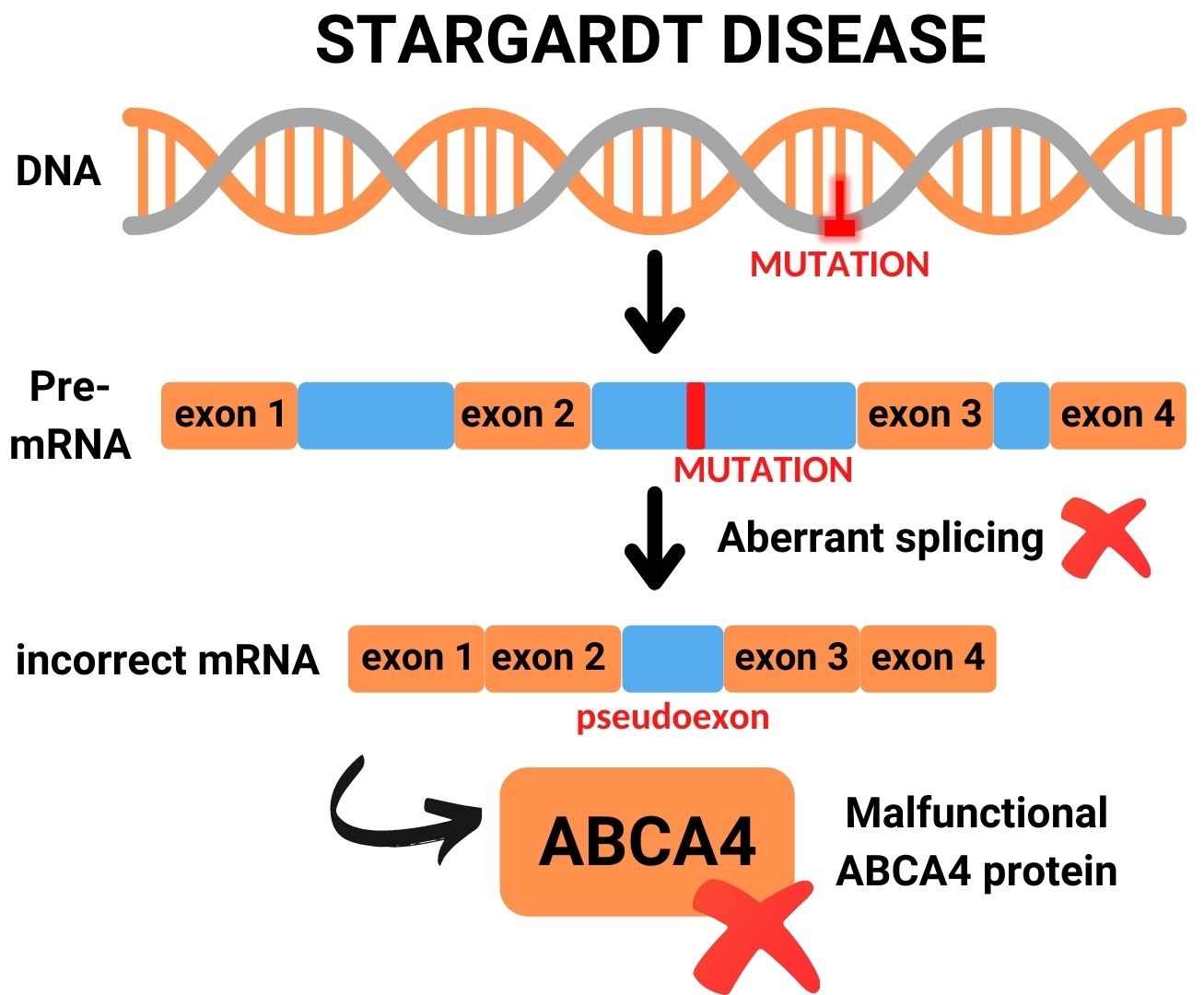
بیماری اشتارگارت یک مشکل مدیریت پسماند است، عامل ایجاد این بیماری نادر جهش در ژن ABCA4 می‌باشد، به این صورت که هم پدر و هم مادر باید حامل این ژن باشند تا به فرزند منتقل شود.

معمولاً در ازدواج‌های فامیلی این جهش مشهود هست. دو نوع جهش ABCA4 موجود است.
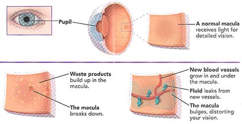
در این جهش‌ها سنتز ویتامین A مختل می‌شود و باعث تجمع پسامد در روی شبکیه شده و گیرنده‌های نوری سطح شبکیه را مختل می‌کند. ویتامین A یک عنصر مهم برای گیرنده‌های نوری سطح شبکیه چشم هستند.

Stargardt Disease: What It Is, Symptoms & Treatment

Stargardt disease is an inherited form of macular degeneration that first appears in childhood or adolescence. It is characterized by progressive vision loss beginning in the macula, the central part of the retina where light falls and visual acuity and color vision are greatest. 
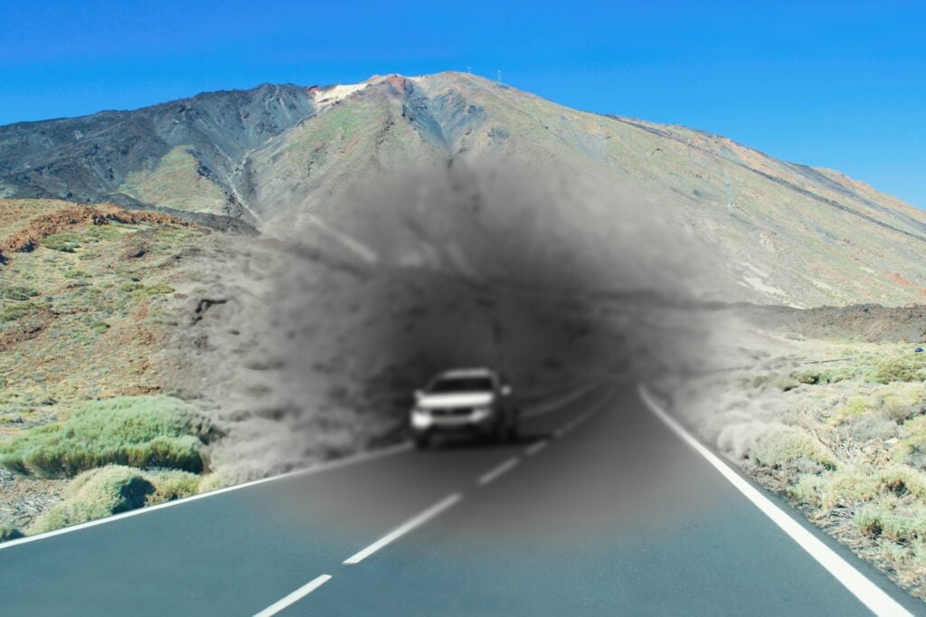
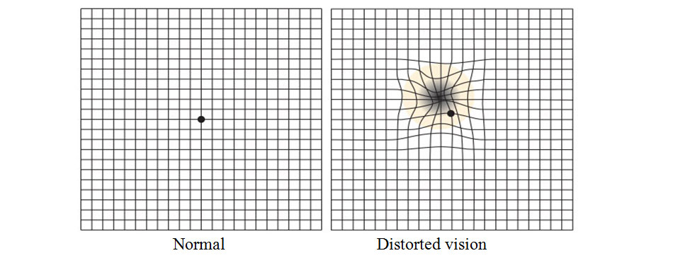
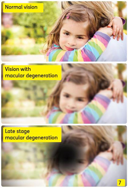
Symptoms include blurred or wavy vision, blind spots, impaired color vision, and difficulty seeing in low light situations. People with Stargardt disease are usually sensitive to glare.[2]

What are symptoms of Stargardts eye disease [3]

  • Someone may initially become aware of an issue with their center vision. It may be distorted, hazy, or have black regions. Side vision (peripheral vision) is frequently unaffected. Colorblindness is a condition in which some individuals have difficulty perceiving colors.
  • When moving between bright and dark environments, eyesight may take longer to adapt than normal.
  • For some patients, Stargardt illness advances slowly at first, then quickly accelerates and finally plateaus. Vision loss may accelerate at roughly 20/40 vision (meaning someone sees at 20 feet what a normal-seeing person sees at 40 feet).
  • While most persons with Stargardt illness eventually lose their central vision, many may have strong side vision for the remainder of their lives. eResearch by Navid Ajamin -- autumn 2024

What can be done if Stargardt disease is diagnosed? There is no cure for Stargardt disease, and there are no treatments.

What devices can help? Since the symptoms are underlying physiology of Stargardt disease are similar to those for other types of macular degeneration, people can usually benefit from the same devices as used for age-related macular degeneration (AMD). These help people retain independence in their homes, school, and jobs. Products include electronic magnifiers and devices that turn text into speech to read aloud mail, bills, books, and other printed materials. Freedom Scientific’s line of video magnifiers and screen magnification software can help.[2]

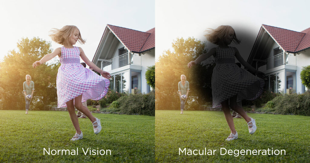
اشتارگات معمولا در کودکان، نوجوانان و بزرگسالان جوان ایجاد می شود. ممکن است شخصی ابتدا متوجه مشکلی در بینایی مرکزی خود شود. این مشکل معمولا تاری دید یا مشاهده نواحی تیره است. در این نوع بیماری چشمی دید جانبی یا محیطی معمولاً تحت تأثیر قرار نمی گیرد. اما برخی از افراد ممکن است در دیدن رنگ ها نیز مشکل داشته باشند. بیماری اشتارگات در برخی افراد ممکن است به کندی پیشرفت کند، سپس سرعت گیرد و به سرعت سطح بینایی را به میزان قابل توجهی کاهش دهد. با توجه به اینکه در بیماری اشتارگات از دست دادن بینایی می تواند به صورت ناگهانی سرعت خود را افزایش دهد، بنابراین در صورتی که با علائم اولیه مانند تاری دید یا مشاهده نواحی تیره مواجه شدید، باید سریعا به پزشک مراجعه کنید.

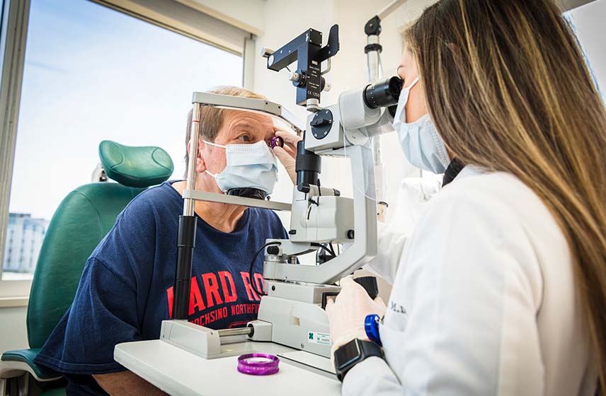
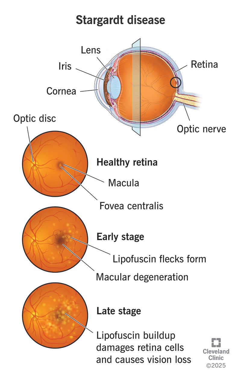
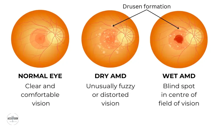
تغییر در بینایی مرکزی Central vision معمولاً منجر به تشخیص اولیه بیماری اشتارگات می شود. یک پزشک متخصص شبکیه چشم در حال معاینه شبکیه یک فرد مبتلا به بیماری اشتارگات ، لکه های زرد رنگ مشخصی را در RPE مشاهده می کند. لکه ها رسوبات لیپوفوسسین هستند که محصول جانبی فعالیت طبیعی سلول های شبکیه می باشند. با این حال، در این بیماری، لیپوفوسین به طور غیر طبیعی تجمع می یابد.

نکته: “توجه داشته باشید که پیشرفت از دست دادن بینایی در بیماری اشتارگات متغیر است. حدت بینایی (قابلیت تشخیص جزئیات و شکل) ممکن است در ابتدا به آرامی کاهش یابد، سپس شتاب بگیرد و دوباره یکنواخت شود. همچنین معمولاً مقداری دید محیطی در فرد مبتلا باقی خواهد ماند.”

? Can people with Stargardts drive

معمولاً بیماری اشتارگات از والدین منتقل می شود. در این بیماری، ژن‌های معیوب (ژن ABCA4) برای داشتن علائم باید از هر دو والدین منتقل شود. هر کودک ۲۵ درصد ممکن است دو نسخه ABCA4 (یک نسخه از هر والدین) را که برای ایجاد این بیماری لازم است، به ارث ببرد. فردی که این ژن را فقط از یکی از والدین دارد، ناقل بیماری اشتارگات خواهد بود، اما علائمی نخواهد داشت. البته سایر اشکال بیماری اشتارگات برای ایجاد علائم تنها به ژن یکی از والدین نیاز دارند، اما این موارد بسیار نادر هستند. برای تشخیص دقیق این بیماری چشم پزشک معمولا از آزمایشی به نام آنژیوگرافی فلورسین استفاده می کند. در این آزمایش یک رنگ به بازوی شما تزریق می شود. از رنگ هنگام گردش در رگ های خونی شبکیه عکس گرفته می شود. در افراد مبتلا به اشتارگات عکس ها ناحیه تیره ای را در بافت شبکیه نشان می دهند. این به چشم پزشک کمک می کند تا بیماری اشتارگات را تشخیص دهد. همچنین در حال حاضر آزمایش ژنتیک برای تشخیص دقیق نوع دژنراسیون ماکولا در دسترس است. این مطمئن ترین راه برای دانستن مبنای ژنتیکی بیماری شما است.

بهترین گزینه برای جلوگیری از ابتلای فرزندان به بیماری اشتارگات انجام آزمایش ژنتیک است که به والدین در تشخیص قطعی بیماری و احتمال خطر ابتلای فرزندان به این بیماری کمک می کند. البته تا به امروز متاسفانه هیچ درمانی برای این بیماری وجود نداشته است. اما با این وجود چندین آزمایش ژن درمانی و دارودرمانی در حال انجام است. در ادامه به برخی از نکاتی که به افراد مبتلا به بیماری اشتارگات کمک می کند.

خوشبختانه اشتارگات یک بیماری ژنتیکی و نادر است که اغلب در کودکان تشخیص داده می‌شود. این بیماری به علت اشکال در ساختارهای استخوانی و بافتی در بدن ایجاد می‌شود. افراد مبتلا به اشتارگات ممکن است دارای قد کوتاهی، مشکلات در مفاصل و اندام‌های حرکتی، اختلالات تنفسی، و مشکلات قلبی باشند. اشتارگات نیازمند مراقبت و مدیریت تخصصی پزشکی است. درمان این بیماری شامل جراحی‌ها، فیزیوتراپی، و مراقبت‌های پزشکی مخصوص می‌شود. ارتقاء کیفیت زندگی افراد مبتلا به اشتارگات از طریق تیم‌های درمانی و پشتیبانی اجتماعی انجام می‌شود تا به افراد این امکان داده شود تا با این بیماری مبارزه کنند و به حیات عادی نزدیک‌تر شوند.

درمان

سلول بنیادی

درمان قطعی برای بیماری اشتارگات در حال حاضر وجود ندارد، اما بیماری به وسیله ژن درمانی و سلول درمانی به وسیله سلول‌های بنیادی قابل کنترل است و احتمال بهبود بیماری با ضریب بیشتری بالا می‌رود، استفاده از سلول‌های بنیادی بستگی به زنده ماندن سلول‌های بنیادی در محیط شبکیه چشم دارد، گاهی نیاز است در مقاطع زمانی مختلف درمان تکرار شود.

ویتامین آ

دانشمندان با جایگزین کردن اتمهای هیدروژن با دوتریوم Deuterium در ویتامین A توانستند Alk-001 را تولید کنند. این محصول که به عنوان ویتامین A دوتره (Deuterated Vitamin A) شناخته می‌شود، اصطلاحاً پاکتر از شکل طبیعی ویتامین A می‌سوزد. دوتریوم (Deuterium)، شکل بی خطری از هیدروژن است که به‌طور طبیعی در بدن انسان تولید شده و غیر رادیواکتیو است. Alk-001 هم‌اکنون در محله سوم کارآزمایی بالینی در بیماری اشتارگارت می‌باشد. . نام علمی Alk-001 عبارت است از C20-D3- retinyl . .acetate

شبکیه مصنوعی

جدیدترین تکنولوژی برای درمان بیماران شبکیه چشم، استفاده از شبکیه مصنوعی یا شبکیه الکترونیکی است. این تکنولوژی در سال ۲۰۱۶ ابداع شد، هرچند کیفیت تصویر به‌دست آمده برای بیمارن چندان واضح نبود، اما پروژه شبکیه مصنوعی در حال ارتقا کیفیت است.

This disease is hereditary and therefore if there is a family history it is wise to be attentive, even though this does not mean that the disease is sure to manifest itself. Approximately 90% of cases are transmitted in an autosomal recessive manner, i.e. both parents must have the affected gene and this is often very hard to establish. In this case, the possibility of a boy or girl having the disease is 25% and it should be remembered that 10% of cases, with a family history, are of dominant inheritance.As it is a recessive gene, the family history of the disease may not be known or available. 
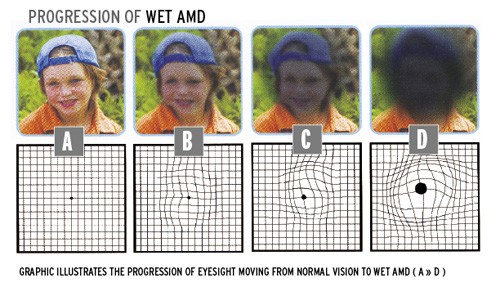
This is why it is necessary to pay special attention to the initial symptoms e.g. if children or adolescents find difficulty in reading or watching the television. At these ages, it is a good idea to explain the pathology to them so that they can be made aware of what will happen to them, can adapt to the situation and can lead a happy life.Stargardt’s disease causes out-of-focus vision that lacks sharpness. This makes it difficult to recognise faces and read both nearby and at a distance. As a result, colours with a similar shade (for example, red and green or blue and yellow) look alike.

A 40-year-old man experiencing decreased vision (visual acuity: 0.8) and dyschromatopsia in both eyes with Stargardt disease. A and B: The fundus photos of the right and left eyes respectively reveal the bull's eye maculopathy characterized by paracentral RPE depigmentation and atrophy, as well as pisiform, round, or dot-like yellow-white flecks. C and D: The red-free fundus images of the right and left eyes. E and F: OCT macula scans of the right and left eyes respectively, highlighting photoreceptor layer disorganization. (Courtesy of J. Khadamy) [5]

STARGARDT FINDINGS. (1A,1B) Fundus photography shows bilateral atrophic macular changes surrounded by diffuse pisciform flecks. (2A,2B) Fluorescein angiography reveals a dark choroid with hyperfluorescent pisciform flecks.[6]

A good knowledge of the disease helps sufferers to understand what is happening to them, adapt their lives to the new situation and take some recommended measures like using sunglasses with u/v protection and avoiding supplements that contain vitamin A.

In the field of research into treatments for this disease, science is progressing. The clinical trials and European projects in which the Barcelona Macula Foundation and the Institut de la Màcula participate in collaboration with leading international research centres are essential and lead to hope that the disease may be treatable in the future.[4]

Reference:

  1. fa.wikipedia.org/wiki/اشتارگات
  2. freedomscientific.com/visionloss/stargardt-disease
  3. lowvisionaids.org/stargardts-disease
  4. barcelonamaculafound.org/en/stargardts-disease-affects-young-people-who-must-face-up-to-a-new-way-of-life
  5. eyewiki.org/Stargardt_Disease/Fundus_Flavimaculatus
  6. aao.org/eyenet/article/diagnosis-management-of-stargardt-disease

See also: Stargardt macular dystrophy and therapeutic approaches

Age-related macular degeneration (AMD) is a medical condition which usually affects older adults and results in a loss of vision in the center of the visual field (the macula) because of damage to the retina. It occurs in “dry” and “wet” forms. It is a major cause of blindness and visual impairment in older adults (>50 years). Macular degeneration can make it difficult or impossible to read or recognize faces, although enough peripheral vision remains to allow other activities of daily life.[1]

Age-related macular degeneration (AMD) is an eye disease that can blur your central vision. It happens when aging causes damage to the macula — the part of the eye that controls sharp, straight-ahead vision. The macula is part of the retina (the light-sensitive tissue at the back of the eye).[9]

Dry macular degeneration, also known as atrophic or non-neovascular age-related macular degeneration (AMD), is a common eye condition that primarily affects the macula, the central part of the retina responsible for sharp, central vision. In dry AMD, there is a gradual breakdown or atrophy of the light-sensitive cells in the macula, particularly the retinal pigment epithelium (RPE) cells. These cells are crucial for supporting the health and function of the photoreceptor cells, including cones, in the macula.

The condition progresses slowly over time and can lead to symptoms such as blurred central vision, distorted vision (metamorphopsia), and difficulty seeing in low light. Unlike wet AMD, which involves abnormal blood vessel growth, dry AMD typically does not involve leaking blood vessels.

There are two main types of dry macular degeneration:

  1. Early and Intermediate Dry Macular Degeneration: In the early stages, small yellow deposits known as drusen may form beneath the retina. Drusen are accumulations of waste materials that can interfere with the normal functioning of the macula. At this stage, individuals may not experience significant vision loss, and the condition may be detected during a routine eye exam.
  2. Advanced Dry Macular Degeneration (Geographic Atrophy): Over time, some individuals with dry AMD may progress to an advanced stage characterized by the development of geographic atrophy. Geographic atrophy involves the loss of RPE cells and photoreceptor cells in discrete patches, leading to the formation of atrophic or “geographic” areas in the macula. This can result in a more significant and irreversible loss of central vision.[8]

Macular Degeneration - Symptoms and Detection
Symptoms of Dry AMD include the presence of drusens that begin to enlarge. Another symptom is blurry areas in central vision. A symptom of Wet AMD is seeing straight lines as wavy ones.

A visual acuity test can help an eye care professional determine if any central vision is lost. An Amsler grid may also be used to detect AMD. In this test you cover one eye and stare at a black dot that has patterns of straight lines. If these lines appear wavy or are missing then AMD could be present.[2]

Prevention

There is growing evidence that by improving your diet, you may also improve the health of your eyes. Research has suggested an association between macular degeneration and a high saturated fat diet.

There is also evidence that eating fresh fruits and dark green, leafy vegetables – foods rich in vitamins C and E, selenium, and carotenoids (including beta-carotene, lutein and zeaxanthin) – may delay or reduce the severity of AMD.

Image result for age related macular degeneration vision


How AMD can affect your sight?

AMD is progressive and it is also painless. While AMD may affect your central vision, most people still retain useful side (or peripheral) vision.
Key symptoms of AMD include:

  • Distortion, where straight lines may appear wavy or bent. For example, lines of tiles in the bathroom appear wavy.
  • Difficulty in reading or doing any other activity which requires fine vision.
  • Difficulty in distinguishing faces.
  • Dark patches or empty spaces, which appear in the centre of your vision.
  • The need for increased illumination, sensitivity to glare, decreased night vision and poor colour sensitivity.[6]

Foods to Enjoy eResearch by Navid Ajamin -- spring 2012

Eat the freshest and brightest fruits and vegetables. Pick the most colorful vegetables and fruits you can find - red, dark green, orange, or yellow. These foods play a key role in keeping your eyes healthy:

Carrots Corn Kiwi Pumpkin Yellow squash Zucchini squash Red grapes Green peas Cucumber Butternut squash Green bell pepper Celery Cantaloupe Sweet potatoes Dried apricots Tomato and tomato products Dark green leafy vegetables Spinach Kale Turnips Collard greens ...

Fish

Eating fatty fish such as salmon, tuna or mackerel two to three times per week can slow the progression of age-related macular degeneration (AMD) according to a study published in the British Journal of Ophthalmology.

This research further confirms earlier studies that suggested eating fish can help reduce the risk of getting AMD and demonstrates that some of those already affected by the disease can benefit as well.

Nuts

Nuts not only contain Omega-3 fatty acids, but also copper which can play a role in preventing age-related eye diseases. Even just a handful of nuts at two or three times a week can reduce your risk of AMD.

Foods to Avoid

A high-fat, high-cholesterol diet can lead to fatty plaque deposits in the macular vessels, which can hamper blood flow and increase the risk of AMD. A diet low in fat promotes good eye health. Skip foods and processed baked goods with high-fat content. In addition recent research has indicated that those consuming red meat (10 times a week or more) were at 47% higher risk for macular degeneration.

Vitamins and Supplements

The National Eye Institute’s Age-Related Eye Disease Study (AREDS) found that taking a specific high-dose formulation of antioxidants and zinc significantly reduces the risk of advanced AMD and its associated vision loss. Slowing AMD’s progression from the intermediate stage to the advanced stage will save the vision of many people.

People who should consider taking the combination of antioxidants plus zinc include those who are at high risk for developing advanced AMD. These people are defined as having either:

  1. Intermediate AMD in one or both eyes. Intermediate AMD is defined as the presence of either many medium-sized drusen or one or more large drusen.

  2. Advanced AMD in one eye, but not the other eye. Advanced AMD is defined as either a breakdown of light-sensitive cells and supporting tissue in the central retinal area (advanced dry form), or the development of abnormal and fragile blood vessels under the retina (wet form) that can leak fluid or bleed. Either of these forms of advanced AMD can cause vision loss. Ask you doctor if taking this special formulation is right for you and where you can obtain the specific formula in your country.[3]

Grapes May Help Prevent AMD

Can eating grapes slow or help prevent the onset of age-related macular degeneration (AMD), a debilitating condition affecting millions of elderly people worldwide? Results from a new study published in Free Radical Biology and Medicine suggest this might be the case. The antioxidant actions of grapes are believed to be responsible for these protective effects.[4]

Home Remedies and Lifestyle Changes [7]

The risk factors for macular degeneration are similar to those of heart disease and stroke. For this reason, lifestyle changes that benefit your heart may also benefit your vision. Lifestyle modifications to consider include:

  • Quitting smoking
  • Making dietary changes, such as limiting foods high in saturated fats (meat, butter, and cheese) and eating a heart-healthy diet full of whole grains, fruits, and vegetables
  • Maintaining weight, since obesity is also a risk factor of dry AMD
  • Managing blood pressure
  • Using sun protection, such as wearing wide-brimmed hats and sunglasses
  • Getting regular exercise (at least 30 minutes of physical activity every day)

As central vision declines in late-stage dry AMD, you can use low-vision tools, such as magnifying tools and handheld computers, to help with daily activities. Low-vision techniques, like using high-lumen light sources, reducing glare, and increasing contrast, can also help compensate for central vision loss.

Discussion. This systematic review and meta-analysis has shown that 8·7% of the worldwide population has age-related macular degeneration, and the projected number of people with the disease is around 196 million in 2020, increasing to 288 million in 2040.[5]

Reference:

  1. en.wikipedia.org/wiki/Macular_degeneration
  2. eyehealthweb.com/diseases/macular-degeneration
  3. amdalliance.org/information_prevention.html
  4. careyeasy.com/eye-care-news/grapes-may-help-prevent-amd
  5. sciencedirect.com/science/article/pii/S2214109X13701451#
  6. vi.ie/supporting-you/everyday-living/eye-conditions/age-related-macular-degeneration-amd
  7. verywellhealth.com/how-dry-amd-is-treated-5192080
  8. rvaf.com/diseases/amddry
  9. nei.nih.gov/learn-about-eye-health/eye-conditions-and-diseases/age-related-macular-degeneration

See also: How Age-Related Macular Degeneration Is Treated

لکهٔ زرد یا ماکولا (به انگلیسی: Macula) بخشی از شبکیه است که بیشترین حساسیت به نور را دارد و موجب دید مستقیم و واضح می‌شود. لکه زرد برای کارهای دقیق مثل خواندن و رانندگی لازم است.

این لکه با شکل بیضوی حاوی رنگدانه‌های زرد و در نزدیکی مرکزشبکیه است. فووه آ (fovea) که بیشترین تراکم سلول‌های مخروطی چشم را داراست در ماکولا قرار دارد. سلول‌های مخروطی گونه‌ای از سلول‌های گیرندهٔ نور هستند که به مغزتوانایی دیدن رنگ‌ها و جزئیات ظریف اشیا را می‌دهند و بیش‌تر در نور قوی تحریک می‌شوند. بنابراین، این بخش که در انتهای چشم و در راستای مردمک قرار دارد، در دقت و تیزبینی چشم نقش دارد. قطر لکهٔ زرد حدود ۲ میلی‌متر است.[1]

Image result for macula

یكی از مهم‌ترین بیماری‌های چشم، استحاله یا تخریب لكه زرد است كه پزشكان به آن «دژنراسیون ماكولا» می‌گویند. تخریب لكه زرد، شایع‌ترین علت كاهش بینایی ناشی ازبالا رفتن سن است. به عبارت دیگر، تخریب یا استحاله ماكولا، علت شایع افت دید در افراد بالای 55 سال است. از آنجایی كه این بیماری فقط دید مركزی را مختل می‌كند، به‌ندرت باعث نابینایی فرد می‌شود؛ با این حال می‌تواند خواندن، رانندگی‌كردن و انجام سایر فعالیت‌های روزمره‌ای كه نیاز به دید مركزی و دقیق دارند را بسیار مشكل یا غیرممكن كند.

تباهی لکه زرد می‌تواند سبب کاهش دید تدریجی یا ناگهانی شود. اگر خطوط مستقیم را موجدار می‌بینید، دیدتان تار است، یا لکه‌های تیره در مرکز دید خود می‌بینید احتمال دارد که دچار علائم اولیه دژنراسیون ماکولا شده باشید. در بسیاری موارد قبل از اینکه بیمار دچار علائم شود چشم پزشک علائم اولیه بیماری را در معاینه تشخیص می‌دهد. این کار معمولاً از طریق تست میدان بینایی صورت می‌گیرد. در دژنرسانس ماکولا دید مرکزی مختل شده و بیمار در مرکز دید خود لکه‌ای سیاه می‌بیند.[4]

دژنراسیون ماكولا بر دو نوع است: خشك (Dry) و تَر (Wet). نوع خشك شایعتر بوده و حدود ۹۰% بیماران به این نوع مبتلا می شوند. نوع تَر معمولا با كاهش دید شدیدتر و جدی تری همراه است.

Related image

زنان بیشتر به این بیماری مبتلا می شوند. اكثر موارد این بیماری با افزایش سن بوجود می آیند. این بیماری می تواند عارضه بعضی داروها نیز باشد. همچنین به نظر می رسد ارث نیز در ابتلا به این بیماری نقش داشته باشد.

علت دقیق این بیماری هنوز شناخته نشده است. نوع خشك ممكن است بر اثر پیر و نازك شدن بافت ماكولا، رسوب رنگدانه ها در ماكولا، و یا تركیبی از این دو ایجاد شود. در نوع تَر عروق خونی جدید در زیر شبكیه رشد كرده و خون و مایع از آنها نشت می كند. این نشت سبب مرگ سلولهای شبكیه شده و باعث ایجاد نقاط كور در دید مركزی می شود.
عواملی كه شانس ابتلا به بیماری را افزایش می دهد عبارتند از: سابقه خانوادگی، مصرف سیگار، فشار خون بالا، دوربینی و چاقی.

دانشمندان در تحقيقاتي جديد اظهار داشته اند سیگاریها چهار برابر بیشتر از کسانی که سیگار نمی کشند در معرض نابینایی حاصل از تخریب لکه زرد چشم قرار دارند.

تحقیقات جدید نشان می دهد: بیش از ۲۵ درصد تخریب لکه زرد و اختلالات بینایی به علت کشیدن سیگار در زمان حال یا گذشته است و درمان موثری برای آن وجود ندارد.

افراد سیگاری در مقابله با افرادی که هرگز سیگار نکشیده اند، چهار برابر خطر ابتلا به عارضه تنفسی دارند و از دیدگاه زیست شناختی، شبکیه به علت تخریب اکسیداتیو حاصل از کشیدن سیگار، صدمه می بیند. eResearch by Navid Ajamin -- autumn 2011

این تخریب توسط رادیکال های آزاد که مولکول های ناپایدار سمی هستند، صورت می گیرد و به حدی شدید است که عوامل حفاظتی مانند وجود مواد ضد اکسید در رژیم غذایی در بهبود آن موثر نیست.

همچنین تخریب لکه زرد باعث از بین رفتن دید مرکزی شده و منجر به ناتوانی در به کارگیری شبکیه جهت فعالیت هایی مانند خواندن، دوختن و حتی رانندگی می شود.

Related image

What is the macula?

The macula is part of the retina at the back of the eye. It is only about 5mm across but is responsible for all of our central vision, most of our colour vision and the fine detail of what we see.

The macula has a very high concentration of photoreceptor cells that detect light and send signals to the brain, which interprets them as images. The rest of the retina processes our peripheral (side) vision. Macular disease causes loss of central vision.[7]

Macular degenerations are retinal diseases of genetic origin that cause an early degeneration of the macula. It affects adults, young people or even children, it all depends on the severity of the damage caused by this genetic anomaly.

The macula deteriorates and collapses affecting vision and causing a loss of vision that may be important and reach legal blindness.

It is a hereditary disease and, therefore, different from age-related macular degeneration (AMD), which is caused by the aging of the body.

This disorder can manifest itself in various forms such as Stargardt’s disease, Best’s vitelliform macular dystrophy and juvenile retinoschisis.

Age-Related Macular Degeneration

What is juvenile macular degeneration?

This disease consists of the deterioration of the macula of the eye. The macula is the part of the retina responsible for the central vision and allows us to see fine and small details clearly.

The macula is much more sensitive to detail than the rest of the retina (peripheral retina, allows lateral vision) and, therefore, if it is affected, the quality of vision is reduced.

When the macula is affected, the central vision is lost and causes unfocused vision and lack of sharpness.

Who can develop this disorder?

It is a hereditary disease, so it is transmitted from parents to children.

  • In the case of Stargardt’s disease, being recessive, it is only inherited if the two parents are affected by the gene.
  • In the case of Best’s vitelliform macular dystrophy, it can be suffered even if only one of the parents is affected.
  • Juvenile retinoschisis is a disease linked to the X chromosome and, therefore, affects males, but it is transmitted from mother to child.

What are the symptoms of juvenile macular degeneration?

Some of the symptoms of juvenile macular degeneration can be:

  • Problems in the central vision that make it difficult to perform activities such as reading small print or traffic signs or threading a needle.
  • Blurred, distorted vision with dark areas.
  • Photophobia (sensitivity to light)
  • Display of black and gray spots.
  • Yellowish flecks around the macula.
  • Impairment of color perception.

Symptoms appear during childhood and / or adolescence and can affect one or both eyes. Lateral vision is not usually affected.

In some cases, patients can maintain useful vision for decades (Best’s vitelliform macular dystrophy) and, in other cases, vision can be lost to legal blindness (Stargardt’s disease).

How is this disorder diagnosed?

Normally, in Stargardt’s disease small yellow spots are visualized in the macula that may extend outwards.

On the other hand, patients with Best’s vitelliform macular dystrophy have a yellowish cyst under the macula, like an egg yolk that, over time, breaks down and looks like a scrambled egg.

Patients with juvenile retinoschisis have a retina that separates into two layers, with spaces between the layers that can be filled with cysts and blood vessels, which can break and cause hemorrhages. This tearing of the layers of the retina can cause a retinal detachment.

What tests are done to diagnose macular degeneration?

  • The ophthalmologist will perform an ocular dilatation test in order to observe the retina and be able to confirm that there are specific signs related to this disorder.
  • The ophthalmologist can also perform a fluorescein angiogram to confirm the diagnosis and, if needed, recommend an ERG (electroretinography) to measure the electrical activity of the retina.
  • More recent tests such as optical coherence tomography (OCT) and autofluorescent retinography provide complementary information of great importance, especially to objectify the progression of the disease.[6]

Age-related macular degeneration in an 82-year-old white male. Colour photographs of the right and left eyes (a, b) show numerous drusen in the posterior pole and fundus tessellation in the macula. Spectral domain optical coherence tomography of the right and left eyes (c, d) shows bilateral drusen. In the left eye (d), there is a large irregular pigment epithelial detachment with hyperreflective contents and adjacent subretinal fluid, consistent with type 1 neovascularization. The choroid is relatively thin in both eyes. Fundus autofluorescence of the right and left eyes (e, f ). There is a hypoautofluorescent geographic focus at the fovea of the left eye (f ) with a curvilinear hyperautofluorescent zone inferior to the fovea, hinting at a legacy of chronic subretinal fluid. Late transit and recirculation fluorescein angiography of the left eye (g) shows irregular hyperfluorescence at the inferior border of the fovea with late, poorly defined staining. There is also extensive staining of drusen. Indocyanine green angiography of the right eye (g) shows no evidence of neovascularization. Indocyanine green angiography of the left eye (h) shows a hyperfluorescent 'plaque' indicating the full extent of the type 1 neovascular lesion.[5]

Treatment for macular degeneration

To this day, there is no treatment to prevent or stop this disorder.

It is important to wear sunglasses with ultraviolet protection, it is recommended to avoid supplements with vitamin A, and it is advisable to perform mobility exercises for patients to adapt to vision loss.[6]

پژوهش ها نشان می دهد: وقتی سیگار ترک شود چشم ها در برابر صدمات بیشتر مصون می مانند و خطر ابتلا به این عارضه در این افراد نسبت به کسانی که تا به حال سیگار نکشیده اند کمی افزایش می یابد.

بر اساس نتیجه این بررسی افراد سیگاری با ترک سیگار می توانند احتمال از بین رفتن سلول های شبکیه چشم خود را کاهش دهند.

بر این اساس ، احتمال ابتلا به این عارضه در افرادی که ۲۰ سال پیش سیگار را ترک کرده اند تا حد یک فرد غیرسیگاری کاهش می یابد.

عارضه تحلیل لکه زرد چشم ، شایع ترین علت نابینایی افراد سالمند است.

ماکولا یا لکه زرد در مرکز شبکیه قرار دارد. شبکیه هم، لایه حساس به نور است که در قسمت خلفی کره چشم قرار دارد. نور روی ماکولا متمرکز می‌شود. در ماکولا میلیون‌ها سلول مخصوص بینایی وجود دارد.

ماکولا یا لکه زرد نور را دریافت و به علائم عصبی تبدیل کرده، به مغز منتقل می‌کند. این روند دید مرکزی نامیده می‌شود.

در واقع دید مرکزی، توانایی رانندگی کردن، خواندن و انجام فعالیت‌هایی که نیاز به دید مستقیم، دقیق و ظریف دارد را فراهم می‌آورد. اگر ماکولا صدمه ببیند، قسمت مرکزی تصویر دیده شده تار می‌شود و تصویر در اطراف منطقه تار شده وضوح بیشتری دارد.

پس اهمیت لکه زرد در این است که در قسمتی از شبکیه که نزدیک عصب بینایی است، سلول‌های اصلی بینایی که مخصوص دید در روز، دید دقیق و دید رنگی هستند، تجمع پیدا کرده‌اند. در واقع انسان به وسیله لکه زرد بهترین و دقیق‌ترین دید را به دست می‌آورد.[2]

روش های درمان دژنراسیون ماکولا

این بیماری درمان قطعی ندارد ولی درمان هایی برای جلوگیری از پیشرفت بیمار و یا حتی بهبود دید وجود دارد. درمان استاندارد این بیماری دربرگیرنده تزریق یک ضد فاکتور رشد اندوتلیوم عروقی است که از پیشروی بیشتر رگهای خونی جلوگیری می کند.

ویتامین ها

در حال حاضر به نظر می رسد بهترین راه برای محافظت از چشم ها از تشدید زودرس ماکولا، داشتن یک رژیم سالم، استفاده از عینک های آفتابی است که چشم شما را از اشعه های مضر UV و اشعه ماوراء بنفش (HEV) محافظت می کند.

ویتامین‌ های آنتی اکسیدان نظیر ویتامین A و ویتامین‌ های C و E ممکن است ماکولا را در برابر تخریب محافظت کنند. مشخص شده است که مصرف ویتامین‌های C و E، ویتامین A و روی، خطر پیشرفت بیماری را در بعضی بیماران تا ۲۸٪ کاهش می‌دهد. از این‌رو به بیماران در خطر ابتلا به نوع پیشرفته توصیه می شود آنتی‌اکسیدان و روی مصرف کنند.

جراحی کاشت لنز تلسکوپی

برای درمان دژنراسیون ماکولا خشک پیشرفته در هر دو چشم، یک گزینه برای بهبود دید ممکن است عمل جراحی کاشت لنز تلسکوپی در یک چشم باشد. لنز تلسکوپی، که شبیه یک لوله پلاستیکی کوچک است، با لنزهای بزرگ برای میدان دید مجهز شده است. ایمپلنت لنز تلسکوپی ممکن است بینایی دور و نزدیک را بهبود بخشد،و همچنین می تواندبرای کمک در شناسایی نشانه های خیابانی مفید باشد.

درمان دارویی با ویزودین (Visudyne)

ویزودین اولین دارویی است که برای نوع تَر بکار می رود. در این روش درمانی، دارو به دست بیمار تزریق شده و سپس با استفاده از یک لیزر غیر حرارتی فعال می شود. فعال شدن دارو سبب ایجاد واکنشی شیمیایی می شود که عروق خونی غیر طبیعی را از بین می برد. از هر 6 بیمار درمان شده یک نفر دید بهتری پیدا می کند و این دو برابر بیمارانی است که از این دارو استفاده نمی کنند.

فیلتراسیون خونی (Blood Filtration)

در این روش که حدود 2 دهه پیش توسط ژاپنی‌ها ابداع شد جریان متفاوت خون در غشاء سبب کاهش مقدار بعضی از پروتئینها و اسیدهای چرب که مقدارشان زیاد است و ممکن است مضر باشند، می‌شود. این تکنیک در بیماریهای مختلفی بکار گرفته شده است ولی نوع جدیدی از آن بنام رئوفرزیس (Rheopheresis) برای درمان نوع خشک AMD تست شده است. رئوفرزیس هنوز توسط سازمان غذا و داروی آمریکا (FDA) تأیید نشده است ولی بصورت تجارتی در کانادا و اروپا در دسترس است.[3]

فوتوکوآگولاسیون با لیزر (Laser Photocoagulation) با تخریب و عایق بندی عروق خونی جدید و جلوگیری از نشت خون و مایع باعث بهبود بیماران مبتلا به نوع تَر می‌شود. در این روش جای لیزر مانند زخمی بر روی شبکیه باقی می‌ماند و باعث به وجود آمدن نقاط کور در دید بیمار می‌شود.[4]

Reference:

  1. fa.wikipedia.org
  2. smokefree.ir
  3. retinclinic.com
  4. wikipedia.org
  5. researchgate.net/figure
  6. icrcat.com/en/eye-conditions/juvenile-macular-degeneration-icr
  7. macularsociety.org/what-macula
  8. fvhospital.com/learn-more/age-related-macular-degeneration

Smoking is prevalent among teenagers now. All that is written on the cigarette is that cigarette smoking is injurious to health. What is not mentioned is that it is very harmful for human sight.

Age-related macular degeneration (AMD) is one of the main socioeconomical health issues worldwide. AMD has a multifactorial etiology with a variety of risk factors. Smoking is the most important modifiable risk factor for AMD development and progression. The present review summarizes the epidemiological studies evaluating the association between smoking and AMD, the mechanisms through which smoking induces damage to the chorioretinal tissues, and the relevance of advising patients to quit smoking for their visual health.

Image result for smoking and macular degeneration

There is growing evidence of the effects of smoking on vision. And these effects can be very significant. Cigarette smoking enhances oxidants which are chemical byproducts in our body. Oxidants can damage cells, including those in the eyes. The links between smoking and eye diseases is strong, including cataracts and age related macular degeneration.

Believe it, smoking increases the chance of your getting blind by up to four times, due to age-related macular degeneration (AMD). AMD is irreversible and is a progressive eye condition in which the central part of the retina (macula) is damaged.

Causes of Macular Degeneration

Once someone has been diagnosed with age related macular degeneration (AMD) or any disease for that matter, the mind starts asking questions like, how did I get this condition, what caused it or perhaps, could I have done something to prevent it?

There are many different factors that contribute to a person developing macular degeneration – some that can be controlled and some that cannot. eResearch by Navid Ajamin -- spring 2011

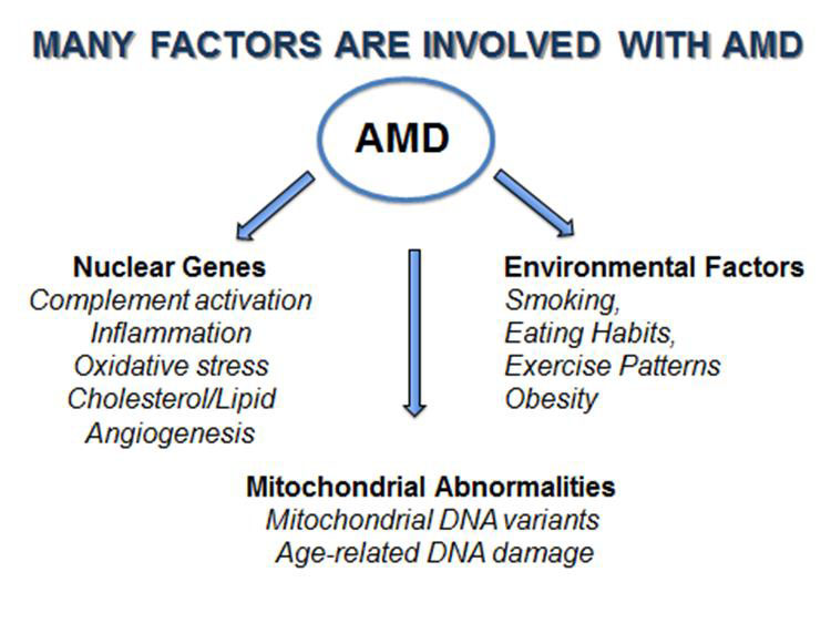
Here are the most common causes and risk factors:

1. Age
Macular degeneration is the number one cause of vision loss in the senior population. The older a person gets, the higher their risk of developing AMD. According to the National Eye Institute, ”a large study found that people in middle-age have about a 2 percent risk of getting AMD, but this risk increased to nearly 30 percent in those over age 75.”

2. Gender
Women are more likely to develop age related macular degeneration than men.

3. Race
It occurs in all races but it is more common in Caucasians.

4. Iris Color
The research shows that there is less pigment in blue eyes, and green eyes for that matter, than there is in brown eyes, so more light is able to penetrate blue eyes. This makes lighter eyes more sensitive to light and is what makes people with blue eyes more likely to have age-related macular degeneration. Macular degeneration is caused when the light sensitive cells in the eyes start to die, which can eventually result in blindness.

5. Heredity
My husband’s father has wet macular degeneration as well as did many of my husband’s aunts and uncles. The lifetime risk of developing late-stage macular degeneration is 50% for people who have a relative with macular degeneration versus 12% for people who’s relatives do not have macular degeneration (4x the risk).

6. Smoking
Smoking is the single most controllable risk factor that contributes to the development of macular degeneration. Every cigarette that is smoked causes damage to one’s vision. Smoking causes vasoconstriction or narrowing of the blood vessels which reduces the blood supply to the eyes. Smoking also creates free radicals which causes cellular damage while decreasing ones levels of antioxidants.

7. Diet Low in Dark Leafy Greens
A Harvard study done by Dr. Johanna Seddon in 1994 concluded that those who ate at least 5 servings of dark leafy greens per week had a 43% lower risk of developing AMD than those who ate small amounts or none at all. These lutein rich greens include kale, collard greens, spinach, Swiss chard, parsley, mustard greens, romaine lettuce and beet greens.

8. Diet Low in Omega 3 Fatty Acids
Thanks again to Dr. Seddon and her researchers at Harvard University, they discovered that people with diets of a 3:1 ratio of omega-6 to omega-3 fatty acids have less macular degeneration. Almost all of our processed foods and vegetable oils contain omega-6 which means that the average American gets way to much omega-6. Because omega 6 competes with omega 3, if we have too much omega-6 then the omega-3 we do take isn’t utilized properly.

9. High Blood Pressure
Dr. Michael A. Samuel the author of Macular Degeneration: A Complete Guide for Patients and Their Families writes “If you have high blood pressure that is fairly well controlled, you have double the risk of wet AMD compared to someone who does not have hypertension at all. If you have uncontrolled high blood pressure that goes above 160/90 you are three times more likely to develop wet AMD.” (Ophthalmology 2003;110: 636-643).

10. Exercise
Those with an active lifestyle were 70% less likely to develop macular degeneration than those who did little or no exercise according to researchers from the University of Wisconsin. The Wisconsin study that began in 1988, tracked almost 4,000 men and women (between the ages of 43-86) over a 15 year period by conducting regular eye tests and recording levels of exercise.

After accounting for other risk factors such as age, sex, history of arthritis, systolic blood pressure, body mass index, smoking, and education, those with a baseline active lifestyle of walking three times or more a week, were 70% less likely to develop macular degeneration than those who did little exercise.

Reference:

  • blurtit.com
  • pmc.ncbi.nlm.nih.gov/articles/PMC3866712
  • enhancedvision.com/low-vision-info/eye-conditions/causes-of-macular-degeneration.html

Healthy habits can lower the risk of age-related macular degeneration. If there is a family history of AMD, you should take necessary precautions like consulting an ophthalmologist. As the disease mostly affects adults above 50, they should make periodical visit to the ophthalmologist. Taking nutritional supplements after consulting a doctor can be very helpful for middle-aged people and those with family history of AMD.

The first symptoms of AMD include:

  • Reduced or blurry vision at the center in one or both eyes
  • Visual distortion – Lines appear wavy
  • Dark spots in the center
  • Colors can fade in a few cases
  • Letters might disappear suddenly when reading
  • Difficulty in adapting to dimly lit places


Quitting smoking, or never starting, is an important way to prevent AMD. Having a healthy lifestyle and lowering cholesterol can help lower your risk for AMD and also help prevent the dry form of the disease from progressing to the wet form, which can cause permanent vision loss.

Researches have already established a direct relation between smoking and AMD. Lutein, an antioxidant, plays a major role in protecting the retina from ultraviolet rays. Tobacco hampers the absorption of lutein. By stopping to smoke, you are not only saving your eyes but also avoid the risk of several other diseases.


People with high blood pressure are more prone to AMD. Regular check up of blood pressure and treating it is very essential for avoiding AMD.

Pollution has damaged the natural protection against sun’s rays. So it is wise to wear sun glasses for protection from ultraviolet rays.

A high fat diet is an invitation for AMD. A diet high in fruits and vegetables and antioxidants can prevent the disease to an extent. Including green vegetables and fish also reduces the risk.

Regular exercise and maintaining an optimum weight are also crucial for preventing AMD.

Check Your Vision Daily To Detect AMD

Amsler Grid can be used to check your vision at home. It is quite similar to a graph paper and has a dark dot in the center. By using the Amsler Grid, you can detect early signs of AMD. Missing area, blurring, wavy or broken links while watching Amsler Grid can alert you against AMD. Early detection of AMD is very vital for controlling it.


An Amsler Grid can be easily printed or you can watch it in your computer screen.

  1. While looking at Amsler Grid, make sure that you wear your glasses if you have one.

  2. Sit or stand about 15 inches away from the grid.

  3. Look at the dark dot in the middle.

  4. While focusing on the dark dot, you should be able to clearly see the lines.

  5. If you see any missing lines or if they are blurred or wavy, immediately contact an ophthalmologist.

  6. If there is no problem when you watched the grid with both eyes, repeat the exercise by closing one eye at a time. This is because AMD can affect one eye but the other eye makes up for it.

Types of Age-Related Macular Degeneration

What Causes Macular Degeneration?

Age-related macular degeneration (AMD) can rob people or their central vision, impacting their ability to read and drive. Fortunately, over the past 10 years research has uncovered some of the clues to what may be causing the disease, and this has helped shape efforts to prevent and treat AMD. eResearch by Navid Ajamin -- winter 2010

Early-Stage AMD

All AMD patients start out with early-stage (early) AMD, which often shows no noticeable symptoms. Eye doctors detect little white spots in the retina called drusen that can occur with advancing age. The retinal cells become less efficient at performing “housekeeping” tasks and small “garbage” deposits develop. The causes of early AMD are thought to involve oxidative stress and inflammation.

Oxidative stress is a disturbance in the balance between the production of very reactive oxygen-containing molecules that can adversely interact with other molecules inside our cells, and our body’s ability to neutralize these molecules. It can be caused by bright light, a poor diet with not enough antioxidants, and too much iron in the retina. The resulting inflammation can contribute to a number of age-related diseases, including age-related macular degeneration. Antioxidants are molecules present in cells that can prevent these harmful reactions.

People who inherit certain genes in the “complement cascade,” which is part of the immune system, have a higher risk for AMD, probably due to inflammatory damage to the retina.

Patients with early AMD may maintain good vision for their entire lives, or they may progress to late AMD. Changes in vision can be monitored at home with an Amsler grid or the ForeseeHome Monitor®.

Related image

Late-Stage AMD

Late-stage (late) AMD comes in two forms: wet AMD, or the dry AMD condition known as geographic atrophy. Wet AMD is always considered a late stage of AMD. In this condition, abnormal blood vessels sometimes grow behind the macula, the central part of the retina. Fluid leaks and vision is distorted.

In advanced dry AMD, there are regions of the retina where cells waste away and die (atrophy). Sometimes these regions of atrophy look like a map to the doctor who is examining the retina, hence the term geographic atrophy.

Geographic atrophy is caused by the death of light-sensitive cells known as photoreceptors, and their support cells known as retinal pigment epithelium cells or RPE. The area of atrophy usually expands slowly over time until it involves the entire central retina (macula). This causes a blind spot in the center of the visual field.

Also, if another part of the retina, called Bruch’s membrane, is damaged, new, abnormal blood vessels can invade the retina in a type of healing response gone wrong. These blood vessels leak blood and fluid into the retina, causing it to become wet. This fluid in the retina immediately disrupts vision and, over time, can lead to damaged retinal scar tissue. The Age-Related Disease Studies found that getting certain vitaminsexternal icon and minerals every day may slow the progression of the disease from the early or middle stages to the later stages. Specifically, combinations of the following vitamins can reduce risk of late AMD by 25%:

  • Vitamin C
  • Vitamin E
  • Beta-carotene
  • Zinc
  • Copper

Green, leafy vegetables have large amounts of many of these vitamins.

If AMD progresses to later stages, your eye doctor may use other therapies, such as injections and laser treatment.

If vision loss does occur, there are vision rehabilitation services and devices to help you use your remaining vision as well as you can. Speak to your eye doctor about new technologiesexternal icon such as magnifiers and telescopic glasses, and ask about resources for low vision in your area.

Ways to prevent age-related macular degeneration:

1. Quit smoking

"Rules 1, 2 and 3 are stop smoking," says Rosenthal. Smoking may double a person's risk of developing AMD. And the habit exposes you to dangerous free radicals, unstable molecules that can cause cellular damage and keep nutrients from reaching the retina. The sooner you can quit, the better.

2. Know your family history

People with a first-degree relative affected by AMD have a much greater risk of developing it, too. Those with family ties to the disease should be vigilant for potential symptoms that include difficulty recognizing faces, struggling to adapt to low light and seeing straight lines that appear wavy.

3. Eat leafy greens

Load up your plate with spinach, kale and Swiss chard, among other green veggies. "They have a lot of antioxidant vitamins," Rosenthal says. Those nutrients help protect against cellular damage from free radicals, which can contribute to eye disease.

4. Take supplements

Patients with deficient diets might consider multivitamins. And people at risk of advanced AMD should ask their doctor about a specialized blend of supplements known as AREDS. The macular degeneration vitamins are "not a treatment or cure but can decrease your risk of getting the more severe forms of AMD," Rosenthal says.

5. Wear sunglasses

Such eyewear offers protection from UV and blue light that can cause retinal damage from repeat exposure (the American Macular Degeneration Foundation recommends wearing a pair with a "UV 400" label). "Lifetime use is probably helpful," Rosenthal says.

6. Maintain a healthy blood pressure and weight

Poor blood circulation due to hypertension can also restrict blood flow to the eyes, thus contributing to AMD. Losing weight is a proven way to lower blood pressure; even small gains help — especially if you already have hypertension.

7. Test yourself with an Amsler grid

The tool that helps doctors detect vision problems related to macular damage can be used at home. If, after staring at the paper grid, you notice that the central part of your vision in one eye has become darker or the grid lines are wavy, call your doctor. Keep your Amsler grid in a place that reminds you to check it daily.

Reference:

  • cl66.org
  • brightfocus.org/macular/causes
  • cdc.gov/visionhealth/resources/features/macular-degeneration.html
  • michiganmedicine.org/health-lab/7-healthy-habits-help-prevent-macular-degeneration
  • myassuredhomenursing.com/age-related-macular-degeneration-symptoms-prevention
nicetoview.blogfa.com
عینک eyewear وبلاگ تخصصی عینک شامل مجموعه مطالب پزشکی است که اطلاعات مفیدی در رابطه با عینک , چشم، لنز، سلامتی چشم و راه های پیشگیری از بیماریهای چشمی، کنترل و درمان آن را در اختیار شما کاربر محترم می گزارد.

Always Be Healthy
همیشه سلامت باشید

Navid Ajamin نوید عجمین
eMail: navid.aj@outlook.com
موضوعات وب
پیوندها

unicef for every child

شمارنده