Preeclampsia and eclampsia are complications of pregnancy. The nurse plays a vital role in helping detect these conditions. Therefore, it’s important to know how to detect this condition in a pregnant patient.
The hormonal changes associated with pregnancy can impact a variety of things, including vision. In some cases, pregnant women may experience blurred vision as a result of high blood pressure. If vision loss is significant, this could be a sign of a serious health issue called preeclampsia. Typically occurring late in pregnancy, this condition can put both mother and child at serious risk if not treated. If you are pregnant and experiencing any significant vision problems, consult with your doctor immediately.
Blurred vision is the most common visual complaint. Focal or generalized arteriolar narrowing is the most common ocular finding in preeclampsia/eclampsia syndrome. Other ocular manifestations include photopsia, visual field defects, sudden inability to focus, and in severe cases, complete blindness.
Causes of Blurred or Distorted Vision
The preeclampsia/eclampsia syndrome is a multisystem disorder that can include cardiovascular changes, hematologic abnormalities, hepatic and renal impairment, and neurologic or cerebral manifestations. It also can affect the eye and visual pathways. Visual symptoms concern up to 25% of patients with severe preeclampsia and 50% of patients with eclampsia. This review discusses the ophthalmic complications of preeclampsia/eclampsia with focus on the hypertensive retinopathy, exudative retinal detachment and cortical blindness.
How common is preeclampsia?
Preeclampsia is a condition unique to pregnancy that complicates between 5% and 8% of all births in the United States. It’s also the cause of about 15% of premature deliveries (delivery before 37 weeks of pregnancy) in the U.S.
Preeclampsia is a serious medical condition that can occur about midway through pregnancy (after 20 weeks). People with preeclampsia experience high blood pressure, protein in their pee, swelling, headaches and blurred vision. But you may have no symptoms.
Treatment is necessary to avoid life-threatening complications. It typically goes away after childbirth.
Preeclampsia is a serious blood pressure condition that develops during pregnancy. People with preeclampsia often have high blood pressure (hypertension) and high levels of protein in their urine (proteinuria). Preeclampsia usually develops after the 20th week of pregnancy.
Preeclampsia can also affect other organs in your body and cause kidney and liver damage, brain injury and other serious side effects. It’s dangerous for both you and the developing fetus. Because of these risks, your healthcare provider will need to monitor your pregnancy closely and recommend treatment right away.
Preeclampsia Vision Changes
Preeclampsia is a hypertensive disorder affecting pregnant women, typically occurring after the 20th week of gestation.
In modern days, preeclampsia remains a leading cause of maternal and perinatal morbidity and mortality worldwide.
The most common symptoms include high blood pressure (hypertension) normally occurring in conjunction with proteinuria (presence of protein in the urine), signs of organ dysfunction, and preeclampsia vision changes.
The extended list of symptoms to look out for includes:
High blood pressure
Vision changes and disturbances
Proteinuria (presence of protein in the urine)
Excessive face & body swelling (edema)
Persistent and severe headaches
Pain or tenderness in the upper right side of the abdomen, just below the ribs
Pain or tenderness in the shoulder
Reduction in urine output (kidney dysfunction)
Severe nausea and vomiting in the second half of pregnancy
Shortness of breath
Another one of the prominent symptoms of preeclampsia is visual disturbances. They often occur during pregnancy and may persist postpartum.
The rise in blood pressure occurring with the condition affects organ systems, including the eyes. Which contributes to a range of visual difficulties. The fluctuations in vision can be alarming and significantly impact a woman's daily life, adding to the already substantial burden of this condition.
Preeclampsia vision changes commonly include blurry vision, light sensitivity (photophobia), and visual disturbances like seeingflashing lightsor floaters.
Preeclampsia vision changes may indicate potential severe complications.
Eye problems are way easier to detect than high blood pressure. So they are quite often the reason a pregnant woman or new mom gets the diagnosis and receives timely medical care.
Blurry vision
The vascular changes and low blood flow to the eyes affect visual function. Blurry vision may occur as a result of changes in the cornea, lens, or retina, leading to a decrease in visual acuity and sharpness. Fluid retention and eye swelling may contribute to blurriness.
Photophobia
Photophobia, as a preeclampsia symptom, makes individuals highly sensitive to light. Thus causing discomfort and a strong aversion to bright light sources. It can further lead to eye strain, headaches, and visual disturbances, adding to the burden of preeclampsia vision changes.
Preeclampsia Flashes
Flashes of light are another ocular discomfort we commonly associate with preeclampsia vision changes. These flashes, often described as brief, bright flickers or streaks of light, can appear suddenly and sporadically in a woman's visual field. Their occurrence is a result of abnormal retinal stimulation, due to vascular alterations.
Preeclampsia Floaters
Preeclampsia floaters are dark spots or specks that appear to "float" in a person's visual field. The causes are tiny protein or cell aggregations in the vitreous humor (the gel-like substance that fills the eye). They may appear as small dots or cobweb-like shapes, often moving with eye movements. Preeclampsia floaters are indicative of abnormal blood flow in the retinal blood vessels.
We protectour skin with sunscreen, but what about our eyes?
Most of us are aware of the dangerous effects ultraviolet (UV) rays have on our skin, but few of us realize the danger imposed on our eyes.
UV radiation, whether from natural sunlight or artificial UV rays, can damage the eye's surface tissues as well as the cornea and lens. UV radiation can burn the front surface of the eye, much like a sunburn on the skin.
UV radiation consists of invisible rays from the sun.
There are three types of UV radiation: UVA, UVB and UVC.
UVC rays do not pose any threat, as they are absorbed by the ozone layer. However, exposure to UVA and UVB rays can have adverse effects on your eyes and vision. Short- and long-term exposure to these dangerous rays can cause significant damage damage. It is important to note that UV radiation can also be given off by artificial sources like welding machines, tanning beds and lasers.
Two types of harmful light rays come from the sun:
ultraviolet A radiation (UVA), and ultraviolet B radiation (UVB).
UVA radiation can cause photoaging, or premature aging of the skin, resulting in wrinkles, uneven pigmentation, and texture changes.
UVB radiation is the main cause of sunburn.
Short-Term Effects of UV Radiation
If you are exposed, unprotected, to excessive amounts of UV radiation over a short period of time, you are likely to experience an effect called photokeratitis. Photokeratitis is an inflammation of the cornea caused by a brief exposure to UV radiation, usually when combined with cold wind and snow. Like a "sunburn of the eye", it may be painful and may create symptoms includingred eyes, a foreign body sensation or gritty feeling in the eyes, extreme sensitivity to light and excessive tearing. Fortunately, this is usually temporary and rarely causes permanent damage to the eyes.
Long-Term Effects of UV Radiation
Long-term exposure to UV radiation can be more serious. Scientific studies and research growing out of the U.S. space program have shown that exposure to small amounts of UV radiation over a period of many years may increase the chance of developing a cataract, and may cause damage to the retina, the nerve-rich lining of the eye that is used for seeing. This damage to the retina is usually not reversible. Cumulative damage of repeated exposure may contribute to chronic eye disease, as well as increase the risk of developing skin cancer around the eyelids. Long-term exposure to UV light is also a risk factor in the development ofpterygium (a growth that invades the corner of the eyes) and pinguecula (a yellowish, slightly raised lesion that forms on the surface tissue of the white part of your eye.)
UV Radiation Protection
It is not yet known how much exposure to UV radiation will cause how much damage, but a good recommendation is to wear quality sunglasses that offer good protection and a wide-brimmed hat when working outdoors, participating in outdoor sports, taking a walk, running errands or doing anything in the sun.
To provide protection for your eyes, your sunglasses should:
block out 99 to 100 percent of bothUV-A and UV-B radiation
screen out 75 to 90 percent of visible light
be perfectly matched in color and free of distortion and imperfection
have lenses that are gray for proper color recognition
If you spend a lot of time in bright sunlight, wrap-around frames can provide additional protection from harmful UV radiation by keeping UV rays from reaching the eyes. Also, remember UV eye protection forchildren and teenagers. eResearch by Navid Ajamin -- summer 2013
They typically spend more time in the sun than adults. Finally,even if you are wearing contact lenses that have UV protection, you still need to wearsunglasses.UV rays will likely affect the eye tissue that is not covered by the contacts. Your eyes will be more comfortable, too, with most of the bright light blocked.
Reference: Vision.about.com Source: American Optometric Association. U/V Protection. 14 Jun 2007.
Headache is one of the most common ailments. But not all headaches are the same — the location of the pain, how severe it is, how long it lasts and how often it occurs, and sometimes what brings on the pain, are some of the variables that doctors use to define different types of headache.
Knowing what type of headache you have can help you and your doctor to manage and treat your headaches.
There are several types of headaches, some common and some complex, resulting in many types of treatments; but for those working specificially with computers may experience a computer eye strain headache. An Eye strain headacheis a common type of tension headache.[1]
eye·strainn. Pain and fatigue of the eyes, often accompanied by headache, resulting from prolonged use of the eyes, uncorrected defects of vision, or an imbalance of the eye muscles.
eyestrainn (Medicine / Pathology) fatigue or irritationof the eyes, resulting from excessive use, as from prolonged reading of small print, or uncorrected defects of vision [4]
Symptoms ofEye Strain
Headaches
Double vision
Tired or sore eyes
Dry eyes
Watery eyes
Itchy eyes
Burning eyes (even when closed)
Heaviness of the eyelids/forehead
Fatigue
Reading problems
Lack of concentration
Back/neck aches
Spasms/twitches around the eyes
Dizziness
Lightheadedness
Car sickness
Nausea
Blurred vision[2]
Tension headaches are by far the most common type of headache. Estimates are that from 70 to 90% of all headaches are tension headaches resulting from muscle spasms in the neck and skull. Common causes like eye strain, muscle fatigue, poor posture, overwork, and stress can bring them on. Anything that can help the body to relax can help relieve the pain such as rest, massage, especially to the skull, neck and shoulders, and exercise. We have developed headache relief exercises for the eyes, using a device specifically designed for the relief from a tension headaches that occur when doing near work such as reading and using the computer.
If you still get headaches after using the eye exercises for a few weeks, the cause may be from one of the following: eResearch by Navid Ajamin -- spring 2013
Hormonal headaches that revolve around the menstrual cycle. Since homones induce the pain response, mens headaches can be prompted by hormones as well.
Vascular headaches such as migraines afflict up to 29.5 million people. Women get 3 times as many migraines than men so hormones may be involved here as well. It is probably tension that causes a constriction of the blood vessel in the brain that produces the visual effect or aura. Shortly thereafter it is replaced with a very severe headache as the involved blood vessel overly dilates to provide increase blood flow to the affected area. Some get physically sick from the severe pain, which is why they have been called sick headaches. There is most likely a genetic component since 4 out of 5 afflicted report family members also get them.
Cluster headaches have been described as the most painful of all headaches. They last around 1/2 hour but may reoccur multiple times during the day. Around 5 times as many men as women suffer this type of pain. Fortunately less than 1% of the population get them.
Sinus headaches occur when the sinuses get inflammed either from an allergy, an infection or a growth.
Organic headaches result in less than 5 % of the cases and are caused from an abnormality in the brain or skull such as a tumor, infection, hemorrhage, aneurysm, hematoma, meningitis, brain abcess or encephallitis.
Remember a headache while at the computer is usually a tension type headache so anything that will help the eye muscles to relax should bring significant relief to an eye strain headache.[3]
If you have visual problems that have not been addressed by prescription glasses or contact lenses, you can get an eye strain headache, which typically causes pain and a heavy feeling around the eyes.[1]
Eye injuries in the workplace are very common. TheNationalInstituteforOccupational Safety and Health (NIOSH) reports about 2,000 U.S. workers sustain job-related eye injuries that require medical treatment each day. However, safety experts and eye doctors believe the right eye protection could have lessened the severity or even prevented 90% of these eye injuries.
Common eye injuries occurring at work can result from chemicals or foreign objects in the eye and cuts or scrapes on the cornea. Other causes of injuries include splashes with grease and oil, burns from steam, ultraviolet or infrared radiation exposure, and flying wood or metal chips.
In addition, health care workers, laboratory and janitorial staff, and other workers may be at risk of acquiring infectious diseases from eye exposure. Some infectious diseases can be transmitted through the mucous membranes of the eye as a result of direct exposure to blood splashes, respiratory droplets generated during coughing, or from touching the eyes with contaminated fingers or other objects.
Two major reasons workers experience eye injuries on the job are because they were:
Not wearing eye protection, or
Wearing the wrong kind of protection for the job.
The Occupational Safety and Health Administration (OSHA) requires the use of eye and face protection whenever there is a reasonable probability of injury that could be prevented by such equipment. Personal protective eyewear, such as goggles, face shields, safety glasses, or full face respirators must be used when an eye hazard exists. The eye protection chosen for specific work situations depends upon the type of hazard, the circumstances of exposure, other protective equipment used, and individual vision needs.
There are four things you can do to protect your eyes from injury:
Know the eye safety dangers at your work.
Eliminate hazards before starting work by using machine guards, work screens or other engineering controls.
Use proper eye protection.
Keep your safety eyewear in good condition and have it replaced if it becomes damaged. eResearch by Navid Ajamin -- spring 2013
گاهی وقت ها سفیدی چشم به رنگ زرددرمی آید. این تغییر رنگ می تواند علل مختلفی داشته باشد، از جمله افزایش ماده ای به نام بیلی روبین در خون. بیلیروبین یکی از رنگدانه هایصفراویاست که از تجزیه هموگلوبینحاصل می شود.زردي چشم ممكنست علل مختلفي داشته باشد. غير از علل عمومي آن در چشم ممكن است به دليل بيماري هاي پلك و يا ملتحمه ايجاد شده باشد كه با معاينه مشخص مي گردد. در غير اين موارد، تابش طولاني مدت آفتاب باعث ايجاد لكه هاي زرد رنگي در اطراف سياهي چشمي مي گردد و در بعضي از موارد بصورت ساختماني و بدون علت بيماري اين حالت ديده مي شود كه درمان خاصي براي آن وجود ندارد.
اگرچه زردی چشم زردی چشم همیشه نشانه ابتلا به هپاتیت نیست بلكه ممكن است فرد به سندرم «ژیلبرت» مبتلا شده باشد.
چند عامل شناخته شده که باعث ایجاد زردی چشممیشوند:
التهاب حاد کبد Acute inflammation of the liver
التهاب مجاری صفراوی Inflammation of the bile ducts
گرفتگی مجرای صفرا Bile duct obstruction
آنمی / کم خونیAnemia - وقتی تعداد زیادی از گلبولهای خونی از بین بروند، میزان بیلی روبین تولیدی نیز افزایش مییابد) و یا کم خونی همولیتیک (افزایش سریع در سطح بیلیروبین به تغییر رنگ چشم منجر میشود. گاهی کل بدن بیمار تحت تاثیر کم خونی همولیتیک قرار میگیرد که این به ظاهر شدن لکههای زرد در تمام قسمتهای بدن میانجامد.
سندروم ژیلبرت- یک بیماری ارثی کبدی که در آن توانایی آنزیمها برای انجام پروسههای آنزیمی کاهش مییابد
بیماری کلستازیس - در این بیماری گردش صفرا در کبد منقطع میشود و به جای دفع در کبد باقی میماند.
تب هموراژیک - ملتحمه لایهی پوستی بسیار ظریفی است و ممکن است بدون هیچ دلیل خاصی بترکد. این ترکیدن میتواند به تغییر رنگ چشم از سفید به قرمز روشن یا زرد منجر شود.بیماران مبتلا به هموراژیک ممکن است در خلال ترکیدن بافت هیچ درد یا اشکالی در بینایی احساس نکنند، اما، خارش و ورم چشم دو مورد از علائم رایج آن هستند.
میخوارگی (اعتیاد به مصرف الکل) - همواره باید از نوشیدن الکل پرهیز کرد چون برای سلامتی بسیار خطرناک است و احتمال هپاتیت، مشکلات قلبی، سیروز، بی اشتهایی، عادات نادرست خواب، و سایر مشکلات و بیماریها را افزایش میدهد.
سیگار کشیدن
تابش زیاد نور آفتاب - در واقع سفیدی چشم ها حالت مات و کدر پیدا می کند که این مسئله ارتباطی با بالا رفتن بیلی روبین خون ندارد.
مصرف برخی داروها - مثل شیمی درمانی، هورمونی و یا مربوط به بدن سازی هپاتیت
بیماری های گوارشی - این بیماری ها صرفا یک علامت ندارند و علامت های زیاد دیگری هم مشاهده می شود
زردی ژنتیکی و نژادی - در این افراد پوست بدن و ملتحمه چشم و حتی زیر زبان به زردی می زند.این افراد هنگام گرسنگی و تشنگی دچار زردی پوست هم می شوند که معمولا گذراست. غلظت بیلی روبین (زردی خون) برخی از افراد به طور ژنتیكی بیش از دیگران است. این سندرم بیشتر در مردان به علت مسائل هورمونی پس از دوران بلوغ بروز می كند. در دوران بلوغ علایم این سندرم ظاهر می شوند البته آنان از بدو تولد به ژیلبرت مبتلا بوده اند.ممکن است زنان هم به ژیلبرت مبتلا باشند ولی چندان دچار زردی نمی شوند.این سندرم، بسیار شایع است به گونه ای كه ۱۰ درصد مردم جهان به آن مبتلا هستند.
گرسنگی های طولانی مدت چند روزه، مصرف برخی از داروهای هورمونی و مربوط به بدنسازی باعث بروز بیشتر زردی چشم می شود. البته سایر آزمایش های افراد مبتلا به ژیلبرت، طبیعی است. مبتلایان به ژیلبرت در مصرف مواد غذایی، فعالیت بدنی و مصرف دارو محدودیتی ندارند و لازم نیست از داروی خاصی به علت ابتلا به این سندرم استفاده كنند.
تب شالیزار (لپتوسپیروز) - رایج در افرادی است که در مناطق گرمتر زندگی میکنند. این عفونت معمولا به خاطر مصرف آب آشامیدنی ناسالم (آلوده به ادرار حیوانات) به وجود میآید. کسانی که به نوشیدن آب از حوضچههای راکد عادت دارند احتمال دارد به تب شالیزار مبتلا بشوند.
لکه زرد اطراف چشم ها بیماری های قلبی می توانند یکی از دلایل به وجود آمدن این لکه ها باشند. این لکه های زردرنگ که اغلب در نزدیکی گوشه داخلی پلک به وجود می آیند، نرم، کوچک و بدون درد هستند و مشکلی در بینایی ایجاد نمی کنند. این مشکل اغلب با تصلب شرایین، دیس لیپیدمی و بیماری عروق کرونر در ارتباط است.
افرادی که دارای چشم حساس اند، بر اثر التهاب مزمن و جذب خون دچار زردی چشم می شوند که با گذشت زمان با استفاده از اشک مصنوعی رفع می شود. eResearch by Navid Ajamin -- spring 2013
افرادی که مبتلا به بیماری هپاتیت هستند نیز به عارضه زردی چشم دچار می شوند.بیماری هپاتیت، بیماری خطرناکی است که بلافاصله بعد از دیدن زردی چشم و رنگ زرد ادرار باید درمان شود.
افرادی که دارای چشم حساس اند به مرور زمان بر اثرالتهاب مزمن با جذب خون و رفع قرمزی چشم به زردی چشم مبتلا می شوند اینعارضه با گذشت زمان و تجویز اشک مصنوعی و ضدعفونی کننده ها توسط پزشک متخصص چشم رفع می شود.
زردی چشم در بعضی از افراد به صورت خال بروز می کند که از طریق عمل جراحی برطرف می شود.
زردی نوزادی
از مواردی كه در روزهای اول پس از تولد باید به دقت مورد توجه والدین قرار گیرد، تغییر رنگ پوست یا ملتحمه چشم نوزاد به زردی است.
بیش از 60 درصد نوزادان در روزهای اول پس از تولد دچار زردی میشوند. بروز این زردی در نوزادانی كه زودتر از موعد به دنیا آمدهاند، بیشتر است.
علت بروز این زردی تجزیه بیشتر گلبولهای قرمز و تولید مادهای به نام بیلی روبین است.
نكته حائز اهمیت آن است كه در نوزادان این ماده به آسانی از سد مغزی - خونی عبور می کند و موجب آسیب سلولهای مغزی میگردد كه در آینده خود را به صورت عقبماندگی ذهنی و معلولیتهای حركتی و ناشنوایی نشان میدهد. بنابراین زردی نوزاد از مواردی است كه باید آن را جدی تلقی کرد و حتما درمان نمود.
10 Home Remedies for Jaundice in Newborns -- parenting.firstcry.com
چنانچه میزان این زردی بالا نباشد، درمان با نور انجام میشود، اما در صورت زردی بالا، جهت جلوگیری از آسیب مغزی، تعویض خون باید صورت گیرد.
One type of discoloration of the front of the eye is conjunctival icterus,
which is the medical term for yellow eyes.(Sometimes, the term scleral icterus also is used to describe yellow eyes.)
The eyes usually start to turn yellow when a compound called bilirubin accumulates in the blood.
This type of yellowing is often referred to as jaundice. Yellowing of the eyes and skin are almost always symptoms of a condition that requires medical treatment. This can prevent serious complications, including organ damage.
Home remedies for yellow eyes include eating a healthful diet high in fiber and lean protein. The best way to get rid of the yellowing is to treat the underlying cause and any other conditions present.
When jaundice is caused by an infection, such as hepatitis C or malaria, a person may need to take antibiotics, antifungals, or antivirals.
When jaundice is the result of alcohol or drug use, a person may need medical assistance to help with quitting or reducing consumption.
If dietary habits are behind jaundice, a person should eat more fruits, vegetables, whole grains, beans, legumes, and lean meats.
Jaundice can also result from organ damage, sometimes caused by:anemia, an injury , cirrhosis ,a blockage, cancer
Depending on the extent of damage and the organs affected, treatments may include surgery, radiation, chemotherapy, or blood transfusions.
Aside from a yellowing of the skin, one of the clearest signs of jaundice in an infant is the yellowing of the eyes.
Jaundice is very common in newborns, and only around 1 in 20 infants affected will require medical treatment.
Neonatal jaundice can usually be resolved by increasing breast-feeding sessions to 8–12 times daily.
The aim is to speed up digestion and bilirubin removal.
When treatment is necessary, a doctor may recommend phototherapy with fiber optic blankets.
Yellow eyes are only one symptom of newborn jaundice.
Jaundice is very common in newborn infants because the liver is still maturing.
New parents should also watch for the following symptoms:
yellow skin, lack of energy, irritability, fever, trouble with eating
Bilirubin often builds up faster than the immature liver of an infant can break it down, causing jaundice to occur frequently.
Some causes of newborn jaundice require further treatment. These include:
Blood incompatibility jaundice: When a mother and a fetus do not have compatible blood types, the mother's body may attack the red blood cells of the fetus while it is in the womb. As the mother's antibodies are already breaking down the infant's red blood cells before birth, this type of jaundice may occur as early as 1 day old.
Jaundice of prematurity: Premature babies are at the greatest risk of jaundice because their livers are highly underdeveloped. Premature babies may have more severe jaundice or jaundice alongside a number of other conditions.
Infections: Some bacterial infections, such as sepsis, can cause newborn jaundice.
Hemorrhage: Internal bleeding can cause jaundice. Premature infants face a particularly high risk of hemorrhages.
This right lateral sclera image shows melanin pigment, the brownish coloured 'splotch' you can see in the sclera. This sign is common as a pigmentation spot in a person with darker skin and brown/hazel eyes, representing a genetic predisposition to liver dysfunction. However it does not present the same pathological circumstance as when the melanin sign appears in a blue eyed, fairer skinned person, which is more important/consequential.When the melanin is located close to the iris, it shows mild to moderate liver hardening with associated bacterial infection. When located in an area/s away from the iris, it represents more severe hardening of liver tissue, with sugar system involvement. A substantial diet and lifestyle change, with ongoing liver cleansing and support, is necessary in this circumstance to avoid the condition and sign worsening over time.
The sclera image shows a very distinct parasite sign – a Protozoa to be specific. The line (in the left of the sclera) is very red and acidic and is shaped like a hook. Attached to this ‘hook’ are multiple enclosed spaces (circles or boxes) which is also an indicator that worms and parasites are present in the body. Parasites play a major role in the disease process, and can be detectable in many different signs within the sclera and the body. Many parasites, worms and viruses are polymorphic - which means they adapt very well to their environment. Some can play a beneficial role within the body when living in healthy, oxygenated tissue, but are also able to take on a negative form, attacking the body and increasing acidity and toxicity levels when their environment is mouldy or necrotic. With correct nutrition and a strict worm and parasite cleanse, these can be addressed and expelled from the body, with the lines eventually fading away.
The Perpendicular is a sign that shows trauma, from either cyst, tumour or physical injury, and is represented as one line joining another – usually from the in- or outside of a semi-circular line. This sign often appears in the uterine and testicular areas, usually present in clients suffering from neoplastic testicular/prostate involvement or cystic ovaries. Some people’s bodies just like to make cysts/tumours, more often than not though they are benign. It is important to look for additional signs that may explain the type of cyst/tumour and other possible related health problems to get a more accurate understanding of the body’s current state of health. To the right of the lower quadrant in the below eye is a Perpendicular located in the prostate. This Perpendicular line is also very obviously thickening toward the iris – which may represent a sign of active neoplasm. It is also important to note that there are worm/parasite pockets and bacteria present in this area, which are very common with this sign.
The sclera image has a very strong, acidic line running into the liver area of the sclera (to the left of the iris). This shows acute congestion of the liver and is simply called the Liver Dysfunction sign. This may be caused from excessive intake of alcohol and drugs, an unhealthy diet high in processed foods or a profession in which airborne smoke-type toxins were regularly inhaled. This can be a common sign, with some sort of liver congestion evident in most sclera’s. With correct nutrition, gentle liver cleansing and ongoing liver support, this congestion can be reduced and the line will fade as a result of improvement in health.
The sclera image is of drug imbedment primarily in the colon – a straight line running parallel to a wavy line. This is a D1, early stages of drug imbedment of simple tissue. This can be reversed and the lines will, in turn, fade away with correct cleansing and nutrition. However, if the issue and general health of the individual is not addressed, it could lead to drug-induced lowered function, loss of function, tissue destruction and eventually drug-induced neoplasm.
There are multiple uneven parallels in the medial quadrant of this right eye. This sign presents as it is named, as uneven parallels (one thicker line parallel to a thinner line), and indicates a degree of fatty buildup within the arterial walls. Although in this circumstance these lines here are are quite faint and thin, they are present throughout both of this client's eyes which may indicate a growing issue that should be monitored. If accompanied by fat metabolism dysfunction markings or liver congestion, it may indicate an issue with this clients ability to digest and metabolise fats, which may be causing the fatty buildup. Diet changes and supplementation should see these lines retreat.
Yellow Central heterochromia
Someone with central heterochromia has different colors within the same eye. Complete heterochromia is when they have two different colored eyes. Heterochromia of the eye is caused by variations in the concentration and distribution of melanin, the pigment that gives color to the skin, hair, and eyes.
What we do know about eye color determination is that it involves two pigments: melanin (brown pigment), and lipochrome (yellow pigment). It also depends on how the iris scatters light. When you see someone with light-blue eyes, it means there is an absence of melanin or brown pigmentation. Conversely, when you see someone with dark-brown eyes, they have an abundance of melanin.
زردی چشم ناشی از پینگوکولا (Pinguecula)
A pinguecula is a yellowish, slightly raised thickening of the conjunctiva on thewhite part of the eye (sclera), close to the edge of the cornea.
Pinguecula treatment depends on how severe the symptoms are. It's especially important for anyone with pingueculae to protect their eyes from the sun, since it's the sun's harmful UV rays that causes pingueculae to develop in the first place and encourages them to keep growing.
To help protect your eyes from pingueculae, shield your eyes from the sun whenever you are outdoors in daylight (even on overcast days because the sun's UV rays penetrate clouds).
Consider purchasingphotochromic lenses, which darken automatically in sunlight and provide 100 percent UV protection. Photochromic lenses also shield your eyes from harmful high-energy blue light. Ask your eye care professional for details.
If a pinguecula is mild but accompanied by dry eye irritation or foreign body sensation, lubricating eye drops may be prescribed to relieve symptoms. Scleral contact lenses sometimes are prescribed to cover the growth, protecting it from some of the effects of dryness or potentially from further UV exposure.
Pingueculae also can lead to localized inflammation and swelling that is sometimes treated with steroid eye drops or non-steroidal anti-inflammatory drugs (NSAIDs). If dry eye is the cause of the pinguecula, eye drops formulated to treat dry eyes also may be prescribed.
Surgical removal of a pinguecula may be considered if it becomes especially uncomfortable, if it interferes with contact lens wear or blinking or if it is cosmetically bothersome.
26 possible conditions:- healthline.com/symptom/yellow-eyes
Jaundice occurs when there is excessive bilirubin in your system.
Hepatitis refers to an inflammatory condition of the liver. It's commonly caused by a viral infection.
A biliary obstruction blocks the bile ducts, which carry bile to the small intestine for digestion and waste removal.
Damage to the liver from excessive drinkingcan lead to ARLD(Alcohol-Related Liver Disease).
Cirrhosis is the severe scarring and poor function of the liver caused by long-term exposure to toxins such as alcohol or viral infections.
Gallstones can block your bile duct and cause abdominal pain.
Thalassemia is a blood disorder in which the body makes an abnormal form of hemoglobin.
G6PD deficiency is a genetic condition caused by a lack of the G6PD enzyme in the blood.
Acute pancreatitisis an inflammation in the pancreas, which causes pain and swelling in the upper left side of the abdomen, nausea, and burping.
AnABO incompatibility reaction can occur if you receive the wrong type of blood during a blood transfusion.
Newborn jaundiceis a yellowing of a baby's skin and eyes.
Red blood cells are normally shaped like discs, which allows them to travel through blood vessels. Sickle cell disease causes red blood cells to be sickle-shaped. (Sickle Cell Anemia)
Drug-induced immune hemolytic anemiais a rare blood disorder.
Liver Cancer
Pancreatic cancer is one of the deadliest forms of cancer and is often difficult to detect.
Breast milk jaundice is associated with breast-feeding.
Yellow feveris a serious, potentially deadly flu-like disease spread by mosquitoes. It's characterized by a high fever and jaundice.
Infectious mononucleosis, or mono, refers to a group of symptoms usually caused by the Epstein-Barr virus (EBV).
Chlamydia is a sexually transmitted infection that may not present any noticeable symptoms. Although sometimes without symptoms.
Calculus of gallbladder with acute cholecystitis occurs when a person has both gallstones and gallbladder inflammation.
Hepatitis B is liver inflammation caused by the hepatitis B virus (HBV).
Thehepatitis E virus is transmitted via the intestinal tract and isn't caused by the hepatitis A virus.
Hepatitis D, also known as the hepatitis delta virus, is an infection that causes the liver to become inflamed.
the different types of hepatitis C.
Hepatitis Ais inflammation of the liver caused by the hepatitis A virus. This highly contagious form of hepatitis can be spread through contaminated food or water.
Weil's disease is a severe form of the bacterial infection leptospirosis.
Treatment of yellow eyes focuses on the underlying medical condition.
Accompanying symptoms might include itchy skin, fullness in the stomach, fatigue, fever, pale stools, dark urine, loss of appetite, nausea and sudden weight loss.The best treatment of yellow eyes is determined by a number of tests, including one that measures the amount of bilirubin in the blood, a complete blood count and other liver tests.
The test results, along with a review of symptoms, medical history, a physical exam and possibly imaging tests, will help determine the proper diagnosis.If the underlying cause of yellow eyes is found to be an infection like hepatitis C or malaria, antibiotics, anti-fungal or anti-viral medications may be prescribed.
If alcohol or drug use are part of the diagnosis, giving up those substances will start the healing process.Diet also can play an important role. The liver processes and metabolizes most digested nutrients, and it works harder when foods are difficult to digest. This includes large amounts of refined sugars, salt and saturated fats.People with jaundice are advised to stay well-hydrated and to eat more liver-friendly foods — fruits and vegetables, whole grains, lean proteins, nuts and legumes.
As the liver begins to heal with treatment, the jaundice and yellow eyes will subside.In some cases, surgery may be necessary to correct a contributing factor like a blocked bile duct.
The following tips may help to reduce the yellowing of eyes:
Stay hydrated.
Consume enough dietary fiber, which can be found in whole fruits, vegetables, beans, legumes, and whole grains.
Eat lean protein, such as that from fish, nuts, and legumes
Avoid processed or packaged foods.
Avoid foods rich in saturated and trans fats.
Avoid refined carbohydrates, which can be found in sugary baked goods and candies.
Do not consume alcohol excessively.
Stop smoking or using tobacco products.
Refrain from using illegal drugs or abusing prescription medications.
we have all been told by someone at some time, “you’ll hurt your eyes if you do that!”
but do you really know what is or is not good for your eyes?
test yourself with the following true or falsestatements and see how much you know about your eyes.
“reading in dim light is harmful to your eyes.” false. using your eyes in dim light does not damage them. for centuries, all nighttime reading and sewing was done by candlelight or with gas or kerosene lamps. however, good lighting does make reading easier and can prevent eye fatigue.
“using computers can damage your eyes.” false. working on computers or video display terminals (vdts) will not harm your eyes. often, when using a vdt for long periods of time, just as when reading or doing other close work, you blink less often than normal. this reduced rate of blinking makes your eyes dry, which may lead to the feeling of eyestrain or fatigue.
try to take regular breaks to look up or across the room. looking at objects farther away often relieves the feeling of strain on your eyes. keep the monitor between 18 to 24 inches from your face and at a slight downward angle. also consider the use of artificial tears. if your vision blurs or your eyes tire easily, you should have your eyes examined by an ophthalmologist.
“wearing the wrong kind of eyeglasses damages your eyes.” false. eyeglasses are devices used to sharpen your vision. although correct eyeglasses or contacts help you to see clearly, wearing a pair with the wrong lenses, or not wearing glasses at all, will not physically damage your eyes. however, children less than eight years old who need eyeglasses should wear their own prescription to prevent the possibility of developing amblyopia or “lazy eye.”
“children outgrow crossed or misaligned eyes.” false. children do not outgrow crossed eyes. a child whose eyes are misaligned may develop poor vision in one eye because the brain will “turn off” or ignore the image from the misaligned or lazy eye. the unused or misaligned eye will not develop good vision unless it is forced to work, usually by patching the stronger eye.
children who appear to have misaligned eyes should be examined by an ophthalmologist. in general, the earlier misaligned eyes are treated, the better. treatment may include patching, eyeglasses, eyedrops, surgery, or a combination of these methods.
“learning disabilities are caused by eye problems.”
false. difficulties with reading, mathematics, and other learning problems in children are often referred to as learning disabilities. there is no strong evidence that vision problems cause learning disabilities or that eye exercises cure learning problems.
children with learning difficulties often need help from teachers and people with special training. before such treatment begins, it is important for the child to have a complete medical eye examination to make certain he or she is seeing as well as possible.
“sitting close to the television can damage children’s eyes.” false. children can focus at close distance without eyestrain better than adults. they often develop the habit of holding reading materials close to their eyes or sitting right in front of the television.
there is no evidence that this damages their eyes, and the habit usually diminishes as children grow older. children with nearsightedness (myopia) sometimes sit close to the television in order to see the images more clearly.
“eating carrots improves your vision.” false. carrots are rich in vitamin a, which is essential for sight, but many other foods also contain this vitamin. a well-balanced diet, with or without carrots, provides all the vitamin a necessary for good vision.
“people with weak eyes should avoid reading fine print.” false. it is said that people with weak eyes or people who wear glasses will “wear out” their eyes sooner if they read fine print or do a lot of detail work.
the concept of the eye as a muscle is incorrect. the eye more closely resembles a camera. a camera will not wear out sooner just because it is used to photograph intricate detail. you can use your eyes without fear of wearing them out.
“wearing eyeglasses will cause you to become dependent on them.” false. eyeglasses are used to correct blurry vision. since clear vision with eyeglasses is preferable to uncorrected vision, you may find that you want to wear your eyeglasses more often. although it may feel as if you are becoming dependent on your eyeglasses, you are actually just getting used to seeing clearly.
“older people who gain ‘second sight’ may be developing cataracts.” true. older individuals who wear reading eyeglasses sometimes find themselves able to read without their eyeglasses and think their eyesight is improving.
the truth is they are becoming more nearsighted, which can be a sign of early cataract development.
“a cataract must be ‘ripe’ before it is removed.” false. with older surgical techniques, it was thought to be safer to remove a cataract when it was “ripe.” with today’s modern surgical procedures, a cataract can be removed whenever it begins to interfere with a person’s lifestyle.
if you are unable to see well enough to do the things you like or need to do, you should consider cataract surgery. surgery is the only way to remove a cataract.
“contact lenses can prevent nearsightedness from getting worse.” false. some people have been led to believe that wearing contact lenses will permanently correct nearsightedness so that eventually they won’t need either contacts or eyeglasses.
there is no evidence that wearing contact lenses produces a permanent improvement in vision or prevents nearsightedness from getting worse.
“eyes can be transplanted.” false. medical science has no way to transplant whole eyes. our eyes are connected to the brain by the optic nerve.
much like a fiber optic cable, the optic nerve is made up of more than one million tiny nerve fibers. this nerve cannot be reconnected once it has been severed. because of this, the eye is never removed from its socket during surgery.
the cornea, the clear front part of the eye, has been successfully transplanted for many years. corneal transplant is sometimes confused with an eye transplant.
“all ‘eye doctors’ are the same.” false. an ophthalmologist is a medical doctor (m.d. or d.o.) with special training to diagnose and treat all diseases of the eye.
to become an ophthalmologist requires a minimum of eight years of medical school and hospital training after college. an ophthalmologist is qualified to provide all aspects of eye care, including cataract, laser, and other eye surgery.
optometrists (o.d.) and opticians are other types of eye care professionals. they are trained and licensed to provide some aspects of eye care, but they are not medical doctors and have not attended medical school and residency training. in most states, they cannot prescribe all medications or perform surgery.
notes
“lazy eye” is often treated by patching the strong eye, forcing the weaker eye to work.
in corneal transplant surgery, a donor cornea (the clear, front part of the eye) replaces a damaged cornea. eResearch by navid ajamin -- spring 2013
Reference:
eyecareamerica.org the foundation of the american academy of ophthalmology
Allergic conjunctivitis is inflammation of the conjunctiva (the membrane covering the white part of the eye) due to allergy. Although allergens differ between patients, the most common cause is hay fever. Symptoms consist of redness (mainly due to vasodilation of the peripheral small blood vessels), oedema (swelling) of the conjunctiva, itching and increased lacrimation (production of tears). If this is combined with rhinitis, the condition is termed allergic rhinoconjunctivitis.
Allergic conjunctivitis occurs when the conjunctiva becomes swollen or inflamed due to a reaction to pollen, dust mites, pet dander, mold, or other allergy-causing substances.
The symptoms are due to release of histamine and other active substances by mast cells, which stimulate dilation of blood vessels, irritate nerve endings and increase secretion of tears.
Treatment of allergic conjunctivitis is by avoiding the allergen (e.g. avoiding grass in bloom during the "hay fever season") and treatment with antihistamines, either topical (in the form of eye drops), or systemic (in the form of tablets). Antihistamines, medication that stabilizes mast cells, and non-steroidal anti-inflammatory drugs (NSAIDs) are safe and usually effective.
Causes eResearch by Navid Ajamin -- winter 2013
The cause of allergic conjunctivitis is an allergic reaction of the body's immune system to an allergen. Allergic conjunctivitis is common in people who have other signs of allergic disease such as hay fever, asthma and eczema.
Among the most common allergens that cause conjunctivitis are:
Pollen from trees, grass and ragweed
Animal skin and secretions such as saliva
Perfumes
Cosmetics
Skin medicines
Air pollution
Smoke
Dust mites
Eye drops
Most cases of seasonal conjunctivitis are due to pollen and occur in the hay fever season, grass pollens in early summer and various other pollens and moulds may cause symptoms later in the summer.
Perennial conjunctivitis is commonly due to an allergy to house dust mite (a tiny insect-like creature that lives in every home).
Giant papillary conjunctivitis is a very rare condition that is mainly caused by an allergic reaction to "debris". Surgery may also cause this type of allergic conjunctivitis.
Contact dermatoconjunctivitis is caused by the rest of the allergens that conjunctiva may come into contact with: cosmetics, medications and so on.
Symptoms
Symptoms may be seasonal and can include:
Intense itching or burning eyes
Puffy eyelids, most often in the morning
Red eyes
Stringy eye discharge
Tearing (watery eyes)
Widened blood vessels in the clear tissue covering the white of the eye
Exams and Tests
Your health care provider may look for the following:
Small, raised bumps on the inside of the eyelids (papillary conjunctivitis)
Positive skin test for suspected allergens on allergy tests
Allergy testing may reveal the pollen or other substances that trigger your symptoms.
Skin testing is the most common method of allergy testing.
Skin testing is more likely to be done if symptoms do not respond to treatment.
Treatment
The best treatment is to avoid what causes your allergy symptoms as much as possible. Common triggers to avoid include dust, mold and pollen.
Some things you can do to ease symptoms are:
Use lubricating eye drops.
Apply cool compresses to the eyes.
Do not smoke and avoid secondhand smoke.
Take over-the-counter oral antihistamines or antihistamine or decongestant eye drops. These medicines can offer more relief, but they can sometimes make your eyes dry. (Do not use the eye drops if you have contact lenses in place. Also, do not use the eye drops for more than 5 days, as rebound congestion can occur).
If home-care does not help, you may need to see a provider for treatments such as eye drops that contain antihistamines or eye drops that reduce swelling.
Mild eye steroid drops can be prescribed for more severe reactions. You may also use eye drops that prevent a type of white blood cell called mast cells from causing swelling. These drops are given along with antihistamines. These medicines work best if you take them before you come in contact with the allergen. Referral to an ophthalmologist before using steroid eye drops should be done since intraocular pressure measurements and a more thorough eye exam (using a slit lamp) is needed.
Reference:
en.wikipedia.org/wiki/Allergic_conjunctivitis
Causes of eye allergies | drkashishgupta.com/- Bathinda India
Allergic conjunctivitis Information | mountsinai.org/ Mount Sinai - New York
See Also:
What Is Allergic Conjunctivitis? What Causes Allergic Conjunctivitis? medicalnewstoday.com
How to cope with the spring conjunctivitis? eyejournal.net
Sinusitis is a very troublesome condition that can be bought about by common cold, bacteria as well as fungal infections. There are a lot of issues you may encounter when dealing with sinusitis. Vision problems, congestion, throbbing headaches, facial, reduced sense of smell or taste, ear pain, fatigue and even bad breath are just a few. While most of these symptoms are manageable, these is one symptom that brings about the greatest concern – vision problems.[1]
Sinusitis and vision problems can be very much related to one another. Many people often find their vision is impeded every time their sinus flares up. Watery eyes, blurred vision, and frequent dull eye pain are all associated with sinusitis. Bacterial sinusitis accounts for more than 15% of all sinus infections and sinusitis vision problems. Depending on the sinus that is infected, multiple symptoms may occur.
The main reason why people experience blurred vision is that all the four sinus regions are located close to the eye. The maxillary sinus is located in the cheek, the ethmoid sinus between the eyes and nose, the sphenoid sinus behind the ethmoid sinus, and the frontal sinus is located in the forehead above the eyes.
Sinusitis is inflammation of the paranasal sinuses, which may be due to infection, allergy, or autoimmune issues. Most cases are due to a viral infection and resolve over the course of 10 days. It is a common condition; for example, in the United States more than 24 million cases occur annually.[2]eResearch by Navid Ajamin -- summer 2012
Eye symptoms
In addition to eye pain or pain behind the eyes, there are other eye symptoms that may be caused by infection-related sinus pressure. These may include:
Eye pain – You may feel pain behind or around the eyes. This may feel like pain in your eyes or a headache behind your eyes.
Eye watering– A chronic infection can lead to watery eyes (epiphora). But these symptoms may also be caused by other conditions. A cold or allergies may cause eye watering and a feeling of stuffiness or pressure. A cluster headache can similarly cause pressure, watery eyes and a stuffy nose.
Swollen eyes– You may also experience eyelid swelling and eye puffiness. This can occur when the sinuses between and below your eyes become inflamed and clogged with mucus. The swelling typically goes away as your condition improves with treatment.
Sinus problems such as chronic sinusitis can also cause blurry vision, vision loss and other problems due to optic nerve damage caused by chronic inflammation, although this is rare.[6]
What are the different types of sinusitis?[3]
Acute sinusitis usually starts with coldlike symptoms such as a runny, stuffy nose and facial pain. It may start suddenly and last 2 to 4 weeks.
Subacute sinus inflammation usually lasts 4 to 12 weeks.
Chronic inflammation symptoms last 12 weeks or longer.
Recurrent sinusitis happens several times a year.
Sinusitis symptoms: [4]
Headache or pressure in the eyes, nose, cheek area, or on one side of the head
Cough with fever, bad breath, and nasal congestion with thick nasal secretions
The common cold, allergic rhinitis (swelling of the lining of the nose), nasal polyps (small growths in the lining of the nose), or a deviated septum (a shift in the nasal cavity)
Thick yellow-green nasal discharge
Postnasal drip, often with a bad taste
Loss of the senses of smell and taste
Facial pain, particularly when leaning forward
Congestion
Toothache
When to see a doctor [5]
Schedule an appointment with your doctor if:
You've had sinusitis a number of times, and the condition doesn't respond to treatment
You have sinusitis symptoms that last more than 10 days
Your symptoms don't improve after you see your doctor
See a doctor immediately if you have the following signs or symptoms, which could indicate a serious infection:
Fever
Swelling or redness around your eyes
Severe headache
Forehead swelling
Confusion
Double vision or other vision changes
Stiff neck
Why Is Your Vision Affected By Colds And Flu? The common cold virus, responsible for head colds will see the most vulnerable parts of the area targeted. As a result, this means that the more delicate tissues in your nasal passages, eyes and back of your throat are at risk of becoming inflamed and infected.
در صورتيكه شما به بيماري ديابت مبتلا هستيد بدن شما نميتواند بدرستي از قند استفاده و آنرا ذخيره كند. ديابت باعث افزايش قند خون، عطش بيش از حد و تكرر ادرار و همچنين تغييراتي در رگهاي خوني بدن ( سرخرگها و سياهرگها) ميشود. ديابت ميتواند به اشكال مختلف روي ديد تاثير بگذارد. باعث ايجاد آب مرواريد ، آب سياه و مهمتر از همه صدمه به رگهاي خوني داخل چشم ميشود.
Diabetic retinopathy is an eye condition that can cause vision loss and blindness in people who have diabetes. It affects blood vessels in the retina (the light-sensitive layer of tissue in the back of your eye). If you have diabetes, it's important to get a comprehensive dilated eye exam at least once a year. Diabetes can lead to swelling in the macula, which is called diabetic macular edema. Over time, this disease can destroy the sharp vision in this part of the eye, leading to partial vision loss or blindness. Macular edema usually develops in people who already have other signs of diabetic retinopathy.
Who is more likely to develop diabetic eye disease?
Anyone with diabetes can develop diabetic eye disease. Your risk is greater with
high blood glucose that is not treated
high blood pressure that is not treated
High blood cholesterol and smoking may also raise your risk for diabetic eye disease.
Some groups are affected more than others. African Americans, American Indians and Alaska Natives, Hispanics/Latinos, Pacific Islanders, and older adults are at greater risk of losing vision or going blind from diabetes.
If you have diabetes and become pregnant, you can develop eye problems very quickly during your pregnancy. If you already have some diabetic retinopathy, it can get worse during pregnancy. Changes that help your body support a growing baby may put stress on the blood vessels in your eyes. Your health care team will suggest regular eye exams during pregnancy to catch and treat problems early and protect your vision.
Diabetes that occurs only during pregnancy, called gestational diabetes, does not usually cause eye problems. Researchers aren't sure why this is the case.
Your chances of developing diabetic eye disease increase the longer you have diabetes.
رتينوپاتي ديابتي چيست؟
رتينوپاتي ديابتي عارضه اي ناشي از ديابت است كه بدليل تغييرات ايجاد شده در رگ هاي خوني رخ مي دهد. پرده شبكيه لايه عصبي در پشت چشم است كه نور را درك ميكند و تصاوير را به مغز ميفرستد. وقتي عروق خوني در شبكيه آسيب ميبينند ممكن است باعث نشت مايع يا خون شده يا منجر به رشد شاخههاي عروقي شكننده و كلافه مانند شده و باعث تخريب شبكيه شود در نتيجه تصويري كه شبكيه به مغز ميفرستد تار شده يا كج و معوج ميشود.
رتينوپاتي ديابتي يكي از علل اصلي كاهش ديد است و كسانيكه ديابت درمان نشده دارند 25 برابر شانس بيشتري براي كوري نسبت به افراد عادي دارند.
هرچه طول بيماري ديابت بيشتر باشد احتمال رتينوپاتي ديابتي بيشتر ميشود. در نزديك به 80% كسانيكه لااقل 15 سال ديابت دارند مقداري صدمه به عروق شبكيه ديده ميشود. در مبتلايان به ديابت نوع يك (نوع جوانان ) احتمال ابتلا به رتينوپاتي ديابتي در سنين پايين تر بيشتر است. چنانچه شما ديابت داريد بايستي بدانيد كه امروزه با بهبود وسائل تشخيصي و درماني، فقط درصد كوچكي از بيماران مبتلا به ديابت مشكلات جدي ناشي از كاهش ديد خواهند داشت، مشروط به اينكه به موقع به چشم پزشك مراجعه نمايند.
انواع رتينوپاتي
Diabetes Type
Duration of Disease
Probability of Retinopathy
Probability of Progression
Type I
10 years
60 to 74%
Unspecified
Type I
15 years
98%
25% proliferative retinopathy
Type I
20 years
100%
50% proliferative retinopathy
Type II
At diagnosis
10 to 20%
Unspecified
Type II
4 years
4 to 29%
Unspecified
Type II
15 years
60 to 80%
5 to 20% proliferative retinopathy
Table1. Incidence of retinopathy relative to duration of Type I and Type II diabetes http://lieyecare.com/diabetic.html
رتينوپاتي زمينه اي مرحله اول رتينو پاتي ديابتي است. در اين مرحله عروق كوچك در پرده شبكيه صدمه ديدهو مايع يا خون از آنها نشت ميكند. مايع نشت كرده باعث تورم پرده شبكيه شده و يا رسوباتي بنام "اگزودا" را ايجاد مينمايد.
با اينكه اين مرحله معمولاً روي ديد تاثيري نميگذارد اما ممكن است بعداً به مراحل شديدتري كه منجر به كاهش ديد ميشود تبديل شود. از اين رو رتينوپاتي زمينه اي به عنوان يك علامت هشداردهنده محسوب ميشود.
گاهي مايعي كه نشت كرده است در مركز ديد جمع ميشود. مركز ديد مسئول ديدن جزئيات ريز اشيا ميباشد (مثلاً حروف يا اعداد). اين مسئله بنام تورم مركز ديد خوانده ميشود و ممكن است سبب شود خواندن يا انجام كارهاي نزديك مشكلتر شود.
رتينوپاتي پروليفراتيو(تكثيري) حالتي است كه رگهاي خوني جديد و غيرطبيعي بروي سطح شبكيه رشد ميكنند. اين پديده "نئوواسكولاريزاسيون - Neovascularization" خوانده مي شود. اين عروق جديد ديواره ضعيفتري داشته و شكننده هستند و ممكن است منجر به خونريزي شوند. زجاجيه ماده شفاف و ژله مانندي است كه مركز چشم را پر ميكند. خون نشت كرده باعث كدر شدن زجاجيه شده و بصورت نسبي عبور نور را از مردمك به پرده شبكيه را مانع ميشود در نتيجه تصوير تار و درهم ميشود . اين رگهاي خوني غيرطبيعي ممكن است تبديل به بافت سفتي شده كه شبكيه را از پشت چشم جدا كنند و باعث جدا شدگي پرده شبكيه شوند كه در صورت عدم درمان ميتواند منجر به كاهش شديد ديد و كوري شود.
رگهاي خوني غيرطبيعي همچنين ممكن است اطراف مردمك ، روي عنبيه (قسمت رنگي چشم) رشد كرده و با افزايش فشار داخل چشم باعث ايجاد آب سياه شود.
رتينوپاتي ديابتي تكثيري (پروليفراتيو) شديدترين نوع بيماري شبكيه ناشي از ديابت ميباشد. حدود 20% افراد ديابتي به آن مبتلا ميشوند و ميتواند باعث كاهش شديد ديد و كوري شود.
معمولاً در مرحله رتينوپاتي زمينه اي علامتي وجود ندارد. اگرچه ممكن است در صورت ايجاد تورم مركز ديد تاري ديد بصورت تدريجي ايجاد شود. شما ممكن است هرگز به تغيير ميزان ديد خود پي نبريد. معاينه چشم پزشكي تنها راهيست كه به كمك آن ميتوان تغييرات داخل چشم شما را پيدا كرد.
وقتيكه خونريزي ايجاد ميشود ديد شما تار شده، لكههايي در آن پيدا ميشود و حتي ممكن است بكلي ديد شما از بين برود. رتينوپاتي ديابتي پروليفراتيو اگرچه بدون درد است اما شكل شديدي از بيماري است و نيازمند توجه پزشكي فوري است.حاملگي و افزايش فشار خون ممكن است رتينوپاتي ديابتي را تشديد كنند.
چگونه رتينوپاتي ديابتي تشخيص داده ميشود؟
بهترين راه براي تشخيص رتينوپاتي ديابتي معاينه چشمي در فواصل منظم ميباشد كه توسط چشم پزشك بايستي انجام شود. رتينوپاتي بسيار شديد ممكن است كاملاً بدون علامت باشد. بيماري را ميتوان با درمان بهبود بخشيد. براي تشخيص رتينوپاتي ديابتي چشم پزشك با استفاده از دستگاهي بنام افتالموسكوپ بداخل چشم شما نگاه ميكند. چشم پزشكي ممكن است قبل از معاينه با استفاده از قطره چشمي مردمك را باز كند.
چنانچه چشم پزشك رتينوپاتي ديابتي را تشخيص دهد ممكن است نياز به عكس رنگي ته چشم يا آزمايش خاصي بنام "آنژيوگرافي با فلوئورسئين" باشد تا مشخص شود كه شما احتياج به درمان داريد يا نه؟ در آنژيوگرافي با فلوئورسئين يك ماده رنگي به داخل رگ شما تزريق ميشود و عكسهاي مخصوصي از چشم شما گرفته ميشود.
چگونه رتينوپاتي ديابتي درمان ميشود؟
براي درمان چشم پزشك مسائل زير را در نظر ميگيرد:
سن شما
تاريخچه پزشكي شما
چگونگي نحوه زندگي شما
چه مقدار شبكيه صدمه ديده است ؟
در بسياري موارد احتياجي به درمان نيست اما بيمار بايد بطور مرتب تحت معاينات چشمي قرار گيرد. در ديگر موارد، درمان براي متوقف كردن صدمات ناشي از رتينوپاتي ديابتي و در صورت امكان بهبود ديد انجام ميشود.
كرايوتراپي(سرد كردن): اگر زجاجيه بدليل وجود خون كدر باشد جراحي ليزر را تا زمانيكه خون جذب شود نميتوان انجام داد. در بعضي موارد خونريزي زجاجيه، كرايوتراپي يا يخ زدن شبكيه ممكن است در كوچك شدن رگهاي خوني غيرطبيعي كمك كننده باشد.
ويتركتيومي (برداشتن زجاجيه): در رتينوپاتي ديابتي پروليفراتيو پيشرفته ممكن است چشم پزشك برداشتن زجاجيه را توصيه كند. اين جراحي ميكروسكوپي در اطاق عمل انجام ميشود. ويتركتيومي زجاجيه پر شده از خون را بر مي دارد و به جاي آن ماده شفافي را جايگزين ميكند. در حدود 70% بيماران بعد از برداشتن زجاجيه بهبودي ديد دارند . گاهي اوقات قبل از انجام عمل برداشتن زجاجيه چشم پزشك ممكن است براي چند ماه يا يكسال صبر كند تا شايد خونريزي خود بخود جذب شود.
ترميم شبكيه: در صورتيكه بافت تخريب شده منجر به جداشدگي شبكيه از پشت چشم شود كاهش شديد ديد يا كوري را باعث ميشود مگر اينكه جراحي براي چسباندن شبكيه بموقع و با موفقيت انجام شود.
نقش بيمار در درمان چيست ؟
مراقبت موفقيت آميز رتينوپاتي ديابتي فقط به درمان اوليه توسط چشم پزشك شما بستگي ندارد. طرز برخورد و توجه شما به درمان داروئي و رعايت رژيم ديابتي ضروري است. شما بايستي ميزان مناسب قند خون خود را حفظ كنيد. از سيگار كشيدن خودداري كنيد و به فشار خون خود نيز توجه داشته باشيد. فعاليتهاي فيزيكي معمولاً براي بيماران مبتلا به رتينوپاتي ديابتي مسئله اي نيست . گاهي در بيماران مبتلا به نوع فعال رتينوپاتي پروليفراتيو محدود كردن فعاليتهاي فيزيكي توصيه ميشود.
كاهش ديد به ميزان زيادي قابل پيشگيري است
رتينوپاتي ديابتي ممكن است بدون هيچ گونه علامتي وجود داشته باشد.
تشخيص اوليه رتينوپاتي ديابتي بهترين روش براي جلوگيري از كاهش ديد است.
بيماران مبتلا به ديابت بايستي حداقل سالي يكبار توسط چشم پزشك معاينه شوند. وقتيكه رتينوپاتي ديابتي ايجاد شد معاينات بيشتر چشم پزشكي با فواصل كمتر ضروري است.
با كنترل دقيق چشم پزشك ميتوانيد درمان را قبل از صدمه ديد شروع كنيد.
ورم ملتحمه نوزادیبا علائم قرمزی ، اشک ریزش ، ترشح چشم ، التهاب ملتحمه و پلک زخم و حتی با سوراخ شدن قرنیه مشخص می شود .
چشم صورتی یا التهاب ملتحمه یک التهاب با علل و عوامل ایجادکنندهٔ مختلف در ملتحمهٔ چشمهاست. در این مطلب میتوانید با التهاب ملتحمهٔ چشم در کودکان، علل، عوامل، انواع و درمان آن آشنا شوید.
Neonatal conjunctivitis, also called ophthalmia neonatorum, typically presents during the first four weeks of life. The infection is usually acquired during delivery and is the most common ocular disease in neonates. Typical symptoms are persistent tearing and a mucoid discharge in the inner corner of the eye.
Is newborn eye discharge normal?
It is normal for a baby to have sticky yellow or white discharge in the corner of one or both eyes and can cause the eyelashes to stick together. This can last for several months.
What is the difference between dacryocystitis and conjunctivitis?
Conjunctivitis is characterized by redness, itching, and discharge from the eye. While it shares the symptom of discharge with dacryocystitis, conjunctivitis typically involves more diffuse redness across the conjunctiva and lacks the localized swelling over the lacrimal sac evident in dacryocystitis.
P39.1
ICD-10 code P39. 1 for Neonatal conjunctivitis and dacryocystitis is a medical classification as listed by WHO under the range - Certain conditions originating in the perinatal period .
Is bacterial pink eye spreadable?
Pink eye caused by viruses and bacteria can easily spread from person to person in different ways. You can get pink eye from: Close personal contact, such as touching or shaking hands. Contact with droplets from the air after an infected coughs or sneezes.
Pneumococcal conjunctivitis is a bacterial eye infection that causes symptoms such as redness, discharge, and crusting of the eyelids. It is caused by a type of bacterium called Streptococcus pneumoniae. Streptococcus pneumoniae can cause many other types of infections, including middle ear infections and sinusitis.[9]
Chlamydia trachomatis is an obligate intracellular parasite and has been identified as the most common infectious cause of neonatal conjunctivitis. The reservoir of the organism is the maternal cervix or urethra.
? Is pink eye contagious
التهاب ملتحمه در کودک
التهاب ملتحمه که کنژنکتیویت و چشم صورتی نیز نامیده میشود، یک التهاب بسیار شایع و قابل درمان ملتحمهٔ چشم یعنی غشای شفافی هست که درون پلکها و سفیدی چشمها را پوشانده است. عروق خونی هنگامی که ملتهب میشوند، مشهودتر میشوند و ویژگی مشخصهٔ این عارضه را که صورتی یا قرمز شدن چشم است ایجاد میکنند. التهاب میتواند توسط عفونت، یک مادهٔ آلرژیزا یا دیگر عوامل محرک ایجاد شود. همچنین التهاب ملتحمهٔ ناشی از عفونتهای باکتریایی و ویروسی بسیار مسری هستند.
علائم التهاب ملتحمه در کودک
اگر سفیدی یک یا هر دو چشم کودکتان و لبهٔ پایین هر کدام از پلکهایش قرمز باشد، احتمال دارد که التهاب ملتحمه داشته باشد. در حینی که سیستم ایمنی بدن کودک برای مبارزه با عفونت تلاش میکند، ممکن است چشمانش اشکریزی داشته باشند، چسبنده شوند یا شوره بزنند. به محض اینکه متوجه علائم التهاب ملتحمه شدید، با پزشک کودک تماس بگیرید.
به خاطر داشته باشید مهم است که فوراً درمان آن را شروع کنید، تا از گسترش ویروسها جلوگیری کنید و از عارضهٔ ثانویهٔ نادر عفونت پلک و بافت نرم دور چشم پیشگیری شود. قرمزی خفیف چشمها و کمی ورم پلک در یک نوزاد ممکن است نوع کوتاهمدتی از التهاب ملتحمه باشد که در واکنش به قطرههای چشمی ایجاد میشود که در هنگام تولد به نوزادان میدهند.
دلایل و عوامل التهاب ملتحمه در کودک
Patient education: Conjunctivitis (pink eye)
التهاب ملتحمهٔ چشم دلایل مختلفی دارد که برخی از چند دلیل محتملتر آن میتواند شامل موارد زیر باشد: ویروس: اگر کودک شما مبتلا به التهاب ملتحمه و همچنین علائم سرماخوردگی است، عفونت به احتمال زیاد ویروسی است. ویروسها شایعترین عامل ایجاد التهاب ملتحمه هستند. باکتری: اگر چشمهای کودکتان ترشحات زرد غلیظی ایجاد میکنند که باعث ورم پلکها یا چسبیدن آنها به یکدیگر میشود، احتمالاً علت آن باکتریهایی مانند استافیلوکوکها، استرپتوکوکها یا هموفیلوسها است. همچنین نوعی جدی از التهاب ملتحمه باکتریایی به نام افتالمیا نئوناتوروم وجود دارد که در نوزادانی که در طول زایمان مادرشان در معرض کلامیدیا یا سوزاک قرار گرفتهاند بروز میکند. آلرژن: واکنشهای آلرژیک در کودکان زیر یک سال نادر است، اما اگر چشمهای کودکتان خارشدار و متورم و دچار آبریزش و خونگرفتگی هستند و یا آبریزش بینی نیز دارد، ممکن است واکنشی آلرژیک به یک عامل محرک مانند گرد و غبار، گرده یا دود باشد. قطرههای چشمی نوزاد: قطرهٔ چشمی که در هنگام تولد برای جلوگیری از عفونت باکتریایی به نوزاد داده میشود میتواند چشمهایش را تحریک کنند. این عارضه گاهی اوقات کنژنکتیویت شیمیایی نامیده میشود. مجاری اشکی مسدود: حداقل ۲۰ درصد از نوزادان در حالی متولد میشوند که یک یا هر دو مجرای اشکی آنها به طور کامل یا جزئی مسدود شدهاند. این انسداد میتواند منجر به علائمی شبیه التهاب ملتحمه مانند ترشحات سفید یا زرد یا یک التهاب ملتحمه تمامعیار شود. عوامل دیگر: هر چیزی که بتواند چشم و پوشش داخلی پلکها را تحریک کند، از مه یا دود، گرفته تا کلر موجود در استخر شنا میتواند باعث ایجاد این التهاب شود. eResearch by Navid Ajamin -- spring 2012
درمان التهاب ملتحمه در کودک
اگر نوزادتان التهاب ملتحمه دارد، بلافاصله با پزشک خود تماس بگیرید. التهاب ملتحمه میتواند برای یک نوزاد عفونتی جدی باشد. پزشک چشمهای کودک را معاینه خواهد کرد و در مورد علائمش سؤال میکند. هرچند درمان با نوع التهاب ارتباط دارد ولی بسیاری از پزشکان توصیه میکنند که برای کمک به پاک کردن هر نوع ترشحاتی در همهٔ انواع التهاب ملتحمه و درمان هر گونه عفونت اولیه یا حتی جلوگیری از عفونت، چند قطره از شیر دوشیدهشدهٔ مادر را چندین بار در روز در چشمهای آسیبدیده بریزید. درمان هر یک از انواع التهاب ملتحمه به شکل زیر است:
التهاب ملتحمهٔ ویروسی: التهاب ملتحمهٔ ویروسی توسط انواعی از ویروسها ایجاد میشود. این نوع کنژنکتیویت معمولاً طی یک هفته یا همین حدود بهبود مییابد. برای درمان نیز پزشک به شما توصیه خواهد کرد که ناحیهٔ درگیر را با شستن چشمهای کودک با آب گرم و پاک کردن ترشحات خشکشده تمیز کنید و لازم است این کار را با ملایمت انجام دهید. اگر چشمهای کودک پس از دو هفته بهبود نیافت، دوباره پزشک را در جریان بگذارید.
گذاشتن کمپرس گرم روی چشم هم ممکن است تسکیندهنده باشد. برای این کار کافی است یک پارچهٔ تمیز را در آب گرم خیس کنید و آن را روی چشمهای کودک خود قرار دهید، برای مثال وقتی در حال شیر خورن است.
التهاب ملتحمهٔ باکتریایی: اگر باکتری عامل بروز التهاب ملتحمه باشد، پزشک پماد یا قطرهٔ آنتیبیوتیکی را تجویز میکند تا برای حدود هفت روز به چشمهای کودکتان اعمال کنید. زدن پماد ممکن است برایتان راحتتر از قطرههای چشمی باشد. برای زدن پماد ابتدا دستهایتان را بشویید و سپس به آرامی پلک پایین کودک را اندکی پایین بکشید و یک خط از پماد را در امتداد آن بمالید. وقتی تیوب را فشار میدهید و کودک چشمهایش را باز و بسته کند، پماد وارد چشمهایش میشود.
اگر هم از قطرهٔ آنتیبیوتیک استفاده میکنید، آن را در گوشهٔ چشم کودکتان بریزید. انجام این کار در زمانی که چشم او بسته است سادهتر خواهد بود. هنگامی که کودک چشمش را باز میکند، دارو وارد چشمش میشود. دستهای خود را قبل و بعد از ریختن دارو در چشمهای کودکتان بشویید. هرگز از داروهای او برای شخص دیگری استفاده نکنید و از قطرهها یا پمادهای قدیمی استفاده نکنید. داروهای قدیمی به احتمال زیاد استریل نیستند و میتوانند عفونت را بدتر کنند.
اطمینان حاصل کنید که کودکتان دورهٔ کامل آنتیبیوتیکهای تجویزشده را حتی بعد از اینکه علائمش از بین رفتهاند، مصرف میکند. در غیر این صورت ممکن است عفونت برگردد. پزشک احتمالاً توصیه میکند چشمهای کودک خود را با آب گرم شستوشو دهید و ترشحات خشکشده را با ملایمت بردارید، زیرا تجمع مایعات عفونی میتواند از اثرگذاری آنتیبیوتیکها بکاهد. گذاشتن کمپرس گرم روی چشم ممکن است تسکیندهنده باشد. یک پارچهٔ تمیز را در آب گرم خیس کنید و آن را روی چشمهای کودک خود قرار دهید، برای مثال در حینی که شیر میخورد.
التهاب ملتحمهٔ آلرژیک: راهحل مقابله با این نوع التهاب این است که مادهٔ آلرژیزا را شناسایی کنید و کودک خود را دور از آن نگه دارید. میتوانید در مورد روشهای مقابله با آلرژی کودک خود را بخوانید. اگر چشمهای کودک او را اذیت میکند، یک کمپرس سرد ممکن است به تسکین التهاب ملتحمهٔ آلرژیک کمک کند.
التهاب ملتحمهٔ شیمیایی: این واکنش به قطرههای چشمی نوزاد است که برای جلوگیری از عفونت به او داده میشود و احتمالاً در حدود ۲۴ تا ۳۶ ساعت طول میکشد.
همچنین به خاطر داشته باشید که التهاب ملتحمهٔ باکتریایی و ویروسی هر دو فوقالعاده مسری هستند. بنابراین، برای جلوگیری از گسترش عفونت، هر بار که مراقبتهای چشم کودک را انجام میدهید، دستهای خود را بشویید. حولهها، لباسها و ملافههای کودک خود را از دیگران جدا کنید و آنها را مرتب بشویید.
شروع ورم ملتحمه می تواند از چند ساعت تا چند هفته بعد از تولد شروع شود .
Pink Eye in Kids: What Every Parent Needs to Know
عوامل ایجاد کننده ورم ملتحمه عوامل باکتریایی یا ویروسی مانند : گونوگوکی ، استاف اورئوس ، کلامیدیا ، هموفیلوس آنفلوآنزا ، پسودوموناس ، استرپتوکوک ، پنوموکوک ، هرپس سیمپلکس می باشند.
در میان این علل سه عامل مهم تر وخطرناک تر است : ورم ملتحمه گونوکوکی ، ورم ملتحمه کلامیدیائی ، ورم ملتحمه با ویروس هرپس سیمپلکس تیپ 2
ورم ملتحمه گونوگوکی :
1-4 روز بعد از تولد شروع می شود و با ترشحات چرکی فراوان ، ورم ملتحمه و تورم پلک ها خود را نشان می دهد از عوارض وخیم وفاجعه آمیز آن زخم وسوراخ شدن قرنیه و و عفونت داخل چشم ( آندوفتالمیت) است . که می تواند به سرعت باعث کوری شود
انواع ورم ملتحمه میکروبی با درمان آنتی بیوتیک قابل درمان است.
ورم ملتحمه کلامیدیائی :
که معمولا 1-2 هفته بعد از تولد شروع می شود با ترشحات چرکی والتهاب ملتحمه و با شدت کمتراز نوع گونوکوکی تظاهر می کند همانند گونوکوک در اثر آلوده شدن چشم نوزاد در هنگام عبور از کانال زایمانی ایجاد می شود و یکی از علل شایع ورم ملتحمه نوزادی است .
جهت پیشگیری از ورم ملتحمه نوزادی در گذشته از نیترات نقره استفاده می شد ولی امروزه برای پیشگیری پماد تتراسیکلین و اریترومایسین به کار می رود .
درمان :
Causes and Treatment for Pink Eye
در نوع گونوکوک ایزوله کردن نوزاد ، درمان وریدی آنتی بیوتیک ، کشت خون وکشت مایع نخاع وکشت از مادر و دادن سفتریاکسون یا سفوتاکسیم و چکاندن قطره های استریل ایزوتونیک نمکی و معاینه دقیق توسط چشم پزشک توصیه می شود.
درنوع پسودومونا ایزوله کردن نوزاد و انجام کشت و درمان داخل رگی سفتازیدیم و جنتامایسین و معاینه دقیق توسط چشم پزشک توصیه می شود.
در نوع استافیلوکوک ایزوله کردن نوزاد وانجام کشت های مختلف و درمان سیستمیک متی سیلین
در نوع کلامیدیا درمان موضعی موثر نیست درمان خوراکی اریترومایسین در 4 دوز به مدت 14 روز چون 20 % عود می کند یک دوره دوم آنتی بیوتیک ممکن است لازم شود .
در سایر باکتری ها چکاندن قطره های موضعی وآنتی بیوتیک های موضعی مثل باسیتراسین ، نئو مایسین ، پلی میکسین ، هر 6 ساعت به مدت 7-10 روز
Conjunctivitis is the term used to describe inflammation of the conjunctiva—the thin, filmy membrane that covers the inside of your eyelids and the white part of your eye (sclera).
Conjunctivitis is most commonly referred to as red or “pink” eye.
The conjunctiva, which contains tiny blood vessels, produces mucus to coat and lubricate the surface of your eye.When the conjunctiva becomes irritated or inflamed, the blood vessels become larger and more prominent, making your eye appear red. Conjunctivitis may occur in one or both eyes.
Bacterial (bak·tee·ree·uhl) conjunctivitis
Symptoms of conjunctivitis include:
inflammation of the eye
increased tearing
soreness of the eye
foreign body sensation
itchiness of the eye
hazy or blurred vision due to mucous or pus
excess mucous (pus)
crusting of eyelashes in the morning.[1]
What causes conjunctivitis?
- Infection is the most common cause. - Allergy is another common cause. For example, many people with hay fever (allergic to pollen) have red and inflamed conjunctiva. - Irritant conjunctivitis sometimes occurs. For example, your conjunctiva may become inflamed after getting some shampoo in your eyes. The chlorine in swimming baths is a common cause of mild irritant conjunctivitis. The rest of this leaflet is about conjunctivitis caused by infection.[2]
What are the most common causes of conjunctivitis in childhood?
Conjunctivitis is an inflammation of the conjunctiva which is usually caused by infection or allergy. It is frequently referred to as pink eye and is the most common acute eye disorder seen by primary care pediatricians and family physicians.
What are the characteristics of allergic conjunctivitis?
Allergic conjunctivitis is characterized by ocular redness and itching. Tearing (clear tears), crusting of the eye lids and photophobia may also be seen. The condition is often recurrent, and seasonal. Children who have allergic conjunctivitis often have a history of other atopic diseases, particularly allergic rhinitis, eczema or asthma.
What are the characteristics of an infectious conjunctivitis?
Infectious conjunctivitis may be bacterial or viral. Bacterial conjunctivitis is twice as common as viral conjunctivitis. Typically in bacterial conjunctivitis the eye is red, there is a purulent discharge, the affected child is often a pre-schooler and there may be an associated otitis media. In viral conjunctivitis there is redness, clear tearing or crusting, usually occurs in an older school age child, and is often associated with pharyngitis.
What organisms are commonly involved in bacterial conjunctivitis?
The most common bacterial organisms causing conjunctivitis are Haemophilus Influenzae and Streptococcus pneumoniae. H. Influenzae conjunctivitis occurs in 40 to 50% of cases and is more likely to be associated with an accompanying otitis media than other organisms. S. Pneumoniae accounts for about 10% of cases and other organisms (Staphylococcus aureus, Bacteroides and Moraxella catarrhalis) account for the remainder.
What is the most common cause of viral conjunctivitis?
Adenovirus conjunctivitis is the most common cause of viral conjunctivitis and may account for up to 20% of infectious conjunctivitis. Outbreaks of adenoviral conjunctivitis have been linked to contaminated equipment in ophthalmology clinics and to swimming pools.
Why is there a need to distinguish viral from bacterial conjunctivitis?
Viral and other non-purulent types of conjunctivitis do not require antimicrobial treatment. Often these children are treated mistakenly for prolonged periods of time with both topical and systemic antibiotics with persistence of the red eye. In some situations the topical antibiotic itself may cause an allergic reaction resulting in a persistent red eye.
What is the pathogenesis of infectious conjunctivitis?
In children the joint communication of the conjunctival sac with the middle ear and nasopharynx probably accounts for the frequent association of otitis media and pharyngitis with acute conjunctivitis.
What is the differential diagnosis of acute conjunctivitis?
In the child with a non-purulent conjunctivitis, one should think of Kawasaki disease, Lyme disease, juvenile rheumatoid arthritis orSteven's Johnson syndrome. When there is decreased vision and light sensitivity the physician must think of uveitis. Trauma and allergic conjunctivitis account for the remainder of the differential diagnosis.
What is the treatment of choice for acute bacterial conjunctivitis?
Acute bacterial conjunctivitis is a self limited condition. However, the use of antibiotic treatment is recommended because it hastens healing considerably and it eradicates the bacterial pathogen allowing children to return to daycare centers and schools within 24 hours of treatment. Topical treatment with polymyxin-bacitracin, garamycin or other suitable topical antimicrobials should be used. There is usually no need to use topical treatment for more than 2 to 5 days when complete resolution should have occurred. Treatment should be applied to both eyes, even if only one eye appears to be infected. Topical application should be applied four times a day.
What approach should be used if the purulent discharge persists despite topical treatment?
If there is persistent eye discharge after Day 4 or 5 of treatment then one needs to consider an alternative diagnosis. The most common occurrence is that of an associated otitis media which has not been recognized or has subsequently developed and requires the use of an oral systemic antibiotic. This occurs most frequently in H. influenzae conjunctivitis. An oral antibiotic which has activity against beta lactamase producing organisms should be used.
Conjunctivitis In Children - Kids Health NZ
What is the treatment for viral conjunctivitis?
Non-purulent viral conjunctivitis requires no treatment.
What is the treatment for allergic conjunctivitis?
Allergic conjunctivitis can be treated with an ophthalmic preparation containing a topical decongestant with or without antihistamine. Prevention of allergic conjunctivitis in susceptible individuals is best treated with topical sodium chromoglycate.[3]
Infectious conjunctivitis is highly contagious, soteach kids to wash their hands well and often with warm water and soap. They also should not share eye drops, tissues, eye makeup, washcloths, towels, or pillowcases.
Be sure to wash your own hands well after touching an infected child’s eyes, and throw away items like gauze or cotton balls after they’ve been used. Wash towels and other linens that the child has used in hot water separately from the rest of the family’s laundry to avoid contamination.
If you know your child is prone to allergic conjunctivitis, keep windows and doors closed on days when the pollen is heavy, and dust and vacuum often to limit allergy triggers. Irritant conjunctivitis can only be prevented by avoiding the irritating causes.
Screening and treating pregnant women for STDs can prevent many cases of pinkeye in newborns. A pregnant woman may have bacteria in her birth canal even if she shows no symptoms, which is why prenatal screening is important.
Conjunctivitis is a common eye condition that affects children, especially under 5 years of age. It can either be caused by an infection or by an allergy. Infectious conjunctivitis is contagious and may spread to other household members. Allergic conjunctivitis is more common in children with allergies such as hay fever.
Neonatal conjunctivitis and dacryocystitis
If your child has conjunctivitis, they may have:
a red or pink eye (or both eyes)
redness behind the eyelid
swelling of the eyelids, making them appear puffy
excessive tears
a yellow-green discharge from the eye which dries when your child sleeps, causing crusting around the eyelids
a gritty feeling (like there is sand in the eye)
itchiness of the eyes and eye rubbing
children with allergic conjunctivitis almost always rub their eyes excessively. They may also have an itchy or runny nose and sneezing
Your child does not need to be excluded from school or childcare if they have conjunctivitis.[8]
You need to contact a doctor or nurse today.
If your child has any of the following:
+ Severe pain in eyes
+ Extreme sensitivity to light (photophobia)
+ Changes in vision including flashing lights (vision can appear blurred or misted because of discharge smeared over the surface of the eye, but this will usually clear on blinking or wiping the eyes)
+ If you notice any redness, swelling or puffiness around the eye or eyelids.
+ Severe headache
+ Persistent vomiting
+ Blisters develop on the skin next to the eye
+ Babies under 28 days with a red eye(s) or lots of pus from their eye(s) - note although a sticky eye due to a blocked tear duct is a very common condition in babies (and does not require medical review), this condition does not cause a red eye
+ Is finding it hard to breathe
+ Seems dehydrated (sunken eyes, drowsy or not passed urine for 12 hours)
+ Is becoming drowsy (excessively sleepy) or irritable (unable to settle them with toys, TV, food or picking up) - especially if they remain drowsy or irritable despite their fever coming down
+ Has extreme shivering or complains of muscle pain
+ Is 1-3 months of age with a temperature of 38°C / 100.4°F or above, or 3-6 months of age with a temperature of 39°C / 102.2°F or above (but fever is common in babies up to 2 days after they receive vaccinations)
+ Continues to have a fever of 38.0°C or above for more than 5 days
+ Is getting worse or if you are worried bacterial conjunctivitis does not improve after 24 hours of antibiotic use
You need urgent help.If your child has any of the following:
- Becomes pale, mottled and feels abnormally cold to touch
- Is going blue around the lips
- Too breathless to talk / eat or drink
- Has a fit/seizure
- Becomes extremely agitated (crying inconsolably despite distraction), confused or very lethargic (difficult to wake)
- Develops a rash that does not disappear with pressure (see the 'Glass Test')
- Is under 1 month of age with a temperature of 38°C / 100.4°F or above
Diabetes is a disease that occurs when the pancreasdoes not secrete enough insulin or the body is unable to process it properly. Insulin is the hormone that regulates the level of sugar (glucose) in the blood. Diabetes can affect children and adults.
What causes diabetic retinopathy? [6]
Glucose, or blood sugar, is a main source of energy — yet too much circulating in the blood can be harmful to the body.
Typically, the pancreas releases the hormone insulin, which helps cells absorb glucose for energy. In the case of diabetes, though, the body doesn’t make enough insulin or doesn’t use it properly. This causes glucose to accumulate in the blood.
Consistent levels of high blood sugar can affect different parts of the body, including the eyes.
Diabetic retinopathy doesn’t only weaken or damage the blood vessels in the eye. It can also cause the development of new abnormal blood vessels in the retina.
The 4 Stages of Diabetic Retinopathy [5]
There are two types of diabetic retinopathy, which progresses in four stages.
The two types of diabetic retinopathy are nonproliferativeand proliferative.
Nonproliferative refers to the early stages of the disease, while proliferative is an advanced form of the disease.
This is the earliest stage of diabetic retinopathy, characterized by tiny swellings/bulges in the blood vessels of the retina. These areas of swelling are known as microaneurysms.
These microaneurysms can cause small amounts of fluid to leak into the retina, triggering swelling of the macula – the back of the retina. Despite this, there are usually no clear symptoms indicating there is a problem.
At this stage, the tiny blood vessels further swell up, blocking blood flow to the retina and preventing proper nourishment. This stage will only cause noticeable signs if there is a build-up of blood and other fluids in the macula, causing vision to become blurry.
Stage 3: Severe nonproliferative diabetic retinopathy
During this stage, a larger section of blood vessels in the retina becomes blocked, causing a significant decrease in blood flow to this area. The lack of blood triggers a signal to the body to start growing new blood vessels in the retina.
These new blood vessels are extremely thin and fragile and cause retinal swelling, resulting in noticeably blurry vision, dark spots and even patches of vision loss. If these vessels leak into the macula, sudden and permanent vision loss may occur. At this stage, there is a high chance of irreversible vision loss.
Stage 4: Proliferative diabetic retinopathy
At this advanced stage of the disease, new blood vessels continue to grow in the retina. These blood vessels, which are thin and weak and prone to bleeding, cause scar tissue to form inside the eye. This scar tissue can pull the retina away from the back of your eye, causing retinal detachment. A detached retina typically results in blurriness, reduced field of vision, and even permanent blindness.
How does diabetes affect the retina?
Patients with diabetes are more likely to develop eye problems such as cataracts and glaucoma, but the effect of the disease on the retina is the main threat to vision. Most patients develop diabetic changes in the retina after approximately 20 years.The effect of diabetes on the eye is called diabetic retinopathy. eResearch by Navid Ajamin -- spring 2012
Over time, diabetes affects the circulatory system of the retina. The earliest phase of the disease is known as background diabetic retinopathy. In this phase, the arteries in the retina become weakened and leak, forming small, dot-like hemorrhages. These leaking vessels often lead to swelling or edema in the retina and decreased vision.
The next stage is known as proliferative diabetic retinopathy. In this stage, circulation problems cause areas of the retina to become oxygen-deprived or ischemic. New, fragile, vessels develop as the circulatory system attempts to maintain adequate oxygen levels within the retina. This is called neovascularization. Unfortunately, these delicate vessels hemorrhage easily. Blood may leak into the retina and vitreous, causing spots or floaters, along with decreased vision.
In the later phases of the disease, continued abnormal vessel growth and scar tissue may cause serious problems such as retinal detachment and glaucoma.
Diabetic retinopathy (DR) is a common complication of diabetes mellitus and is a major cause of vision loss in middle-aged and elderly people. One-third of people with diabetes have DR. Severe stages of DR include proliferative DR, caused by the abnormal growth of new retinal blood vessels, and diabetic macular oedema, in which there is exudation and oedema in the central part of the retina. DR is strongly associated with a prolonged duration of diabetes, hyperglycaemia and hypertension. It is traditionally regarded as a microvascular disease, but retinal neurodegeneration is also involved. Complex interrelated pathophysiological mechanisms triggered by hyperglycaemia underlie the development of DR. These mechanisms include genetic and epigenetic factors, increased production of free radicals, advanced glycosylation end products, inflammatory factors and vascular endothelial growth factor (VEGF). Optimal control of blood glucose and blood pressure in individuals with diabetes remains the cornerstone for preventing the development and arresting the progression of DR. Anti-VEGF therapy is currently indicated for diabetic macular oedema associated with vision loss, whereas laser photocoagulation prevents severe vision loss in eyes with proliferative DR. These measures, together with increasing public awareness and access to regular screening for DR with retinal photography, and the development of new treatments to address early disease stages, will lead to better outcomes and prevent blindness for patients with DR.[4]
SIGNS AND SYMPTOMS
The affect of diabetic retinopathy on vision varies widely, depending on the stage of the disease. Some common symptoms of diabetic retinopathy are listed below, however, diabetes may cause other eye symptoms.
Blurred vision (this is often linked to blood sugar levels)
Floaters and flashes
Sudden loss of vision [1]
Diabetic retinopathy can be divided into non-proliferative and proliferative diseases.
Non-proliferative disease:Tiny blood vessels in the retina are damaged by diabetes that begin to leak and bleed. This leakage of fluid will cause the macula to swell and vision to be lost.
Proliferative disease: In this advanced stage of diabetic retinopathy, abnormal and fragile blood vessels grow along the retina and in the vitreous. Without timely treatment, these new vessels bleed causing scar tissue to grow and can lead to blindness.[3]
DETECTION AND DIAGNOSIS
Diabetic patients require routine eye examinations so related eye problems can be detected and treated as early as possible. Most diabetic patients are frequently examined by an internist or endocrinologist who in turn work closely with the ophthalmologist.
The diagnosis of diabetic retinopathy is made following a detailed examination of the retina with an ophthalmoscope. Most patients with diabetic retinopathy are referred to vitreo-retinal surgeons who specialize in treating this disease.[1]
TREATMENT
Treatment for diabetic retinopathy depends on the stage of the disease and is directed at trying to slow or stop the progression of the disease.
In the early stages of Non-proliferative Diabetic Retinopathy, treatment other than regular monitoring may not be required. Following your doctor's advice for diet and exercise and keeping blood sugar levels well-controlled can help control the progression of the disease.
Some bleeding into the vitreous gel may clear up on its own. However, if significant amounts of blood leak into the vitreous fluid in the eye, it will cloud vision and can prevent laser photocoagulation from being used. A surgical procedure called a vitrectomy may be used to remove the blood-filled vitreous and replace it with a clearfluid to maintain the normal shape and health of the eye.
Persons with diabetic retinopathy can suffer significant vision loss. Special low vision devices such as telescopic and microscopic lenses, hand and stand magnifiers, and video magnification systems can be prescribed to make the most of remaining vision.[2]
PREVENTION
Researchers have found that diabetic patients who are able to maintain appropriate blood sugar levelshave fewer eye problems than those with poor control. Diet and exercise play important roles in the overall health of those with diabetes.
Diabetics can also greatly reduce the possibilities of eye complications by scheduling routine examinations with an ophthalmologist.
Many problems can be treated with much greater success when caught early.[1]
سوزش، سرخ شدن، خارش و اشكي شدنچشم ميتواند در صورت عدم درمان منجر به مشكلات جدي در فرد شود.
پنج دليل عمده سوزش چشم عبارت است از: عوامل محيط زيستي، بيماري، عفونت، عوامل شيميايي و اسيدي سوزش آور و جراحت در چشم. عوامل محيط زيستي همچون آلودگي هوا،دود سيگار يا گرد و غبار است كه موجب سوزش و آلرژي ميشوند. عوامل سوزش آور چشم مواردي از قبيل مصرف مواد شيميايي، صابون، استفاده از لنز آلوده و يا استفاده دائم از لنز را شامل ميشود.
عفونت مانند pinkeye ميتواند ويروس، باكتري و يا از نوع قارچي باشد. شما ممكن است به طور تصادفي چشم خود را بخارانيد و موجب سوزش چشم شويد. چشم شما ميتواند حتي هنگامي كه بيش از حد در معرض نور خورشيد قرار ميگيرد نيز بسوزد، شما ممكن است يك عفونت ويروسي و يا باكتريايي، سرماخوردگي يا آلرژي داشته باشيد كه باعث سوزش چشم شود و خشكي مزمن و ادواري چشم نيز ممكن است منجر به سوزش چشم شود.
هنگامي كه يك سوزش مزمن يا مكرر را تجربه ميكنيد و هيچ چيز به التيام آن كمك نميكند، هنگامي كه دچار يك سوختگي با مواد شيميايي ميشويد و يا چشم شما قرمز ميشود و زماني كه چشم شما ترشحاتي را توليد كرده و يا خوني ميشود، بايد به پزشك مراجعه كنيد.
همچنين اگرمتوجه تغييراتي در بينايي و درد غيرقابل تحمل شديد، هنگامي كه شما يك آلرژي سختي را تجربه ميكنيد، اگر بيماريهايي چشمي همانند آب مرواريد يا آب سياه داريد، اگر سوزش چشم شما همراه با سر درد، تب و يا ميگرن است و يا اگر مبتلا به ديابت هستيد بايد حتما براي معاينه و درمان به چشم پزشك مراجعه كنيد.
تشخيص و درمان:
اگر چشمان شما به طور دورهاي يا دائم خشك ميشود شما ميتوانيد با نظر پزشك از قطره و پماد استفاده كنيد.
يك چشم پزشك ميتواند به طور دقيق آزمايشاتي را انجام دهد كه مشخص ميكند شما در خطر ابتلا به خشكي مزمن چشم هستيد و اين آزمايشات ميتواند نوع درمان مناسب با چشم شما را مشخص كنند.
هنگامي كه در معرض نور خورشيد قرار ميگيرد از عينكآفتابياستفاده كنيد و يا هنگامي كه در آب كلردار مشغول شنا هستيد حتما از عينكهاي مخصوص استفاده كنيد.
در هنگام كار با مواد شيميايي همچون شويندهها و سفيدكنندهها بايد از عينكهاي ايمنيآزمايشگاهي استفاده كرد و نيز در هنگام شستشو مراقب باشيد كه صابون به داخل چشم نرود و از دست زدن و يا ماليدن چشم خودداري كنيد.
عمل گذاشتن لنز درچشم بايد با رعايت نكات بهداشتي و با توصيه چشم پزشك صورت گيرد، لنزهاي آلوده ميتواند باعث عفونت و سوزش چشم شود.(۱)
Sometimes it's easy to tell what's causing an eye to burn. For example, your eyes might burn if you get chemicals in them, such as shampoo ingredients, chlorine from a swimming pomoisturizersol, or sunscreen. Other common irritants that can make your eyes burn include makeup, skin moisturizers, soapand cleaning products.
Burning eyes can have many causes. A trip to the eye doctor is the best way to get relief.
Wearing contact lenses for long periods of time also can make your eyes burn.
Burning eyes also can stem from environmental irritants like smog, smoke, dust, mold, pollenor pet dander. If you are allergic to any of these substances, they are even more likely to make your eyes burn. However, even "clean" air can cause your eyes to burn, especially when it's particularly hot, cold or dry.
Although getting something in your eyes can cause them to burn, burning eyes sometimes signal a serious eye condition. For example, conditions such as ocular rosacea, dry eyesand blepharitiscan cause symptoms of burning eyes.
In fact, anything that causes inflammation can create a burning sensation. Eye allergies, as well as bacterial and viral eye infections, can cause inflammation that leads to burning eyes. Even a common cold or the flu can cause eyes to burn.
In rare instances, burning eyes can be a sign of a serious sight- or life-threatening condition such as uveitisor orbital cellulitis.
Often, burning eyes occur alongside other symptoms that can give your eye doctor clues about the root cause of your discomfort. For example, when burning eyes occur with itching, it may signal allergies; or if you have burning and eye discharge, this could mean an infection.
How To Get Relief From Burning Eyes If a household product gets in your eyes and causes burning, the first thing you should do is check the product label for specific instructions. In many cases, you will be able to safely rinse your eyes to alleviate the burning sensation.
For example, children and adults often get sunscreen in their eyes during the warmer months. Though the burning or stinging may initially be significant, rinsing your eyes gently with clean water often will provide quick relief.
If you are an allergy sufferer, your doctor may prescribe specific eye drops that can minimize the burning you might usually experience during allergy season. These drops differ from oral allergy medicines, which can sometimes cause eyes to burn by drying them out.
If you are taking an allergy medication, or any other medication that you believe is causing your eyes to burn, make sure you discuss your concerns with your doctor before discontinuing use.
Burning eyes caused by a dry eye condition usually can be relieved with frequent use of lubricating eye drops (also called artificial tears). When selecting a brand of artificial tears, consider one that is preservative-free — particularly if you plan to use the drops frequently. If your discomfort continues, let your doctor know, since there are other dry eye treatments that may be more effective and also help relieve your burning eyes.[3]
Many underlying problems and conditions can cause itchy eyes.
These underlying causes include:
Eyestrain: Caused from staring at a computer screen for too long or driving long distances.
Allergies: Contact with a foreign substance that causes itchy, red eyes, such as makeup, pollen, and dander.
Dry eye: A condition where the eye does not get enough lubrication throughout the day.
Atopic dermatitis: A type of eczema that causes red, itchy skin as well as dry eyes.
Conjunctivitis: A highly contagious infection often referred to as pink eye that causes red, itchy, and burning eyes.
Blepharitis: Inflammation of the eyelid caused by the follicle becoming blocked.
Meibomian gland dysfunction: When the meibomian glands are blocked and do not produce enough liquid to lubricate the eyes.
Some medications: Common culprits that can cause dry eyes include antihistamines and blood pressure medication.
Diagnosis People with persistent itchy eyes should see a doctor to diagnose the cause.
A doctor will likely start by reviewing the person's medical history and symptoms. The doctor will then probably do a physical exam, which includes checking the person's eyes and eyelids. If there is any discharge on the person's eyelids, the doctor may take a sample of the discharge with a swab and send it to a lab for testing.
If a doctor suspects that an allergy is causing the itchy eyes, they may do a patch test. They might also recommend a follow-up visit with an eye doctor.
Home remedies
Using a humidifier may help to prevent the eyes and skin from becoming dry and itchy.
Some home remedies can help treat and prevent itchy eyes including:
applying warm and cool compresses
keeping the eye area clean
using a humidifier[4]
avoiding allergens
using eye drops
following the 20-20-20 rule
Rinse your eyelids with lukewarm water. Rinsing can remove allergens and irritants from your eye, reducing inflammation and dryness.[5]
Mix a small amount of baby shampoo with warm water. Dip a cotton swab into the water, and then use it to clean the base of your eyelashes. This method unclogs oil glands and minimizes inflammation.
Drink more waterto increase eye moisture and reduce dryness. Dry eyes can trigger stinging, burning, and irritation.
Step away from the computer and give your eyes a break.Staring at a bright computer screen for hours may contribute to irritation and burning.
Wear sunglasses to protect your eyes from the sun and wind.
Eat more omega-3 fatty acids to alleviate dry eyes and burning. Good sources of omega-3 include salmon, tuna, anchovies, and sardines. You can also receive omega-3s from flaxseeds if you’re a vegan or vegetarian. Talk to a doctor to see if supplements are right for you.
Apply cucumber slices over the affected eye to reduce inflammation, swelling, puffiness, and burning.
آلرژی یعنی این که بدن نسبت به برخی از عوامل خارجی واکنش بیش از حد نشان دهد. در حالت معمولی وقتی بدن در شرایط خاصی قرار می گیرد ممکن است پوست، بافت های مخاطی، بینی و چشم به عوامل محرک آن محیط واکنش نشان دهند که این امری طبیعی است، اما گاهی اوقات این واکنش بدن به آن عوامل خارجی از حد طبیعی بیشتر است که به آن افزایش حساسیت گفته می شود لذا در واقع آلرژی واکنش افزایش حساسیتی نسبت به عوامل محرک خارجی است. آلرژی دارای طیف وسیعی است و از خفیف شروع می شود و تا شکل شدید آن که به شکل آسم است ادامه پیدا می کند. آلرژی می تواند به شکل تحریکات و حساسیت های پوستی مانند قرمزی، خارش، سوزش، تغییر رنگ پوست، حتی کهیر تظاهر کند. eResearch by Navid Ajamin -- spring 2012
در آلرژی چشمی، چشم دچار قرمزی، خارش، سوزش و ریزش اشک می شود که باید آن ها را جدی گرفت. در چشم ملتحمه چشم در تماس مستقیم با مواد حساسیت زای موجود در هوا قرار می گیرد.
در ملتحمه تعداد زیادی رگ، سلول های ایمنی و مواد دفاعی وجود دارد که باعث حفاظت لایه های داخلی تر کره چشم در برابر عفونت ها و عوامل خارجی می شود. هرگاه کنترل طبیعی این واکنش های دفاعی به هم بخورد واکنش های حساسیتی ایجاد می شود لذا چشم ها به شدت می خارد، پلک ها متورم و سنگین می شوند و ممکن است به نور هم حساسیت ایجاد شود.
هنگامی که ذرات معلق در هوا پخش می شوند، در حالت معمولی چشم همه افراد قرمز می شود اما اگر چشم قدری بیشتر از حالت طبیعی واکنش نشان دهد این حالت آلرژی چشمی است. حال ممکن است در مرحله ای فرد هیچ گونه واکنش آلرژی نداشته باشد یا ممکن است در ابتدای طیف آلرژی باشد. لذا این افراد علاوه بر خارش و سوزش در بافت های مخاطی آبریزش بینی دارند و به دفعات زیاد عطسه می کنند، حتی ممکن است این افزایش حساسیت، پوست را هم درگیر کند و اختلالات تنفسی مانند آسم را ایجاد کند.
آلرژی چشمی منجر به آسم نمی شود اما ممکن است فردی که در ابتدای طیف واکنش های آلرژی زاست دچار آلرژی چشمی شود اما هیچ وقت آلرژی تنفسی پیدا نکند. به عبارت دیگر اگرچه آلرژی چشمی در ابتدای واکنش آلرژیک است اما ممکن است این عوامل محرک یا همان آلرژن ها برای هر فردی متفاوت باشد و هر فرد نسبت به یک ماده خاص حساسیت داشته باشد و عوامل محرک برای همه افراد یکسان نباشد.
برای بسیاری از بیماران این سوال پیش می آید آیا می توان عامل حساسیت زا را از بین برد تا بدن نسبت به آن واکنش نشان ندهد، جواب این سوال این است که خیر، تا زمانی که عامل محرک خارجی وجود داشته باشد می تواند روی چشم اثر بگذارد و علائم بالینی در فرد تظاهر کند. از طرفی چون نمی توانیم عامل محرک را در محیط حذف کنیم لذا به طور کامل نمی توانیم از ابتلاء به این بیماری پیش گیری کنیم اما می توانیم تا حدودی با عوامل ایجاد آلرژی چشمی مقابله کنیم.استفاده از عینک آفتابی یکی از مواردی است که به کمک آن می توان تا حدودی از تماس مستقیم عوامل محرک با چشم جلوگیری کرد. سلول هایی در داخل عروق خونی وجود دارند که در شرایط خاص مانند وجود عوامل محرک در محیط تحریک می شوند. این سلول ها پس از تحریک شدن، آزاد و از عروق خونی بدن خارج می شوند. خارج شدن این سلول ها از عروق خونی سبب ایجاد واکنش های آلرژی زا می شود که به صورت خارش، قرمزی و سوزش در بدن تظاهر می کند. هنگامی که این سلول ها در پوست آزاد شوند پوست دچار کهیر و هنگامی که در ناحیه تنفسی آزاد شوند به صورت آسم پدیدار می شود. یکی از علامت های بارز آلرژی، خارش است و وقتی نقطه ای از بدن که احساس خارش در آن جا وجود دارد خارانده می شود موجب می شود که عروق خونی گشادتر و تعداد بیشتری از سلول های عروق خونی که در بالا ذکر شد آزاد می شود وقتی هم که تعداد بیشتری از این سلول ها آزاد شود فرآیند خارش تشدید می شود.
آماری در زمینه مبتلایان به آلرژی ها وجود ندارد اما تعداد این مبتلایان بسیار زیاد است افرادی هم که زمینه آلرژی دارند بیشتر در معرض خطر هستند و واکنش آلرژی چشمی در این افراد بیشتر است.
از عوامل شیمیایی می توان به ذرات معلق پراکنده در محیط کارخانه هاو یا ذرات آلوده کننده هوا اشاره کرد که سبب تحریک چشم ها و مخاط می شود. عوامل فیزیکی هم عواملی مانند گرده گل ها و گیاهان، ذرات خاک، مو، کرک و پر حیوانات هستند که ممکن است یک چشم را و یا هر دو چشم را درگیر کنند.
ممکن است آلرژی از دوران کودکی شروع شود اما زمان اوج ابتلا به آلرژی در دوران نوجوانی و جوانی است که بدن بیشترین شدت واکنش آلرژی را دارد و سپس در سن میانسالی خودبه خود کاهش پیدا می کند.
همیشه پیش گیری آسان تر از درمان است اما چون نمی توانیم عوامل حساسیت زا را از محیط حذف کنیم می توانیم با استفاده از عینک آفتابی تا حدودی از ورود عوامل محرک به چشم جلوگیری کنیم. در صورت شدت بیماری نیز می توان با مراجعه به پزشک متخصص چشم و طبق نظر ایشان داروهایی را استفاده کرد که پاسخ بدن به عوامل محرک را کاهش داد. در صورتی که آلرژی چشمی درمان نشود فرد دچار مشکل و ناراحتی می شود و کیفیت زندگی وی پایین می آید ضمن این که در صورت خاراندن چشم، ممکن است عوامل عفونی و آلودگی نیز وارد چشم شود و مشکلات بیشتری را برای بیمار ایجاد کند. اگرچه آلرژی معمولا روی دید افراد تاثیر ندارد اما در حال حاضر در مقالات جدید عنوان شده است افرادی که در سن اوج ابتلا به آلرژی چشمی هستند ممکن است در اثر خاراندن چشم دچار قوز قرنیه چشم نیز شوند.(۲)
این بیماری به افتادگی پلک فوقانی اطلاق شده و می تواند یک یا هر دو پلک را در گیرد . در فرم خفیف بیماری افتادگی پلک در حد بالای مردمک چشم می باشد ولی در موارد شدیدتر افتادگی پلک منجر به پوشاندن قسمتی از مردمک شده به طوری که میدان بینایی فوقانی را کاهش می دهد .
پتوز می تواند یک یا هر دو چشم را درگیر کند. وراثتی باشد.در بدو تولد بروز کند.در سنین بالاتر اتفاق بیفتد.
بارزترین علامت پتوز یا بلفاروپتوز خود افتادگی پلک است. سایر علائم عبارتند از مشکل در باز کردن چشم یا نیاز به کج کردن سر به عقب برای بهتر دیدن. خستگی چشم، انحراف چشم، یا دوبینی نیز میتوانند به همراه پتوز ( افتادگی پلک فوقانی ) اتفاق افتند.
علل افتادگی پلک چشم گوناگونند. برخی ممکن است با افتادگی چشم موروثی متولد شوند، که به آن پتوز (افتادگی پلک فوقانی) مادرزادی گفته میشود. اکثر موارد پتوز اکتسابی با بالا رفتن سن و در نتیجه کشیده شدن و شل شدن تاندونی که پلک را باز نگه میدارد، ایجاد میشوند.
Eyelid Structures
SKIN
The eyelid skin is the thinnest in the body.
The skin contains fine hairs, sebaceous glands, and sweat glands.
EYELID FOLDS
In non-Asians, the levator palpebrae superioris muscle has some attachments to the upper border of the tarsus, which forms a superior eyelid fold.
In non-Asians, the levator palpebrae superioris muscle does not have these attachments, so the superior eyelid fold is minimal or absent.
SUBCUTANEOUS CONNECTIVE TISSUE
The significant eyelid swelling seen in conditions such as preseptal cellulitis (shown above) is caused by the accumulation of fluid in the loose connective tissue.
various layers of the eyelid :
Skin
Subcutaneous connective tissue (the Oculoplastics BCSC book lumps the skin and subcutaneous tissue into one layer, as clinically they are fairly indistinct)
Orbicularis oculi muscle
Orbital septum
Levator palpebrae superioris muscle (not present in the lower eyelid)
Müller muscle (inferior tarsal muscle in the lower eyelid)
Tarsus
Conjunctiva
TYPES OF PTOSIS [7]
According to the onset of ptosis can be:
• intrinsic • acquired
According to the cause it can be:
Aponeurotic ptosis: It is most often involuntive as a result of the aging process. The upper eyelid is lowered on both sides and asymmetrically with worsening ptosis throughout the day. The upper eyelid levator muscle function is usually normal.
Myogenic ptosis: It is caused by a disorder of the upper eyelid levator muscle itself or by a disorder at the neuromuscular junction. It is most often the first sign of myasthenia gravis disease. The eyelids are lowered on both sides and asymmetrically, worsening during the day and recovering after rest.
Neurogenic ptosis: Most often it is unilateral, due to impaired brain nerve function responsible for eyelid movement or sympathetic nerves.
Mechanical ptosis: It is caused by impaired motility of the upper eyelid with orderly nerve and muscle function.
Traumatic ptosis is a consequence of the upper eyelid injury.
What is congenital ptosis[8]
Congenital ptosis also called congenital blepharoptosis, is a drooping eyelid that is present at birth or within the first year of life. Congenital ptosis is usually present at birth but may manifest within the first years of life. In ptosis, the upper eyelid falls to a position that is lower than normal. In severe cases of ptosis, the drooping eyelid can cover part or all of the pupil and interfere with vision, resulting in amblyopia.
Amblyopia may result from obscuration of the vision directly or from development of astigmatism indirectly. Development of amblyopia is an indication for immediate surgical correction.
Occlusion amblyopia
Astigmatism from the compression of the droopy eyelid
Ocular torticollis
In most cases of congenital ptosis, the problem is isolated and does not affect the vision. Any ptosis that develops over a period of days or weeks can signal a serious medical problem and needs further neurologic and physical evaluation.
Although not all patients with congenital ptosis need surgical intervention, patients need to be closely monitored for the possible development of deprivational amblyopia. Since amblyopia may not be reversed after age 7-10 years, appropriate and timely medical and surgical treatment of congenital ptosis is critical to preserve the child’s vision.
Scarless ptosis surgery before and after pictures
Ptosis is a drooping of the upper eyelid. A droopy eyelid can cause blurring of vision or watering of the eye due the weight of the eyelid pressing on the front of the eye.
Congenital ptosis causes
In most cases of congenital ptosis, the cause is idiopathic (unknown).
Histologically, the levator muscles of patients with congenital ptosis are dystrophic 1). The levator muscle and aponeurosis tissues appear to be infiltrated or replaced by fat and fibrous tissue. In severe cases, little or no striated muscle can be identified at the time of surgery. This suggests that congenital ptosis is secondary to local developmental defects in muscle structure.
Congenital ptosis may occur through autosomal dominant inheritance. Common familial occurrences suggest that genetic or chromosomal defects are likely.
Other potential causes of congenital ptosis include:
Blepharophimosis syndrome: This condition consists of short palpebral fissures, congenital ptosis, epicanthus inversus, and telecanthus.
Third cranial nerve palsy: Signs of aberrant regeneration are usually present. The pupil may be paradoxically small and nonreactive.
Horner syndrome: Ipsilateral findings of mild ptosis, miosis, and anhidrosis characterize this syndrome. The ipsilateral lower eyelid may be elevated. Also, because of the lack of sympathetic innervation to the iris melanocyte development, a difference in the iris color between the eyes may result (called heterochromia).
Marcus Gunn jaw-winking syndrome: The motor nerve to the external pterygoid muscle is misdirected to the ipsilateral levator muscle. Lid elevation occurs with mastication or with movement of the jaw to the opposite side.
Birth trauma
Duane syndrome: In this condition, the sixth cranial nerve fails to innervate a lateral rectus muscle. Then, the muscle acquires an innervation of the third cranial nerve. Although the synkinesis produced does not involve lid innervation, enophthalmos with apparent ptosis may result. In Esotropic Duane syndrome, the upper eyelid droops further and the lower lid elevates when the eye is adducted because of a co-contraction of the horizontal rectus muscles.
Periorbital tumor: Neuroblastoma, plexiform neuromas, lymphomas, leukemias, rhabdomyosarcomas, neuromas, neurofibromas, or other deep orbital tumors may produce ptosis or proptosis.
Kearns-Sayre syndrome: This mitochondrial deletion disorder is characterized by progressive external ophthalmoplegia, heart block, retinitis pigmentosa, and central nervous system manifestations. This condition begins in childhood but is rarely present at birth. The conditions are most likely to become symptomatic in the first or second decade of life. Bilateral ptosis is a prominent feature of this syndrome.
Myotonic dystrophy: Patients with this condition may present with polychromatic cataracts, gonadal atrophy, or premature thinning and/or loss of hair. Myotonic dystrophy is an autosomal dominant disorder that is characterized clinically by myotonia and progressive muscular weakness. Frontal balding and temporalis muscle wasting are also clinically evident.
Myasthenia gravis: A defect at the neuromuscular junction produces relative unresponsiveness to released acetylcholine, resulting in ptosis.
Pseudotumor of the orbit: Patients with this condition may present with ptosis due to inflammation and edema of the eyelid.
Pseudoptosis: Less tissue in the orbit (eg, unilateral smaller eye, fat atrophy, blowout fracture) produces the appearance of ptosis secondary to the decreased volume of orbital contents.
افتادگی پلک همچنین میتواند در اثر تروما، جراحی، یا آسیب به عضلاتی که پلک را بالا میکشند یا اعصابی که این عضلات را کنترل میکنند، اتفاق افتد. تومورها، اختلالات عصبی، بیماریهای سیستمیک، و برخی داروهای خاص نیز میتوانند باعث به وجود آمدن پتوز (افتادگی پلک فوقانی) شوند.
بسته به علت ایجاد آن، پتوز (افتادگی پلک فوقانی) میتواند به صورتهای زیر دستهبندی شود:
پتوز (افتادگی پلک فوقانی) نوروژنیک: به حالتی گفته میشود که اعصاب متصل به عضلات تحت تأثیر قرار بگیرند.
پتوز (افتادگی پلک فوقانی) میوژنیک: به حالتی گفته میشود که افتادگی پلک در اثر مشکلی در عضله ایجاد شده باشد که پلک را بالا میبرد.
پتوز (افتادگی پلک فوقانی) آپونوروتیک: به حالتی گفته میشود که تاندون مسئول لیفت پلک کشیده شده و ضعیف شده باشد.
پتوز (افتادگی پلک فوقانی) مکانیکال: به حالتی گفته میشود که سنگینی پلک برای عضلات خیلی زیادتر از آن است که بتوانند بلندش کنند.
Extra-ocular muscles responsible for eye movement
عوامل متعددی ممکن است امکان ابتلای شما به پتوز (افتادگی پلک فوقانی) را افزایش دهند.
از قبیل:
بالا رفتن سن
زمینه خانوادگی ابتلا به پتوز (افتادگی پلک فوقانی)
آسیبهای ناشی از تولد
جراحی چشم برای برداشتن آب مروارید
فلج فیبرهای عصبی پلکها
دیابت
ضربه
سندرم هورنر
ترومای سر یا پلک
تومور مغزی
تحلیل و فساد عضلانی
میاستنی گراویس
در برخی موارد، برای تعیین علت پتوز (افتادگی پلک فوقانی) اکتسابی، آزمایشات پزشکی انجام میپذیرد. از آنجا که چند دلیل برایافتادگی پلک وجود دارد، شدیداً توصیه میشود که حتی اگر علائم خفیف باشند، برای تشخیص به پزشک مراجعه کنید.
پتوز در کودکان پتوز مادرزادی به معنای وجود پتوز در بدو تولد می باشد. اگر کودکی با پتوز متوسط تا شدید متولد شود، ممکن است برای تکامل بینایی نیازمند درمان فوری باشد. پتوز مادرزادی اغلب در اثر نقص در تکامل عضله بالابرنده پلک فوقانی که عضله لواتور نام دارد، اتفاق می افتد. اگرچه معمولا یک اختلال مجزا می باشد، ولی در موارد مادرزادی، ممکن است همراه با اختلالات زیر باشد:
- اختلال حرکات چشمی - بیماری های عضلانی - تومورهای پلک یا سایر تومورها - اختلالات عصبی - عیوب انکساری - پتوز مادرزادی معمولا با گذشت زمان بهبود نمی یابد.
چرا این بیماری رخ می دهد ؟ این بیماری می تواند در زمان تولد تظاهر کند که افتادگی پلک مادر زادی اطلاق می شود همچنین زمانی که عضلات بالا برنده پلک تشکیل نشده باشد افتادگی پلک مادر زادی است .در نوع ناشی از افزایش سن، تاندون عضلات با لا بر پلک از محل اتصا لشان بر روی پلک جدا شده و در نتیجه افتادگی پلک رخ می دهد که معمولا در سنین بالای ۰ ۵- ۴۵ سال رخ می دهد و در اثر پوست اضافی در پلک می باشد . علائم و نشانه های پتوز کدامند؟ شایعترین علامت آشکار پتوز در کودکان، افتادگی پلک می باشد. در پتوز مادرزادی، اغلب عدم تقارن چین پلک فوقانی وجود دارد. کودکان مبتلا به پتوز ممکن است برای دیدن مجبور شوند گردن خود را به عقب خم کنند یا ابروها را به سمت بالا بکشند. این مانورهای سر و صورت نشان می دهند که کودک سعی می کند برای دیدن از هر دوچشم استفاده نماید. بعد از گذشت چند سال، این وضعیت های غیر طبیعی سر ممکن است منجر به اختلالات ظاهری سر و گردن شوند.
مشکلات ناشی از پتوز در کودکان کدامند؟ شایع ترین اختلال جدی مرتبط با پتوز دوران کودکی، آمبلیوپی (تنبلی چشم) می باشد. به دو علت پتوز می تواند ایجاد تنبلی چشم کند:
انسداد مسیر بینایی در موارد پتوز شدید
ایجاد آستیگماتیسم و تاری دید ناشی از آن
بعلاوه پتوز ممکن است در صورت وجود انحراف چشم آن را مخفی نماید و این مورد نیز می تواند منجر به تنبلی چشم شود.
Causes & Risk Factors [2]
While most often caused by aging, there are a variety of underlying health conditions and other complications that can cause ptosis.
Age
Injury or trauma
Infection or tumor of the eyelid
Tumor inside the eye socket
Side effect of cataract surgery
Side effect of corrective eye surgery, like LASIK, PRK, LASEK, RLE and others
Levator muscle problems
Eye tumor
Diabetes
Myasthenia gravis, a rare and progressive muscle weakness disorder.Eyelid drooping is often a first sign of this condition.
Muscle disease like muscular dystrophy
Brain tumor
Stroke
Brain aneurysm
Horner’s syndrome
Cancer of the nerves
Bell’s palsy
Side effect of a Botox injection
about Ptosis Repair
Ptosis repair by external levator advancement should be performed in the operating room under monitored anesthesia care. As the patient's cooperation is required for portions of the procedure, excessive sedation should be avoided.
(a,b) Upper eyelid ptosis repair with knotted sutures (Genova et al., 2005)
Management of Complications
Undercorrection: Undercorrection of eyelid ptosis is one of the most common complications in ptosis repair. If noted within the first 3 to 4 days and edema is minimal, the patient can be taken back to the operating room for an adjustment. If undercorrection is noted after 4 days, the patient should be followed at least 6 months before a second operation is performed.
Overcorrection: Overcorrection of eyelid ptosis presents as lagophthalmos and can lead to corneal exposure and dry eyes; it should at minimum be treated with ophthalmic saline drops 3 to 4 times during the day and with ophthalmic ointment at bedtime. If the lagophthalmos is mild, you can treat conservatively with eyelid massage several times throughout the day. A temporary tarsorrhaphy suture may be utilized while the swelling resolves and the tissues are softened with scar massage. If the overcorrection is severe or if corneal injury is present or symptoms fail to improve within 1 to 2 weeks with conservative treatment, surgical correction is required.
Corneal abrasion: Corneal abrasions can occur as a result of injury to the eye during surgery or post-operatively as a result of incorrectly placed sutures that irritate the eye. This is treated with ophthalmic antibiotic drops or ointment four times per day until fully healed. If the corneal abrasion is due to an incorrectly placed suture, the suture should be adjusted to avoid further corneal injury. If the corneal abrasion is severe, significant scarring can lead to decreased vision.
Infection: Infections of the eyelid after surgery are uncommon but should be treated with oral antibiotics. If an abscess is present, incision and drainage of the site is necessary.
Retrobulbar hematoma: Retrobulbar hematomas are extremely rare but can lead to blindness if not diagnosed and treated early. This is caused by bleeding into the post-septal compartment of the eye, resulting in acute onset of severe pain, proptosis, decreased vision and extraocular movements, dilated pupils, scotomas, and increased ocular pressure. The treatment of this condition is emergent and requires surgical exploration to evacuate the hematoma and obtain hemostasis. A lateral canthotomy under local anesthesia can be performed at the bedside while the patient is waiting to be transferred to the operating room.
Key Principles
The upper eyelid margin normally lies 0.5 to 2 mm below the superior corneal limbus. Ptosis describes the descent of the upper eyelid margin below the normal anatomical position and is classified as mild (1–2 mm), moderate (3–4 mm), and severe (>4 mm).
Hering's test: The Hering's test attempts to expose hidden contralateral ptosis. Manually elevating the ptotic eyelid with a cotton-tip applicator to normal position will normalize the increased neural input and unmask hidden ptosis of the contralateral eyelid, if it exists.The law proposes that conjugacy of saccades is due to innate connections in which the eye muscles responsible for each eye's movements are innervated equally. The law also states that apparent monocular eye movements are actually the summation of conjugate version and disjunctive (or vergence) eye movements.
Fasanella-Servat or conjunctival-Mullerectomy procedures are indicated in patients with mild ptosis and moderate to good levator muscle excursion.
Levator advancement procedure is indicated in patients with attenuation or dehiscence of the levator aponeurosis associated with mild to severe ptosis and moderate to good levator muscle excursion.
Frontalis sling procedure is indicated in ptosis patients with poor levator function and intact frontalis muscle. This is commonly performed in children with congenital ptosis.[4]
پتوز مادرزادی چگونه درمان می شود؟ در اغلب موارد، پتوز کودکان با جراحی درمان می شود. اگر تنبلی چشم هم وجود داشته باشد، ممکن است درمان با قرار دادن پوشش بر روی چشم سالم، استفاده از عینک یا قطره چشمی ضروری باشد. تشخیص لزوم انجام جراحی و انتخاب روش مناسب،
بر اساس عوامل زیر صورت می گیرد: - سن کودک - درگیری یک یا هر دو پلک - شدت پتوز - قدرت عضلات بالا برنده و پایین آورنده پلک - وضعیت حرکات چشمی
در اوایل دوران زندگی معمولا ضرورتی به انجام جراحی برای موارد خفیف تا متوسط پتوز وجود ندارد. هم در موارد مذکور و هم موارد شدیدی که تحت عمل جراحی قرار گرفته اند، باید به طور منظم از نظر تنبلی چشم، عیوب انکساری و حالت های وابسته توسط چشم پزشک بررسی و در صورت نیاز تحت درمان قرار گیرند. eResearch by Navid Ajamin -- spring 2012
علل پتوز بالغین چیست؟
شایعترین علت پتوز در بالغین، کشیدگی یا جداشدگی تاندون عضله لواتور از روی پلک می باشد. وقوع این فرایند ممکن است ناشی ازعلل زیر باشد: - افزایش سن - به دنبال جراحی کاتاراکت (آب مروارید) یا سایر جراحی های چشمی - ضربه به چشم پتوز بالغین همچنین ممکن است عارضه ای از سایر بیماری ها مانند بیماری های عصبی یا عضلانی و در موارد نادر تومورهای حدقه چشم باشد که عضله بالابرنده پلک یا عصب آن را درگیر می کنند پتوز چشم راست- بعد از عمل
پتوز بالغین چگونه درمان می شود؟ چشم پزشک می تواند بررسی جامعی از مشکل شما به عمل آورده و راجع به روش های درمانی مختلف و عوارض وخطرات احتمالی آن ها اطلاعات لازم را در اختیار شما قرار دهد. برای تشخیص علت پتوز و انتخاب بهترین روش درمانی ممکن است نیاز به انجام آزمایش خون، عکسبرداری های مخصوص و یا سایر آزمایشات باشد. در صورت یافتن علت زمینه ای باید ابتدا این مورد اصلاح گردد، اما در اغلب موارد، درمان نهایی جراحی است که نوع جراحی نیز بر اساس شدت پتوز و سایر یافته های بالینی تعیین می گردد.
Before surgery
A thorough examination should be performed preoperatively to exclude certain diseases for which surgery is not indicated.
Before the surgery, the following should be done / prepared: • complete blood count • small coagulogram • allergy list (pay special attention to medication allergies) • list of medications the patient is consuming (special attention should be paid to blood thinners such as: Aspirin, Andol, Martefarin, Warfarin, Heparin, etc.).
During the surgery
The surgery is performed under local anesthesia in adults. It lasts from 45 to 90 minutes, depending on whether surgery involves both eyelids. The surgeon makes an incision in the anterior ocular fold of the eyelid. Stitches are inserted into the muscle to strengthen its attachment to the eyelid and adjust the height of the eyelid.
After the surgery
After the ptosis surgery, it is necessary to limit certain activities for two weeks. It is also recommended to avoid eye rubbing or do anything that may cause eye irritation. The eyelid may be slightly swollen after the surgery and the eye may not be able to close completely within the first few of days.[7]
درمان این بیماران با عمل جراحی صورت می گیرد جراحانی که در این عمل مهارت دارند می توانند جراحی را تحت بیحسی موضعی انجام دهند ولی گاهی تحت بیهوشی عمومی هم صورت می گیرد. در طی این عمل اتصال عضله بالا برنده پلک با محل اتصالشان بر روی پلک مجددا برقرار می شود ولی به طور شایعتر در نوع مادر زادی عضله را کوتاه می کنند عمل اصلاح افتادگی پلک عمل بسیار ظریفی است و مدت زمان آن برای هر چشم ۴۵ دفیفه تا یک ساعت می باشد . در افتادگی پلک ناشی از افزایش سن اکثرا نیاز به عمل بلفاروپلاستی ( برداشتن پوست اضافه پلک ) نیز وجود دارد .
خطرات جراحی پتوز کدامند؟ خطرات جراحی پتوز شامل عفونت، خونریزی و کاهش بینایی می باشد، ولی وقوع این عوارض بسیار نادر است. بلافاصله بعد از عمل، ممکن است چشم بطورکامل بسته نشود، ولی این عارضه معمولا مقطعی است. در طی این مدت می توان از قطره و پماد برای محافظت چشم استفاده نمود. اگرچه ظاهر پلک بطور قابل توجهی اصلاح می شود، اما ممکن است پلک ها کاملا قرینه به نظر نرسند. در اغلب موارد بعد از جراحی در نگاه به پایین پلک عمل شده نسبت به طرف مقابل مختصری بالاتر قرار می گیرد و شب ها مختصری باز می ماند که این دو عارضه مشکل آفرین نیستند. در موارد نادر، حرکات کامل پلک قابل برگشت نیست. در برخی موارد، ممکن است بیش از یک بار عمل جراحی لازم باشد.
توصیه ׃ بیمار میبایست دو هفته قبل از عمل جراحی داروهایی که شانس خونریزی حین عمل یا پس از عمل جراحی را افزایش می دهند مثل آسپیرین، دیکلوفناک، بروفن، وارفارین ، …… را قطع نماید .
خلاصه پتوز در بالغین و کودکان با انجام جراحی قابل درمان است و این درمان علاوه بر اصلاح وضعیت ظاهری می تواند باعث بهبود بینایی نیز شود. در پتوز کودکان، انجام معاینات منظم چشم پزشکی در سال های اول زندگی برای پیشگیری و درمان تنبلی احتمالی چشم از اهمیت ویژه ای برخوردار است.
به طور كلی رنگ چشم بستگی به مقدار ماده ای به نام ملانیندارد. ملانین رنگدانه ی قهوه ای تیره است كه در عنبیه افراد وجود دارد. چشم آبی نشان دهنده میزان كم ملانین است، در حالی كه چشم قهوه ای حاوی مقدار زیادی از این ماده است. در نتیجه انسانهایی كه پوست و موی تیره دارند ، دارای مقدار زیادی ملانین هستند، بنابراین استعداد داشتن چشم قهوه ای را دارند ولی افرادی كه مو و پوست روشن دارند مقدار كمی از این رنگدانه دارند و رنگ چشم اكثر آنها روشن است.
به همین علت است كه بیشتر نوزادان با چشم آبی به دنیا می آیند ولی به مرور زمان وقتی بدن آنها شروع به تولید ملانین می كند ، رنگ چشم آنها هم عوض می شود. eResearch by Navid Ajamin -- winter 2011
وقتی كه یك انسان مقدار متفاوتی ملانین در هر یك از عنبیه هایش باشد، رنگ دو چشم او متفاوت خواهد بود.
"Heterochromia Iridium"( اصطلاح علمی برای داشتن رنگ چشم های متفاوت در یك جاندار) تقریباً در انسانها نادر ، ولی در بسیاری از حیوانات مثل اسب، گربه و بعضی از نژادهای سگ رایج است.
There are many types and causes of heterochromia. An infant can be born with it or develop it soon after birth. In these cases, it is called congenital heterochromia.
heterochromia het·ero·chro·mia -ˈkrō-mē-ə
: a difference in coloration in two anatomical structures or two parts of the same structure which are normally alike in color
In most cases, children born with heterochromia will experience no other symptoms. They do not have any other problems with their eyes or general health. However, in some cases heterochromia can be a symptom of another condition.
Causes of heterochromia in infants can include:
Horner’s syndrome
Benign heterochromia
Sturge-Weber syndrome
Waardenburg syndrome
Piebaldism
Hirschsprung disease
Bloch-Sulzberger syndrome
von Recklinghausen disease
Bourneville disease
Parry-Romberg syndrome
When a person gets heterochromia later in life, this is called acquired heterochromia.
Young Boy With Different Colored Eyes
Causes of acquired heterochromia include:
Eye injury
Bleeding in the eye
Swelling, due to iritis or uveitis
Eye surgery
Fuchs' heterochromic cyclitis
Acquired Horner’s syndrome
Glaucoma and some medications used to treat it
Latisse, a repurposed glaucoma medication used cosmetically to thicken eyelashes
Pigment dispersion syndrome
Ocular melanosis
Posner-Schlossman syndrome
Iris ectropion syndrome
Benign and malignant tumors of the iris
Diabetes mellitus
Central retinal vein occlusion
Chediak-Higashi syndrome
این خصوصیت ( داشتن دو رنگ چشم متفاوت در یك فرد) نتیجه تغییرات در یكی از ژنهایی است كه رنگ چشم را كنترل می كنند كه می تواند ارثی باشد. البته صدمات و یا بعضی از داروهای خاص در افزایش و یا كاهش مقدار رنگدانه ها در یكی از عنبیه ها تاثیر می گذارد و یا حتی بعضی از سندرم ها مانند:Syndrome Waardendurg می تواند باعث بروز این خصوصیت درفرد شود. البته درحالیكه گروهی از این افراد از لنزهای رنگی برای تطابق دو چشم خود استفاده می كنند، عده ای دیگر به این خصوصیت بارز خود می بالند!
دلیلرنگی بودن چشم
the lack of melanin causes
چشم شما به شکل کره است، که در جلو يک برآمدگی دارد و نور از همان جا وارد چشم می شود. این برجستگی را قرنيه نامند ، که بی رنگ است. پشت آن به ترتيب مردمک و عنبيه چشم قرار دارند ، که اولی بر حسب مقدار نور، فراخ يا تنگ ميشود. دومي به شکل عدسي است که شکست نور را سبب ميشود. اگر به چشم افراد مختلف نگاه کنيد، آنها را در رنگهای مختلفي خواهيد ديد . چرا در افراد مختلف رنگ چشم متفاوت است؟ در داخل عنبيه ملانين (رنگدانهها) وجود دارند همان مادهای که پوست را رنگ مي کند . سلول های کوچک ملانين قهوه ای پررنگ هستند. اگر تعداد آنها زياد باشد. رنگ چشم ها قهوه ای سير خواهد بود ، وبر عکس کم بودن آنها باعث ميشود،که چشم ها قهوه ای پررنگ باشند.
البته رنگ چشم نمي تواند به طور کامل سياه باشد ،اما دليل آبي بودن برخي چشم ها را مي توان چنين توضيح داد : چشم هايي ميتوانند آبي باشند ، که ملانين کمتری دارند . در اين گونه چشم ها توده هاي کوچکي از ماده خاصي وجود دارند، که قابل رويت نيستند آنها از هفت رنگ تشکيل دهنده نور سفيد (قرمز- نارنجي – زرد- سبز- آبي – نيلي – بنفش)،که دريافت مي کنند، همه را جذب کرده، فقط آبي را پخش مي کنند . پس رنگ آبي چشم ، بازتاب ملانين هايي است، که در عنبيه قرار دارند.
Types of eye heterochromia
The different types of heterochromia of the eye include:
Central heterochromia
Central heterochromia is characterized by having two different colors in the same iris. Usually, the outer ring of the iris is one color while the inner ring is another.
The inner ring often seems to have "spikes" of different colors that radiate from the pupil or the black circle at the center of the iris. Eyes that have this pattern may be referred to as "cat eyes." The outer color is considered to be the true iris color in people with central heterochromia.
Central heterochromia tends to occur in irises that have low levels of melanin.
Complete heterochromia
People with this condition have two different-colored eyes. For example, they may have one blue eye and one brown eye.
Sectoral heterochromia(Cat eye)
In people with sectoral heterochromia, also known as partial heterochromia, one part of the iris is a different color from the rest. Sectoral heterochromia often resembles an irregular spot on the iris of the eye and does not form a ring around the pupil.
Identifying heterochromia
Heterochromia of the eye is easy to identify. The person will have two different colored eyes or color differences within one or both eyes.
Color differences may be slight and may only become apparent under certain lighting conditions or in photographs.
Aside from variations in eye color, there are usually no other signs and symptoms of heterochromia. However, if a medical condition or trauma is responsible for the heterochromia, other signs and symptoms may be present.
تعریف: اورژانس های چشم و بینائی به مواردی گفته میشود که بایستی در اسرع وقت و فوری اقدام به درمان نمود و عدم درمان به موقع باعث وارد شدن صدمات جبران ناپذیر به چشم و سیستم بینائی می شود.
What to Do in an Eye Emergency
An eye emergency occurs any time you have a foreign object or chemicals in your eye, or when an injury or burn affects your eye area.
Remember, you should seek medical attention if you ever experience swelling, redness, or pain in your eyes. Without proper treatment, eye damage can lead to partial loss of vision or even permanent blindness.
Preventing eye injury Eye injuries can happen anywhere, including at home, work, athletic events, or on the playground. Accidents can happen during high-risk activities, but also in places where you least expect them.
What are 5 ophthalmic emergencies?
All ocular emergencies, including a penetrating globe injury, retinal detachment, central retinal artery occlusion, acute angle-closure glaucoma, and chemical burns, should be referred immediately to the emergency department or an ophthalmologist.
Careful eye examination and simple tests can help primary care physicians make decisions about appropriate treatment and referral. All patients with eye problems should be tested for visual acuity and ocular movements. Confrontation visual field examination, pupillary examination, and direct ophthalmoscopy of both eyes also should be performed. Ocular injury from high-velocity trauma or from chemicals may be easily misdiagnosed. After a chemical burn, thorough eye washing for at least 30 minutes or until the pH of the eye is within physiologic range is critical to prevent further damage. Use of an eye shield is required in patients with a ruptured globe to protect the injured eye and preserve the patient's vision.
There are things you can do to decrease your risk of eye injuries, including:
Wear protective eyewearwhen you use power tools orengage in high-risk sporting events. You’re at an increased risk any time you’re around flying objects, even if you’re not participating.
Follow the directions carefully when working with chemicals or cleaning supplies.
Keep scissors, knives, and other sharp instruments away from young children.
Teach older children how to use them safely and supervise them when they do.
Don’t let your children play with projectile toys, such as darts or pellet guns.
Childproof your home by either removing or cushioning items with sharp edges.
Use caution when cooking with grease and oil.
Keep heated hair appliances, like curling irons and straightening tools, away from your eyes.
Keep your distance from amateur fireworks.
To decrease your chances of developing permanent eye damage,you should always see an eye doctor after you experience an eye injury.
Symptoms of eye injury
Eye emergencies cover a range of incidents and conditions, each with their own distinct symptoms.
You should contact your doctor if it feels like you have something in your eye, or if you experience any of the following symptoms:
loss of vision
burning or stinging
pupils that are not the same size
one eye is not moving like the other
one eye is sticking out or bulging
eye pain
decreased vision
double vision
redness and irritation
light sensitivity
bruising around the eye
bleeding from the eye
blood in the white part of the eye
discharge from the eye
severe itching
new or severe headaches
If there’s an injury to your eye, or if you have sudden vision loss, swelling, bleeding, or pain in your eye, visit an emergency room or urgent care center.
Serious complications can occur from an eye injury. You shouldn’t attempt to treat yourself.
Although you may be tempted, be sure not to:
rub or apply pressure to your eye
try to remove foreign objects that are stuck in any part of your eye
use tweezers or any other tools in your eye (cotton swabs can be used, but only on the eyelid)
put medications or ointments in your eye
If you wear contact lenses, don’t take them out if you think you’ve suffered an eye injury. Attempting to remove your contacts can make your injury worse.
The only exceptions to this rule are in situations where you have a chemical injury and your lenses didn’t flush out with water, or where you cannot receive immediate medical help.
The best thing you can do in an eye emergency is to get to your doctor as soon as possible.
? What conditions are commonly misdiagnosed
Chemical injuries to the eye
Chemical burns result when cleaning products, garden chemicals, or industrial chemicals get into your eyes. You can also suffer burns in your eye from aerosols and fumes.
If you get acid in your eye, early treatment generally results in a good prognosis. However, alkaline products like drain cleaners, sodium hydroxide, lye, or lime can permanently damage your cornea.
If you get chemicals in your eye, you should take the following steps:
Wash hands with soap and water to remove any chemicals that may have gotten on your hands.
Turn your head so the injured eye is down and to the side.
Hold your eyelid open and flush with clean cool tap water for 15 minutes. This an also be done in the shower.
If you’re wearing contact lenses and they’re still in your eye after flushing, try to remove them.
Get to an emergency room or urgent care center as quickly as possible. If possible, continue to flush your eye with clean water while you’re waiting for an ambulance or traveling to the medical center.
Small foreign objects in the eye
If something gets in your eye, it can cause eye damage or a loss of vision. Even something as small as sand or dust can cause irritation.
Take the following steps if you have something small in your eye or eyelid:
Try blinking to see if it clears your eye. Don’t rub your eye.
Wash your hands before touching your eye. Look into your eye to try to locate the object. You may need someone to help you with this.
If necessary, look behind your lower lid by pulling it down gently. You can look under your upper lid by placing a cotton swab on the lid and flipping the lid over it.
Use artificial tear eye drops to help rinse out the foreign body.
If the foreign object is stuck on one of your eyelids, flush it with water.
If the object is in your eye, flush your eye with cool water.
If you cannot remove the object or if the irritation continues, contact your doctor.
Large foreign objectsstuck in your eye
Glass, metal, or objects that enter your eye at high speed can cause serious damage. If something is stuck in your eye, leave it where it is.
Do not touch it, do not apply pressure, and do not attempt to remove it.
This is a medical emergency and you should seek help immediately. Try to move your eye as little as possible while you wait for medical care. If the object is small and you’re with another person, it may help to cover both eyes with a clean piece of cloth. This will reduce your eye movement until your doctor examines you.
Sudden Blurry Vision or Vision Loss
Blurry vision can happen if you have a refractive error like myopia or hyperopia, but sudden blurred vision is concerning. Not all instances of sudden blurry vision are emergencies but seek treatment regardless. Only your eye doctor can determine the cause of your blurry vision.
Sudden blurry vision can be a symptom of:
Detached retinaTreating A Child In An Emergency
Concussion
Stroke
Hyphema
Age-related macular degeneration
Macular hole
Eye infection
Migraine
Eyestrain
If you ever experience sudden vision loss, seek immediate medical attention. Don’t assume this issue will resolve itself—protect your vision by seeing your optometrist.
Corneal Abrasion
A corneal abrasion is a scratch on your cornea. This injury can happen because of contact with dirt, dust, sand, wood, metal, contact lenses, and other materials. You may have a corneal abrasion if you’re experiencing:
Pain
A gritty feeling in your eye
Watery eyes
Redness
Light sensitivity
Headache
You require an assessment after scratching your cornea. In the meantime, you can rinse your eye with clean water to remove as much material as possible and blink several times to get rid of smaller particles.
اورژانس های چشم عبارتند از:
گلوکوم با زاویه بسته AACG
انسداد ورید و شریان مرکزی چشم
سوختگی های شیمیائی
سوختگی های تشعشعی
سوختگی های حرارتی
اجسام خارجی در چشم
پارگی های قرنیه و صلبیه
صدمات اوربیت
نارسائی پیوند چشم
1) گلوکوم
اصطلاحی است برای گروهی از بیماری های چشم به دلیل بالا رفتن فشار داخلی چشم (IOP) آسیب میبیند و در نتیجه بینائی شخص کاهش میلبد و اگر درمان نشود شخص بینائی خود را از دست میدهد.این افزایش IOP به دلیل گیر افتادن مایع زلالیه که بین عدسی و عنبیه جریان دارد می باشد(مایع زلالیه عدسی و قرنیه را تغذیه میکند).
چندین نوع گلوکوم وجود دارد اما یکی از موارد خطرناک گلوکوم که یک اورژانس چشمی است گلوکوم با زاویه بسته می باشد. که عنبیه راه خروج مایع زلالیه را میبندد در نتیجه مایع زلالیه که دائما در حال تولید می باشد.در چشم تجمع پیدا میکند و فشار داخل چشم را به سرعت بالا میبرد.
علائم:
کاهش سریع و پیشرونده بینائی و تاری دید،درد پیرامون چشم،پر خونی ملتحمه(چشم شخص قرمز به نظر میرسد)و احتقان درد ممکن است با تهوع و برادیکاردی(کاهش ضربان قلب) توام باشد،کاهش دید مرکزی،مردمک ها به شکل بیضی عمودی در می آیند و در نیمه متسع تثبیت می شوند و به نور واکنش نمی دهند و قدرت تطابق خود را از دست میدهند.در صورت عدم درمان شخص ظرف ۱-۲ روز بینائی خود را از دست میدهد.
درمان:
جراحی لیزری فوری برای یاز کردن راه خروجی مایع زلالیه و همچنین از داروهای کاهنده IOP مثل پیلوکاربین استفاده کرد. چشم دیگر هم باید بوسیله پیلوکاربین یاجراحی درمان شود تا از حملات بعدی جلوگیری شود.
اگر شخص پیوند چشم زده باشد و دچار نشانه های بالا شد باید هر چه سریعتر به چشم پزشک مراجعه کند.
درمان: تجویز فوری کورتیکوستروئید های موضعی به صورت ساعتی و تزریق کورتیکوستروئیدها در پیرامون چشم می باشد.تمام تلاش ها متوجه جلوگیری از حفظ پیوند است که ممکن است در موارد شدید و مقاوم رد پیوند از عوامل تضعیف کننده سیستم ایمنی بدن استفاده شود.
۳)انسداد ورید و شریان مرکزی چشم:
علائم:دید چشم بطور ناگهانی بدون احساس هیچ دردی از بین میرود.از علت های آن می توان بیماری های دریچه ای قلب،رکود خون وریدی،افزایش غلظت خون و….را نام برد.این بیماری معمولا در افراد با فشار بالا و یا افرادی که دچار ارترواسکلروزیس هستند اتفاق بیافتد.اگر از بین رفتن بیماری به دلیل انسداد ورید و شریان مرکزی چشم باشد و وقت آن از ۱ ساعت بگذرد دیگر کاری نمیتوان انجام داد و بینائی برای همیشه از بین میرود،پس در این گونه موارد فوت وقت اصلا جایز نیست و باید شخص را هر چه سریعتر به چشم پزشک رسانید.
۴ )سوختگی های شیمیائی:
شامل سوختگی های اسید و باز می باشد که هر دو خطرناک است ولی باز از اسید خطرناک تر است و در میان بازها آمونیاک از همه خطرناکتر. بازها چون میتوانند بیشتر در چشم نفوذ کنند و تا مدت بیشتری به آسیبهای خود ادامه دهند.و iop را افزایش دهند.اسیدها به نسبت بازها خطر کمتری دارند زیرا پروتئین های مربوط به بافت های آسیب دیده رسوب کرده و به مانعی در برابر نفوذ بیشتر اسید تبدیل می شوند.
اساس درمان در این مواقع شتشوی چشم تا حدی است که ph چشم نرمال و ph قرنیه به حدود۷/۳-۷/۶ برسد.بهترین مایع برای شتشو محلول نرمال سالین و یا رینگر و اگر نبود آب استریل و یا آب جوشیده سرد شده و یا آب معمولی.باید پشت پلک ها را هم شست و سریع مصدوم را به مرکز تخصصی چشم انتقال داد.
۵)سوختگی های تشعشعی:
در این نوع سوختگی ها معمولا در اثر قرار گرفتن طولانی مدت قرنیه چشم در معرض اشعه های مضر مانند اشعه ماورای بنفش اتفاق میافتد و باعث سوختگی و زخم قرنیه میشود.اهم آن سوختگی در جوشکاران بدون عینک محافظ،اسکی بازان و سوختگی ناشی از لامپ های خورشیدی است.
علائم:درد شدید در چشم،حساسیت شدید به نور و ریزش اشک زیاد می باشد.
اساس درمان در این نوع سوختگی بستن چشم همراه با استراحت و استفاده از مسکن جهت کنترل درد و استفاده از قطره های آنتی بیوتیک و سیکلوپلژیک ها(برای اتساع مردمک چشم)که البته با تجویز پزشک انجام میشود.
اگر لایه های عمقی قرنیه در گیر نشده باشد معمولا ۲۴-۴۸ ساعت بهبودی حاصل میشود.از قطره های تتراکائین منحصرا در بیمارستان برای بیحسی موقت چشم برای عمل جراحی،معاینه استفاده میشود.و در حد یکی دو بار هم مصرف نمیگردد(شخص در هنگام استفاده از این قطره ها نباید چشم خود را بمالد چون ممکن است به قرنیه آسیب بزند.شروع اثر دارو ۲۰ ثانیه تا ۱ دقیقه و اثر آن ۱۰-۲۰ دقیقه باقی میماند.استفاده ی بیش از یکی دو بار به شدت به سلولهای قرنیه آسیب میرساند و زمینه را برای زخم وعفونتهای قرنیه فراهم میسازد،که نتیجه ی نهائی(ایجاد یک بافت جوشگاه)در قرنیه و افت شدید بینائی است.معمولا جوشکارها و تراشکارها ی بدون محافظ که دچار سوختگی قرنیه شده اند از این دارو سر خود و به دفعات زیاد مصرف میکنند و در نتیجه دچار مشکل میشوند.این افراد حتی بدون درمان ۶-۸ ساعته خوب میشوند و درد آنها تسکین می یابد.این افراد باید در طول ۶-۸ ساعت چشمها را بسته و از مسکنهایی مثل استامینوفن با یا بدون کدئین استفاده نمایند و استراحت کنند.
۶)سوختگی های حرارتی:
در سوختگی های حرارتی به علت بسته شدن غیر ارادی پلک ها اغلب تنها پلک ها آسیب میبینند و قرنیه آسیبی نمیبیند.تورم و قرمزی پلکها از علائم مهم این عارضه است.
کمک های اولیه شامل شستن چشم ها با آب فراوان است تا حرارت چشم گرفته شود و بعد باید چشم را پانسمان کرد و شخص را به مرکز درمانی مربوطه رساند.
اساس درمان در سوختگی های چشم مانند درمان سوختگی های سایر نقاط بدن است.یعنی استفاده از مسکنهای سیستمیک مانند مرفین و استفاده از پمادهای آنتی بیوتیک مانند تتراسیکلین،و یکی از مهمترین اقدامات دوختن پلک های بالا به پلک های تحتانی با نخ بخیه ۴ صفر سیلک است.تا از بروز چروکیدگی پلک و برگشتن پلک ها به سمت خارج جلوگیری شود.
۷)اجسام خارجی در چشم:
چنانچه اجسام خارجی در سطح قرنیه یا ملتحمه باشد و فرو نرفته باشد میتوان آن را از سطح قرنیه برداشت ولی اگر احتمال فرو رفتن آن باشد نباید دستکاری کرد و باید یک لیوان کوچک یا کاپ چشمی روی چشم گذاشت و بعد چشم را با باند بست.این عمل به این خاطر است که پانسمان روی چشم فشار وارد نکند و باعث تخلیه محتویات چشم نشود.
اجسامی مثل آهن،استیل،مس،و سبزیجات میتوانند موجب واکنش التهابی شدید شوند.اگر اجسامی مثل میخ یا چاقو در چشم فرو رفته باشد باید ابتدا در دو طرف آن ۲ باند گذاشت و یا با باند امامه ای آن را فیکس کرد و بعد ۱ لیوان روی آن گذاشت تا به چاقو یا جسمی که در چشم فرو رفته فشار نیاید و بیشتر در چشم فرو نرودبعد لیوان را با باند فیکس کرد.
در تمام موارد بالاباید هر دو چشم بست زیرا حرکت چشم سالم چشم آسیب دیده را هم تکان میدهد و باعث آسیب بیشتر آن میشود.در صدمات نافذ چشم آسپیرین منع مصرف دارد زیرا باعث تداوم خونریزی میشود.در صدمات نافذ چشم در صورت عدم حس نور تخلیه ی اولیه کره ی چشم انجام میشود که طی آن محتویات چشم با مقداری عصب بینائی خارج میشود. این عمل باید ظرف ۲ هفته از صدمه ی اولیه انجا م شود و گرنه باعث بروز افتالمی سمپاتیکی (ophthalmia sympathetic) میشود که التهاب ناشی از صدمه چشم مبتلا به چشم سالمتر نیز سرایت میکند و آن را نابینا میگرداند.
۸ )پارگی های قرنیه و صلبیه:
بسیار خطرناک هستند زیراقسمت های درونی چشم در معرض عفونت قرار میگیرند و در نتیجه دچار آسیب میشوند.در هنگام وارد شدن ضربه های سنگین و احتمالیه پارگی قرنیه و صلبیه نباید به چشم فشار آورد یا آن را پانسمان کرد زیرا ممکن است محتویات چشم در اثر فشار بیرون بریزد پس باید به شیوه ای که در بخش اجسام خارجی گفته شد با یک لیوان کوچک یا کاپ چشمی چشم را بست.
درمان شامل دوختن پارگی و پیشگیری از عفونت است که توسط پزشک انجام میشود.
۹)صدمات اوربیت:
در صدمات اوربیت چون صدمه به جمجمه خورده است اول باید علائم مغزی راچک کرد و وضعیت او را پایدار کرد بعد بینائی را چک نمود.بعد از پایداری وضعیت مصدوم برای وسعت صدمات چشم در اولین فرصت بینائی شخص را کنترل کرد که در چه حدی است.کره چشم باید از نظر صدمات بافت نرم مورد نظر قرار گیرد.
در ضربات کند مثل ضربه مشت ،زانو،آرنج،توپ گلف، و تنیس چون یک مرتبه اثر این ضربات فشار داخل اوربیت زیاد میشود استخوان کف اوربیت که کمترین مقاومت را دارد میشکند. در صدمات اوربیت نیز اگر خونریزی دارد باید هر چه سریعتر مصدوم را به مرکز تخصصی چشم انتقال داد.
پیشگیری از صدمات چشم:
هنگام سرخ کردن غذا در ماهی تابه کاملا آن را به روغن آغشته کنید تا از پاشیده شدن روغن به بیرون جلوگیری شود.
هنگام استفاده از مواد شیمیائی قوی از عینک ایمنی استفاده کنید و بعد از استفاده دست های خود را خوب بشورید.
برای حفاظت چشم در برابر اشعه های نور خورشید از عینک استفاده نمائید.
۴-در باغ یا جنگل مراقب برخورد شاخه درختها با چشم و صورت خود باشید.
به کسی اجازه ندهید جلو یا کنار ماشین چمن زنی بایستد.سنگها را از جلوی ماشین چمن زنی بردارید زیرا ممکن است با برخورد و یا از زیر ماشین در برود و صدمات جدی و شدیدی را به چشم وارد کند
برای مشاهده ی نور ماشین در شب از چراغ قوه استفاده نمائید و از کبریت یا شمع استفاده نکنید.
در هنگام استفاده از کابل پرشی در استارت ماشین از عینک محافظ استفاده نمائید.ابتدا اطمینان پیدا کنید که شخص با ماشین در تماس نیست،مطمئن شوید که کابل های مورد استفاده با هم اتصال ندارندو هرگز کابل را به پایانه ی منفی باطری وصل نکنید.
در ورزشهای تنیس،اسکواش و از این قبیل مراقب برخورد توپ با چشم باشید
در هنگام آتش بازی به کودکان اجازه ندهید آتش را شعله ور کنند و مواد محترقه در آتش بریزنند.سعی نکنید خاکستر را دوباره شعله ور کنید و در پایان روی آن آب بریزید.
A good education for your child means good schools, good teachers and good vision. Your child's eyes are constantly in use in the classroom and at play. So when his or her vision is not functioning properly, learning and participation in recreational activities will suffer.
The basic vision skills needed for school use are:
Near Vision. The ability to see clearly and comfortably at 10-13 inches.
Distance Vision. The ability to see clearly and comfortably beyond arm's reach.
Binocular coordination. The ability to use both eyes together.
Eye movement skills. The ability to aim the eyes accurately, move them smoothly across a page, and shift them quickly and accurately from one object to another.
Focusing skills. The ability to keep both eyes accurately focused at the proper distance to see clearly and to change focus quickly.
Peripheral awareness. The ability to be aware of things located to the side while looking straight ahead.
Eye/hand coordination. The ability to use the eyes and hands together[1]
Your child's vision is essential to his success in school. When his vision suffers, chances are his schoolwork does, too.
Vision problems are common among school-age kids. According to Prevent Blindness America, one in four school-age children have vision problems that, if left untreated, can affect learning ability, personality and adjustment in school.
School-age children also spend a lot of time in recreational activities that require good vision. After-school team sports or playing in the backyard aren't as fun if you can't see well.
Warning Signs of Vision Problems in Kids
Refractive errors are the most common cause of vision problems among school-age children. Parents, as well as teachers, should be aware of these 10 signs that a child's vision needs correction:
Consistently sitting too close to the TV or holding a book too close
Losing his place while reading or using a finger to guide his eyes when reading
Squinting or tilting the head to see better
Frequent eye rubbing
Sensitivity to light and/or excessive tearing
Closing one eye to read, watch TV or see better
Avoiding activities which require near vision, such as reading or homework, or distance vision, such as participating in sports or other recreational activities
Complaining of headaches or tired eyes
Avoiding using a computer, because it "hurts his eyes"
The eye depends on the flow of tears to provide constant moisture and lubrication to maintain vision and comfort. Tears are a combination of water, for moisture; oils, for lubrication; mucus, for even spreading; and antibodies and special proteins, for resistance to infection. These components are secreted by special glands located around the eye. When there is an imbalance in this tear system, a person may experience dry eyes.
When tears do not adequately lubricate the eye, a person may experience:
Pain
Light sensitivity
A gritty sensation
A feeling of a foreign body or sand in the eye
Itching
Redness
Blurring of vision
Sometimes, a person with a dry eye will have excess tears running down the cheeks, which may seem confusing. This happens when the eye isn't getting enough lubrication. The eye sends a distress signal through the nervous system for more lubrication. In response, the eye is flooded with tears to try to compensate for the underlying dryness. However, these tears are mostly water and do not have the lubricating qualities or the rich composition of normal tears. They will wash debris away, but they will not coat the eye surface properly.
Anatomy of lacrimation, showing a) Lacrimalgland b) Superior lacrimal punctum c) Superior lacrimal canal d) Lacrimal sac e) Inferior lacrimal punctum f) Inferior lacrimal canal g) Nasolacrimal canal
What Causes Dry Eyes? eResearch by Navid Ajamin -- summer 2011
In addition to an imbalance in the tear-flow system of the eye, dry eyes can be caused by situations that dry out the tearfilm. This can be due to dry air from air conditioning, heat, or other environmental conditions. Other conditions that may cause dry eyes are:
The natural aging process, especially menopause.
Side effects of certain medications such as antihistamines and birth control pills.
Diseases that affect the ability to make tears, such as Sjogren's syndrome, rheumatoid arthritis, and collagen vascular diseases.
Structural problems with the eye lids that don't allow them to close properly.
How Are Dry Eyes Treated?
Though dry eyes cannot be cured, there are a number of steps that can be taken to treat them. You should discuss treatment options with an ophthalmologist.
Treatments for dry eyes may include:
Artificial tear drops and ointments. The use of artificial teardrops is the primary treatment for dry eye. Artificial teardrops are available over the counter. No one drop works for everyone, so you might have to experiment to find the drop that works for you. If you have chronic dry eye, it is important to use the drops even when your eyes feel fine, to keep them lubricated. If your eyes dry out while you sleep, you can use a thicker lubricant, such as an ointment, at night.
Temporary punctal occlusion. Sometimes it is necessary to close the ducts that drain tears out of the eye. This is first done via a painless test where a plug that will dissolve over a few days is inserted into the tear drain of the lower eyelid to determine whether permanent plugs can provide an adequate supply of tears.
Permanent punctal occlusion. If temporary plugging of the tear drains works well, then silicone plugs (punctal occlusion) may be used. The plugs will hold tears around the eyes as long as they are in place. They can be removed. Rarely, the plugs may come out spontaneously or migrate down the tear drain. Many patients find that the plugs improve comfort and reduce the need for artificial tears.
Restasis. In 2002, the FDA approved the prescription eye drop Restasis for the treatment of chronic dry eye. It is currently the only prescription eye drop that helps your eyes increase their own tear production with continued use.
Other medications. Other medications, including topical steroids, may also be beneficial in some cases.
Surgery. If needed, the ducts that drain tears into the nose can be permanently closed to allow more tears to remain around the eye. This is done with local anesthetic on an outpatient basis. There are no limitations in activity after having this surgery.
Dry eyes can be diagnosed through a comprehensive eye examination. Testing, with emphasis on the evaluation of the quantity and quality of tears produced by the eyes, may include:
Patient history to determine the patient's symptoms and to note any general health problems, medications or environmental factors that may be contributing to the dry eye problem.
External examination of the eye, including lid structure and blink dynamics.
Evaluation of the eyelids and cornea using bright light and magnification.
Measurement of the quantity and quality of tears for any abnormalities. Special dyes may be put in the eyes to better observe tear flow and to highlight any changes to the outer surface of the eye caused by insufficient tears.
You can take the following steps to reduce symptoms of dry eyes:
Remember to blink regularly when reading or staring at a computer screen for long periods of time.
Increase the humidity in the air at work and at home.
Wear sunglasses outdoors, particularly those with wraparound frames, to reduce exposure to drying winds and the sun.
Nutritional supplements containing essential fatty acids may help decrease dry eye symptoms in some people. Ask your optometrist if taking dietary supplements could help your dry eye problems.
Avoiding becoming dehydrated by drinking plenty of water (8 to 10 glasses) each day.
Related to:
Dry eyes due to smoking and radiations
Dry eyes due to hormoneimbalance
Dry eyes due to alcoholusage
Dry eyes due to sun exposure
Dry eyes due to lasik
Dry eyes due to bell'spalsy
Dry eyes due to allergies
Dry eyes due to sjogren's syndrome
Dry eyes due to contacts
Dry eyes due to lowhumidity
Dry eyes due to dryair
Dry eyes due to glacuma
Dry eyes due to hypothyroidism
Dry eyes due to lack of sleep
Dry eyes due to dehydration
Dry eyes due to thyroid
Dry eyes due to inflammation
Dry eyes due to sinus infection
Dry eyes due to vitamin D deficiency
Dry eyes due to computer , LCD/LED , Lamp , tablet , phoneset or any digital devices
Dry eyes due to some medications, including antihistamines, nasal decongestants, birthcontrolpills, and antidepressants
یوئیت(Uveitis)به التهاب بافت يووهآ(uvea) چشم گویند. يووهآ لایهٔ میانی چشم است و شامل عنبیه، جسم مژگانی و مشیمیه میباشد. بسیاری از انواع یووئیت مزمن هستند.
uvea /ˈjuːvɪə/
the pigmented layer of the eye, lying beneath the sclera and cornea, and comprising the iris, choroid, and ciliary body.[5]
علائم یوئیت:
علائم یووئیت عبارتند از:
تاري ديد
حساسيت به نور
ريزش اشك از چشم
اجسام شناور در ميدان بينايي
درد و قرمزي (قرمزي دور قرنيه)
البته علائم یوئیت بستگي به محل درگيري التهاب در سيستم يووهآ دارد.
اهميت يووهآ(uvea)چيست؟
بيشتر عروق خوني تغذيه كننده چشم در يووهآ ميباشد. التهاب يووهآ ميتواند بر قرنيه, شبكيه, صلبيه و قسمتهاي حياتي ديگر چشم تأثير گذارد. از آنجايي كه يووهآ در مجاورت بسياري از قسمتهاي مهم چشم قرار دارد لذا التهاب اين لايه از چشم ممكن است ديد را تهديد نموده و از التهابهاي شايعتر لايههاي بيروني چشم شديدتر باشد.
یوئیت قدامی Anterior : به التهاب عنبیه به تنهایی و یا به التهاب عنبیه و جسم مژگانی یوئیت قدامی اطلاق میشود.
یوئیت میانی Intermediate : به التهاب جسم مژگانی یوئیت میانی گفته میشود.
یوئیت خلفی Posterior : به التهاب مشیمیه گفته میشود. یوئیت میانی و یوئیت خلفی معمولاً بدون درد هستند. نشانههای یوئیت خلفی و یوئیت میانی شامل، کاهش دید و مگس پران (دیدن سایهای متحرک شبیه مگس در میدان بینایی) بوده و معمولاً دو طرفهاست. بیشتر بیماران دچار یوئیت میانی، در سنین طفولیت یا جوانی مبتلا میشوند.
یوئیت منتشر Panuveitis : به التهاب در همهٔ قسمتهای یوه یوئیت منتشرگفته میشود. یوئیت قدامی شایعترین نوع یووئیت است. نشانههای یوئیت قدامی ممکن است تنها در یک چشم بروز کند. بیمار ممکن است درد خفیف تا شدید، قرمزی چشم و حساسیت به نور داشته باشد. ممکن است دید بیمار نیز دچار کاهش شده باشد.
بیماری یووئیت منتشر مجموعهای از نشانههای همهٔ انواع را دارد.
علل يوئيت بسيار گوناگون است از جمله:
ويروس (مانند شينگل، مامپس يا هرپس)
قارچ(مانند هيستوپلاسموزيس)
پارازيت (مانند توكسوپلاسموزيس)
ممكن است باعث ايجاد يوئيت شوند. در بسياري از موارد علت ابتلا به بیماری یوئیت ناشناخته است. همچنين يووئيت ممكن است در نتيجه آسيب به چشم يا ناشي از بيماري باشد كه در نقاط ديگر بدن رخ داده است (مانند آرتريت). ايجاد التهاب در يك چشم ممكن است در نتيجه آسيب شديد چشم ديگر باشد(يووئيت همدرد Sympalhetic).
عوارض بیماری یوئیت چشم:
ممکن است بیماری یووئیت منجر به برخی عوارض از جمله آب مروارید، کاهش یا افزایش فشار چشم، آب سیاه، مشکلات شبکیه و کراتوپاتی باند شکل (Band keratopathy) شود. هر یک از این عوارض ممکن است منجر به کاهش دید شود. بیماری یوئیت چشم ممکن است در هر سنی بروز کند.
چگونگی تشخیص بیماری یوئیت چشم:
انجام معاينه دقيق چشم توسط چشم پزشك به هنگام بروز علائم از اهميت ويژهاي برخوردار است. التهاب درون چشمي ممكن است براي هميشه روي بينايي اثر بگذارد يا گاهي به نابينايي منجر شود. چشم پزشك از وسايل ويژهاي استفاده كرده، درون چشم را معاينه ميكند و غالباً بر همان اساس تشخيص بيماري را ميدهد.
در برخي شرايط انجام آزمايشهاي خوني، آزمايشهاي پوستي عكس از سينه و گاهي حتي نمونهبرداري به طريقه جراحي از چشم ممكن است در تشخيص مفيد باشند از آنجايي كه بیماری یوئیت ميتواند با وجود بيماري در قسمت هاي ديگر بدن همراه باشد، لذا ارزيابي و درك سلامتي عمومي بيمار حائز اهميت است. اين كار ممكن است نيازمند انجام مشاوره با متخصصين پزشكي ديگر باشد.
بیماری يوؤيت جدى است كه مى تواند عوارض دائمى روى چشم بگذارد، بنابراين بايد بلا فاصله بعد از تشخيص، درمان بیماری یوئیت چشم را شروع شود.
معمولا استروئيد (كورتون) به شكل قطره به همراه قطره سيكلوپلژيك مثل تروپيكاميد جهت كاهش درد و التهاب تجويز ميشود. بسته به نوع و شدت بيماري ممكن است استروئيد به صورت تزريق اطراف چشم ويا قرص خوراكى نيز تجويز گردد. یوئیت قدامي بدليل گرفتاري قسمت هاي قدامي چشم، معمولا با قطره قابل درمان است.
گاهي در درمان یوئیت از داروهاي ديگري كه باعث كنترل التهاب مي شوند و اصطلاحا به آنها ايمونوساپرسيو (سرکوبگر سیستم ایمنی) گفته مي شود، استفاده مي گردد. اين داروها و همچنين استروئيدها ممكن است عوارضي ايجاد كنند كه شامل گلوكوم، كاتاراكت، افزايش قند خون، افزايش فشار خون، مشكلات كليوي و پوكي استخوان مي باشند.
بنابراين بسيار مهم است كه فردی که دارای بیماری یوئیت است دستورات دارويي پزشك را بدقت اجرا کند و بيمار بطور منظم تحت معاينه و پيگيري قرار گيرد، تا پيشرفت بيماري و يا عوارض ناشي از داروهای یوئیت تحت كنترل باشد. گاهي در يوئيت هاي مزمني كه لازم است سالها دارو مصرف كنند، دارو را به صورت مخازني در داخل چشم كاشته، و دارو به مدت چند ماه يا سال بتدريج در داخل چشم آزاد مي گردد.[4,5]
Uveitis (pronounced you-vee-EYE-tis) is basically an inflammation of the eye. The condition involves all inflammatory processes of the middle layers of the eye, also called the uveal tract or uvea. The uvea includes the iris (colored part of the eye), choroid (a thin membrane containing many blood vessels) and ciliary body (the part of the eye that joins these together).
The uvea is very important because its many veins and arteries transport blood to the parts of the eye that are critical for vision.
Symptoms of uveitis may include: eResearch by Navid Ajamin -- summer 2011
Eye redness and irritation
Blurred vision
Eye pain
Increased sensitivity to light[1]
Floating spots before the eyes
Alteration of the color of the iris
Photophobia - abnormal sensitivity to light
Eye redness
General vision problems
Floaters - debris in the eye make you see tiny rods, chains of transparent bubbles floating around in your field of vision
Cloudy vision
Headaches
A small pupil[3]
Uveitis may develop rapidly, and it is very important that you see your ophthalmologist for a complete eye exam if you develop these symptoms, especially if a painful, red eye does not clear up quickly.Left untreated, uveitis may permanently damage your vision.[1]
About half of all uveitis cases — with most occurring in the anterior uvea — don't have an obvious cause. Symptoms of anterior uveitis include light sensitivity, decreased visual acuity and eye pain.
Intermediate and posterior uveitis usually are painless. Symptoms of these types of uveitis include blurred vision and floaters, typically in both eyes. Most people who develop intermediate uveitis are in their teens, 20s or 30s.
Diffuse uveitis has a combination of symptoms of all types of uveitis.[2]
Uveitis has many potential causes, including infection with a virus, fungus, bacteria or parasite, inflammatory disease affecting other parts of the body, or injury to the eye.
There are four types of uveitis:
Iritisis the most common form of uveitis. It affects the iris and is often associated with autoimmune disorders such as rheumatoid arthritis. Iritis may develop suddenly and may last up to eight weeks, even with treatment.
Cyclitisis an inflammation of the middle portion of the eye and may affect the muscle that focuses the lens. This also may develop suddenly and last several months.
Retinitisaffects the back of the eye. It may be rapidly progressive, making it difficult to treat. Retinitis may be caused by viruses such as shingles or herpes and bacterial infections such as syphilis or toxoplasmosis.
Choroiditisis an inflammation of the layer beneath the retina. It may also be caused by an infection such as tuberculosis.
Because uveitis is serious, treatment needs to begin right away. For uveitis not caused by an infection, your ophthalmologist may prescribe eye drops containing steroids to reduce swelling and drugs to relieve pain. Antibiotics are used in patients with infectious uveitis.
Dark glasses will help with light sensitivity.
Complications of uveitis may include glaucoma, cataracts,abnormal growth of blood vessels in the eyes that interfere with vision, fluid within the retina, and vision loss. Early diagnosis and treatment by your ophthalmologist is critical.[1]
New evidence shows that smoking also appears linked to development of uveitis, according to a study reported in the March 2010 issue of Ophthalmology.
"Cigarette smoke includes compounds that stimulate inflammation within the blood vessels, and this may contribute to immune system disruption and uveitis," said University of California San Francisco (UCSF) researcher and ophthalmologist Dr. Nisha Acharya.[2]
Types of Uveitis
There are four types of uveitis: anterior, intermediate, posterior and panuveitis.
Anterior uveitis (also called iritis) affects the front section of the uvea, including, the tissue between the iris and the cornea, the iris itself, and the ciliary body. This type is usually the easiest to treat.
Intermediate uveitis affects the middle of the uvea, including the retina, the blood vessels behind the lens and the vitreous (the transparent, gel-like substance in the center of the eye).
Posterior uveitis affects the back of the uvea, including the retina or the choroid (the tissues that make up the wall in the back of the eye. This type is difficult to treat and is most likely to cause severe complications.
Panuveitis affects all three layers of the uvea at once. This is the rarest type of uveitis.[6]
What are the risk factors of uveitis? [7]
Patients over 60, those with long-term uveitis, and some who have uveitis in the middle or back of the eye might be at increased risk of complications. If left untreated, uveitis can cause long-term problems such as:
Cataracts, which cloud the lens of the eye
Damage to the optic nerve that can lead to vision loss
Detachment of the retina
Glaucoma, when fluid can’t drain from the eye leading to increased pressure in the eye
Macular edema, or swelling of the retina
Retina scarring in spots, which can lead to vision loss
یکی از عناصر اصلی انجام عمل جراحی یا معالجات پزشکی که شکل بنیادین و مهمی به خود گرفته و در واقع به گونه ای با حیات و تغییر در وضعیت موجود بیمار مرتبط است ، اخذ رضایت از بیمار می باشد . رضایت در معالجه و درمان به لحاظ تبعات و پیامدهایی که به دنبال دارد ، در قلمرو حقوقی جایگاهی ماندگار یافته است و در زمره یکی از مقولات مهم و به مثابه رویکردی نوین در حقوق پزشکی برشمرده می شود . رضایت به درمان مبین این واقعیت است که بیمار از آنجا که به منظور معالجه و درمان ، مدتی ولو بسیار اندک ، جسم خویش را در اختیار پزشک و معالج قرار می دهد ، در نتیجه باید بر این امر به صورت آگاهانه و با اراده آزاد رضایت داشته باشد . چنین مساله ای از یک سو ناشی از رابطه پزشک و بیمار و وجود حس اعتماد بیمار به پزشک بوده و از سوی دیگر یکی از عوامل سقوط مسئولیت پزشکی نسبت به بیمار محسوب می گردد . بدیهی است که این نظریه دامنه مفهومی وسیعی داشته و صرفاً محدود به حوزه رضایت بیمار نمی باشد ، بلکه در مواردی هم که بیمار در وضعیت حیات نباتی است یا به سن رشد و بلوغ نرسیده ، نیز با اخذ رضایت از اولیاء قانونی وی این مهم محقق می گردد . در این رهگذر حقوق پزشکی از مسائلی همچون مفهوم رضایت ، تنوع گونه های آن اعم از کتبی و شفاهی یا صریح و ضمنی ، آگاهانه و آزادانه بودن رضایت ، تفاوت رضایت با برائت ، نحوه اثبات و احراز وجود یا عدم رضایت ، استثنائات وارده بر رضایت به ویژه در فوریت ها و موارد ضروری و اورژانسی مورد مطالعه قرار می گیرد .
منشور حقوق بيمار
1 ـ بيمار حق دارد در اسرع وقت درمان و مراقبت مطلوب موثر و همراه با احترام كامل را بدون توجه به عوامل نژادي ، فرهنگي و مذهبي از گروه درمان انتظار داشته باشد . 2 ـ بيمار حق دارد محل بستري ، پزشك ، پرستار و ساير اعضاي گروه معالج خود را در صورت تمايل بشناسد. 3 ـ بيمار حق دارد در خصوص مراحل تشخيص، درمان و سير پيشرفت بيماري خود اطلاعات ضروري را شخصاً و يا در صورت تمايل از طريق يكي از بستگان از پزشك معالج درخواست نمايد . به طوري كه در فوريت هاي پزشكي اين امر نبايد منجر به تاخير در ادامه درمان و يا تهديد جاني بيماري گردد. 4 ـ بيمار حق دارد قبل از معاينات و يا اجراي درمان، اطلاعات ضروري در خصوص عوارض احتمالي و يا كاربرد ساير روش ها را در حد درك خود از پزشك معالج دريافت و در انتخاب نهايي درمان مشاركت نمايد . 5 ـ بيمار حق دارد در صورت تمايل شخصي و عدم تهديد سلامتي آحاد جامعه طبق موازين قانوني رضايت شخصي خود از خاتمه درمان را اعلام و يا به ديگر مراكز درماني مراجعه نمايد . 6 ـ بيمار حق دارد جهت حفظ حريم شخصي خود از محرمانه ماندن محتواي پرونده پزشكي، نتايج معاينات و مشاوره هاي باليني جز در مواردي كه بر اساس وظايف قانوني از گروه معالج اعلام صورت مي گيرد، اطمينان حاصل نمايد . 7 ـ بيمار حق دارد از راز داري پزشك و ديگر اعضاي تيم معالج خود و به طور باليني افرادي كه مستقيماً بر روند درمان شركت ندارند، موكول به كسب اجازه بيمار خواهد بوده. مطلع باشد. 8 ـ بيمار حق دارد از دسترسي به پزشك معالج و ديگر اعضاي اصلي گروه معالج در طول مدت بستري انتقال و پس از ترخيص اطمينان حاصل نمايد. 9 ـ بيمار حق دارد با كسب اطلاع كامل از نوع فعاليت هاي آموزشي و پژوهشي بيمارستان كه بر روي سلامتي و درمان او موثرند، تمايل و رضايت شخصي خود به مشاركت درماني را اعلام و يا در مراحل پژوهش از ادامه همكاري خودداري نمايد. 10 ـ بيمار حق دارد در صورت ضرورت اعزام و ادامه درمان از ساير مراكز درماني قبلاً از مهارت گروه معالج، ميزان تعرفه ها و پوشش بيمه اي خدمات در مركز درماني مطلع گردد.
Did you know that staring at your computer for even a few hours at a stretch can strain your eyes so much that it can cause blurred vision?
Blurry vision is the loss of sharpness of eyesight, making objects appear out of focus and hazy. The primary causes of blurred vision are refractive errors — nearsightedness, farsightedness and astigmatism — or presbyopia. But blurry vision also can be a symptom of more serious problems, including a potentially sight-threatening eye disease or neurological disorder.
Common causes of blurry vision:
Astigmatism
Concussion
Corneal abrasion
Cataracts
Conjunctivitis
Dry eye syndrome
Diabetes
Eye Strain
Eye infection
Glaucoma
High blood pressure
Hyperopia
Myopia
Macular degeneration
Migraine
Optic neuritis
Presbyopia
Pregnancy
Retinal detachment
Refractive error
Stroke
When do I seek medical help for blurred vision? You should call your local emergency services and get immediate medical attention if your blurred vision comes on suddenly and you have any of these symptoms:
severe headache
difficulty speaking
loss of muscle control on one side of your body
facial drooping
trouble seeing
Blurred vision can affect both eyes, but some people experience blurry vision in one eye only.
Cloudy vision, where objects are obscured and appear "milky," is very similar to blurry vision. Cloudy vision usually is a symptom of specific conditions such as cataracts.
Blurry vision and cloudy vision both can be symptoms of a serious eye problem, especially if they occur suddenly.
To determine whether you have blurry vision and what is causing it, see an eye doctor for a comprehensive eye exam.
While the simplistic explanation for this condition is the inability to see properly, doctors define it as a loss of sharp vision. One can develop blurry vision in one or both eyes and may come on suddenly or gradually. Apart from all the other factors like staring at your computer screen, blurred vision may also be a normal sign of aging.
Depending on the cause, blurred vision can begin suddenly and disappear as quickly, or you may have severe episodes within a given period. Digital eye strain is also one of the reasons why you experience blurred vision. Here are some causes of blurred vision that would need medical intervention:
Eye Conditions causing blurred vision
1. Myopia: Myopia, or better known as nearsightedness, is an eye condition where you can see objects near you very clearly, but far away objects seem hazy or blurry. This condition is usually diagnosed at the age of 8 to 12 years of age. Eyestrain from long hours on the computer or too much reading are also risked factors for myopia, even in adults. Nearsightedness can be corrected using contact lenses or glasses.
2. Cataract: Clouding of the lens of the eye is called cataract. Cataracts develop slowly, so there usually are no problems with your vision in the early stages. With time, they may cause blurred vision. You may also experience sensitivity to light and see a halo around lights; you many also have double vision. Surgery is the safest and best way of dealing with cataracts.
3. Glaucoma: Is an eye condition that is the result of optic nerve damage, which leads to eventual loss of vision. The most common type of glaucoma that causes blurry vision is acute angle-closure glaucoma (where a person loses sight from the corners of the eyes first and then completely loses the ability to see). The other symptoms of this eye condition are reddening of the eye, eye pain, nausea and vomiting and formation of a halo around the light.
Other Diseases and Disorders
Many medical conditions may cause blurry vision. Some of the most common ones are:
4. Diabetes: Diabetes is a metabolic disorder that everyone now is familiar with. There is always someone in your family or someone among your friends or colleagues who is suffering from this disease. If someone is diabetic for a long time, there are high chances that they might develop vision problem called diabetic maculopathy. Diabetic maculopathy affects the retina (the nerve layer lining the back of the eye). Diabetes causes the blood vessels of the retina to leak fluid and protein. This makes the retina to swell and when the centre of the retina, called macula, is swollen, vision blurs.
5. Low blood sugar: Your vision may blur if your blood sugar levels drop drastically, say below 70mg/dL. You are likely to have episodes of low blood sugar or hypoglycaemiaif you have diabetes. The other signs are a rapid heartbeat, sudden mood changes, unexplained fatigue, headache, difficulty sleeping, tingling sensations, and trouble concentrating.
6. Mini stroke: Medically termed transient ischemic attack, a mini stroke causes blurring of vision. During a mini stroke, blood stops flowing to the brain although it does not kill the brain cells as a real stroke does. However, get immediate medical attention to prevent the risk of an actual stroke. During a mini-stroke, you may experience numbness in an arm or leg, fatigue, lack of balance, trouble speaking, and sudden rise in blood pressure; you may lose consciousness or suffer from temporary memory loss. And of course, you may also see hazy.
7. A brain aneurysm – This is a condition in which an artery in the brain balloons or bulges. A brain aneurysm becomes a potentially life-threatening condition if the artery bursts and leaks blood into the brain. This is called haemorrhagic stroke. Although sudden, extremely severe headache is the key symptoms of the condition, it also causes blurred or double vision, nausea, and vomiting, sometimes loss of consciousness, seizure, stiffness in the neck, and sensitivity to light.
8. Certain autoimmune diseases – Someautoimmune diseasestoo can cause blurry vision. These include multiple sclerosis, a disease that affects the brain and spinal cord;myasthenia gravis, a neuromuscular disease causing weakness of muscles; and systemic lupus erythematosus, in which the body’s immune system attacks its own healthy cells.
Medication
Another probable reason for your blurry vision is certain medicines you might be taking for your illness/disorder. This includes:
9. Antihistamines: Theantihistaminesyou are taking to relieve your allergy symptoms can rapidly increase the pressure inside the eye, especially if you suffer from glaucoma, causing blurry vision. Apart from blurred vision, you may also have other symptoms such as eye pain, headache, nausea, and vomiting. Glaucoma is basically of two types – open angle and narrow-angle. If you have narrow angle glaucoma, your doctor may ask you to avoid antihistamines that could dilate your pupils.
10. Corticosteroids: Corticosteroids and more specifically prednisone is another drug that can cause blurry vision. Prednisone is used to treat conditions like arthritis, asthma and other breathing disorders, ulcerative colitis, psoriasis, and severe allergies. The drug can increase eye pressure in some people and is a significant risk for developing glaucoma or cataract.
11. Antipsychotic drugs: The antipsychotic drugs chlorpromazine and thioridazine are other drugs that may cause cataract in some people. Both of them, especially thioridazine, can lead to blurred vision as toxic substances are deposited in the retina of the eye. You may also lose night vision.
12. Breast cancer treatment drug: Tamoxifen used in breast cancertreatment can also cause retinal changes and decrease color vision. Get your eyes tested when you start the drug and at least every two years as long as you are on the drug. Isotretinoin used, rarely, to prevent skin or pancreatic cancers, but primarily used as a treatment for cystic acne can cause blurred vision and decreased night vision.
13. Erectile dysfunction drugs: Viagra and other erectile dysfunction drugs, too, may sometimes cause blurry vision. Other than blurred vision, men may also see a temporary blue tinge in objects and become light sensitive.
14. Tuberculosis drugs: Ethambutol, an antibiotic to treat tuberculosis, and isoniazid, another TB drug have shown to be associated with optic nerve problems. People taking this drug have reported loss of visual acuity, blurring of vision, and difficulty seeing certain colors.
There are of course many other causes for blurry vision such as anaemia and migraine, but whatever the cause seek immediate medical attention if blurry vision occurs after a head trauma or if blurry vision is accompanied by symptoms such as:
- You become unconscious or your level of alertness changes
- Slurred speech, inability to speak, or difficulty understanding speech
- High fever
- Seizure
- Numbness or paralysis on one side of the body
- Memory loss
- Sudden weight loss
There can be many causes of blurred vision. Examples of common causes are:
refractive errors, such as near-sightedness, far-sightedness, or astigmatism
abrasions to the cornea
age-related macular degeneration
cataracts
corneal opacification, or scarring
infectious retinitis
migraine
optic neuritis
retinopathy, such as diabetic retinopathy
stroke
trauma or injury to the eyes
Do not ignore your blurred vision.
Take timely action and save your eyes and your health.
VisualFatigue Syndrome(VFS) is caused by focusing on objects, such as computers, that are 1 to 3 feet away for extended periods of time.The symptoms of VFS are not only bothersome, they can also be painful, reduce the enjoyment of your day, and reduce the quality of your work.
Stress impacts us mentally and physically, but did you know it can affect our vision?
How Stress Affects Your Vision: The Mind-Eye Connection
When we are severely stressed and anxious, high levels of adrenaline in the body can cause pressure on the eyes, resulting inblurred vision. People with long-term anxiety can suffer from eye strain during the day on a regular basis. If you become highly sensitised to any slight movement, over time the strain from other senses can cause muscular tensions and headaches.
SYMPTOMS OF STRESS-RELATED VISION IMPAIRMENT:
Tunnel vision. You may lose some of your peripheral vision and feel like you can only see straight in front of you.[4]
Sensitivity to light and movement; light may hurt your eyes or make it difficult for you concentrate, and focus.
Eye twitching; eyes can randomly spasm, with no pain but discomfort.
Very dry or very weteyes;both can be a symptom, however, can also be caused by other issues.
Blurry vision; finding it hard to concentrate, or focus. If you have additional symptoms, visit your local GP.
Eye strain; discomfort and minor pain as your eyes feel tight and swollen.
Eye floaters; tiny spots that swim across your vision.
If you have any of these symptoms with no other medical issues, the best option for you is to get enough rest, eat healthily, use meditation, or any stress relief exercises that help you to relax. Taking at least a few minutes to consciously relax will help your body calm down.[3]
Risks and consequences of oxidative stress. The eye is an organ that is predisposed to great levels of oxidative stress. The eye is constantly exposed to factors such as radiation, chemicals, oxygen, drugs, which induce the formation of reactive oxygen species (ROS) that can ultimately damage cells. This figure is modified from Flammer J. Glaucoma, Glaucoma A Guide for Patients. An Introduction for Care-Providers. A Quick Reference. 3rd ed. Cambridge: Hogrefe & Huber; 2006. Figure S1.29; p 222.
In recent years there has been a shift in the way we use our vision. Instead of using our eyes to see most things at distance, we spend most of the day viewing objects that are within arms reach. These items include the computer, television, cell phone, PDAs, even books are now available in digital format. Both the real world and written word have now been replaced by a constant barrage of illuminated, digital pixels. This new visual environment commonly induces visual fatigue. eResearch by Navid Ajamin -- spring 2011
Users of digital media may experience eyestrain, blurred vision, tired eyes, dry eyes, neck and back pain. Even those who can see 20/20 and those who do not normally wear glasses may experience visual fatigue. Studies have shown us that 83 percent of all individuals experience one or more symptoms of Visual Fatigue Syndrome.
Unfortunately most of those affected by visual fatigue are not getting the help they need.The reason is poor education. Most people do not understand why they are experiencing problems and, in many cases, eye doctors are not properly trained to recognize the symptoms of visual fatigue. Even when an eye doctor is well informed, he or she may lack the proper tools necessary to help patients combat symptoms.[2]
Driving is a complex task, requiring full concentration and a calm attitude. Heightened emotions such as stress, anger or upset are a form of cognitive distraction that can significantly impede drivers’ ability to spot and respond to hazards. Research has found that drivers who suffer from work-related stress are more likely to speed and take other risks while driving and more like to be involved in serious crashes[5]
Blinking, a normal involuntary reflex, occurs, on average, 12 times per minute or 17,000 times a day, Professor Alan Hedge of Cornell University states. Sometimes children begin blinking so frequently it seems that they're blinking 17,000 times a minute, rather than a day. Parents often get concerned over the causes of rapid eye blinking, but most blinking is benign and self-limiting, according to a 2001 study reported by lead author David Coats of Baylor College of Medicine in the journal "Ophthalmology."
Causes
Causes for frequent blinking vary greatly. Frequent blinking can occur because a child has visual problems such as convergence insufficiency, which is troublefocusing both eyes on an up-close object. Double or blurred vision caused by convergence insufficiency can lead to excessive blinking, Baylor College of Medicine explains. Problems with the eyelids or the anterior segment of the eye, which consists of the cornea, iris, anterior chamber and lens, accounted for 37 percent of excessive blinking, Coats reported in the 2001 "Ophthalmology" study. Uncorrected refractive errors caused 14 percent of cases, he added.
Habit tics accounted for 23 percent of excessive blinking in Coats' report. Tics may also occur in children with Tourette's syndrome, a type of tic disorder that usually begins between ages 3 and 10, Family doctor.org reports. Movement disorders, called dystonias, can also cause excessive blinking, the National Institute of Neurological Disorders and Stroke reports. Allergic reactions can also cause excessive blinking, pediatrician Jeffrey Hull states. Rapid eye blinking may also occur as part of epilepsy, notes the Milton S. Hershey Medical Center.
Excessive Blinking in Children
Characteristics
In 89 percent of children with excessive blinking, both eyes are affected. Boys with excessive blinking outnumber girls two to one, Coats reported.
Diagnosis
History and clinical examination are usually adequate to make a diagnosis, Coats says, with neurological examination and neuroimaging usually not being required.
Treatment
Treatment depends on the cause. Tics often require nothing more than ignoring the symptoms until they disappear, Hull advises. Medications may help relieve specific symptoms of Tourette's. Correcting refractive errors and vision problems helps stop blinking caused by trouble seeing. Anticonvulsants are used to treat epilepsy. Dystonias may also respond to medications such as anticonvulsants as well as drugs that correct neurotransmitter abnormalities.
Considerations
?Why Is My Toddler Blinking a Lot
Excessive blinking rarely causes serious problems and often resolves without treatment. Blepharospasm, a type of dystonia that causes first one eye and then both, in most cases, to close forcefully and repeatedly, can cause functional blindness even though both eyes see normally, by causing the eyelids to eventually remain completely closed, the National Institute of Neurological Disorders and Stroke warns. Conditions such as epilepsy and dystonia require close medical evaluation and follow-up.eResearch by Navid Ajamin -- spring 2011
Excessive blinking can be caused by problems with the eyelids or anterior segment (front surface of the eye), habitual tics, refractive error (need for glasses), intermittent exotropia or turning out of the eye, and stress. It is very rare for excessive blinking to be a sign of an undiagnosed neurologic disorder.
A pediatric ophthalmologist will be able to diagnose the cause of the symptoms. A thorough exam will be performed. If there is a problem such as an ingrown eyelash, corneal abrasion (scratch on the front surface of the eye), conjunctivitis (pink eye), foreign body in the eye, allergies affecting the eye or eye dryness, this can easily be diagnosed by performing an examination with an instrument called a slit lamp. This is a special microscope used to magnify the eye. If glasses are needed, this can also be easily detected. Any strabismus (in turning or out turning of the eye) will be diagnosed when the ophthalmologist examines the eye movements.
A pediatric ophthalmologist will be able to diagnose the cause of the symptoms. A thorough exam will be performed. If there is a problem such as an ingrown eyelash, corneal abrasion (scratch on the front surface of the eye), conjunctivitis (pink eye), foreign body in the eye, allergies affecting the eye or eye dryness, this can easily be diagnosed by performing an examination with an instrument called a slit lamp. This is a special microscope used to magnify the eye. If glasses are needed, this can also be easily detected. Any strabismus (in turning or out turning of the eye) will be diagnosed when the ophthalmologist examines the eye movements.If an abrasion or conjunctivitis is diagnosed, eye drops, or ointment may be given.
Glasses may be prescribed if the excessive blinking is caused by blurry vision.
بلفارواسپاسم Blepharospasmبه معنای پلک زدن غیر ارادی یا بسته شدن غیر ارادی پلکها است.
بلفارواسپاسم یک عارضه پیش رونده عصبی است که بیشتر در زنان میانسال و سالمند اتفاق میافتد.
علت ایجاد بلفارواسپاسم چیست؟
به نظر می رسد كه بلفارواسپاسم ناشی از نوعی از اشكال در عملكرد “عقدههای قاعده ای مغز” باشد. عقده های قاعده ای بخشی از مغز هستند كه در تنظیم حركات هماهنگ عضلات دخالت دارند. البته هنوز به طور دقیق نمی دانیم كه چه مشكلی باعث بروز بلفارواسپاسم می شود.
در بیشتر موارد بلفارواسپاسم به صورت خودبهخودی و بدون هیچ علت مشخصی ایجاد می شود.
البته در بسیاری از افراد، خشكی چشم عامل محركی باشد كه در افراد حساس باعث شروع بلفارواسپاسم شود.
گاهی بلفارواسپاسم یا انواع دیگر دیستونی در افراد یك خانواده بروز می كند، بنابراین شاید زمینه های ارثی و ژنتیكی در بروز آن مؤثر باشند.
توصیههای بیماری بلفارواسپاسم [3]
۱. آموزش بیمار و اطرافیان و حمایت موثر افراد خانواده از فرد مبتلا به بلفارواسپاسم میتواند نتایج مفیدی در کنترل علائم داشته باشد.
۲. استفاده از عینکهای آفتابی تیره به دو علت به مبتلایان بلفارواسپاسم کمک میکند. اول آنکه عینک آفتابی با جلوگیری از تابش نور شدید به چشم، مانع از تحریک چشم شده و تا حدی از بروز حمله بلفارواسپاسم جلوگیری میکند.
ثانیاً عینک تیره با پنهان کردن چشم، مانع از آن میشود که سایرین متوجه پلک زدن غیر عادی فرد شوند، در نتیجه حضور در اجتماع را برای فرد آسانتر میکند.
۳. استفاده از قطرههای اشک مصنوعی و مرطوب کنندههای چشم برای درمان خشکی چشم و درمان التهاب پلک (بلفاریت) با کاهش تحریک سطح چشم ممکن است به کنترل حملات بلفارواسپاسم کمک کند.
?Why do we blink so much
لرزش پلک چشم چیست؟ [4]
لرزش چشم یا پرش پلک که با نام میوکیمیا نیز شناخته می شود، یکی از اسپاسم های ناخواسته عضلات پلک چشم است که معمولا در پلک بالا رخ می دهد، البته میوکیمیا ممکن است در پلک پایین هم رخ دهد. در این حالت پلک چشم می پرد و ممکن است این حرکت آنقدر خفیف باشد که شخص متوجه آن نشود و در حالتی دیگر نیز ممکن است آنقدر لرزش پلک چشم شدید باشد که باعث کلافه شدن فرد شود. دلیل عمده این نوع پرش ها، انقباضات غیر ارادی ماهیچه ها است که با روش هایی می توان از این انقباضات ماهیچه ای جلوگیری کرد.
دلایل
همانگونه که گفته شد، لرزش های پلک چشم معمولا به دلیل اسپاسم های عضلات چشم و پلک است که این اسپاسم ها می تواند به دلایل ذیل رخ دهند.
خستگی و سوزش چشم ها
کشیدگی عضلات اطراف چشم
کارهای سنگین فیزیکی
میزان استرس بالا
مصرف برخی از داروها
مصرف کافئین، تنباکو و الکل? pet allergens -- Does allergies cause blinking
در واقع موارد ذکر شده می توانند در بوجود آمدن و تشدید اسپاسم های عضلانی پلک بسیار موثر باشند. اما به جز موارد گفته شده،دلایل عصبی و مغزی دیگری نیز وجود دارند که باعث ایجاد عارضه پریدن پلک می شوند که این دلایل عبارتند از:
فلج صورت یکی از مشکلاتی است که ممکن است باعث لرزش غیر طبیعی پلک شود.
دیستونی که یکی از عواملی است که باعث ایجاد اسپاسم ناگهانی عضلات می شود.
بیماری پارکینسون می تواند منجر به لرزش پلک های چشم شود.
سندرم تورتس نیز می تواند باعث حرکت های ناخواسته و پرش پلک شود.
خراش های موجود بر روی قرنیه چشم نیز ممکن است باعث پرش پلک شود.
بیماری سیستم عصبی موسوم به مولتیپل اسکلروز نیز می تواند منجر به اختلالات حرکتی در پلک ها شود.
برای درمان لرزش پلک چشم، ابتدا باید عاملی که در به وجود آمدن لرزش پلک تاثیر داشته است را شناسایی کرده و آن را برطرف نمایید. اما راهکارهایی وجود دارند که به صورت کلی می توانند باعث درمان این عارضه شوند.
As cold winter months arrive, many people will live in dry, indoor conditions with the heat on all day, which contributes to the season’s being the most common time people complain about dry, itchy and watery eyes.
Dry eye occurs when there is low tear production, or when the tear quality is poor and the tears evaporate too quickly. When people blink, tears spread evenly over the eye, keeping them smooth, healthy and clear. This is an important step for healthy, comfortable vision. However, it is estimated that 4.88 million Americans age 50 and older have dry eye, and suffer from irritated, burning and scratchy eyes.
In most cases, dry eye can be managed successfully; but colder weather is a common dry eye irritant of which people may be unaware. Dry eyes are often a common complaint for contact lens wearers during the colder, winter months.
Winter brings some of the most popular holidays of the year, but the season also comes with extreme temperatures and precipitation. Whether you love the season for its festivities or hate the season for its weather, you have to take steps to protect yourself from the potential hazards of winter conditions.
Cold can Affect Your Eyes & Vision
While you probably remember to drive more carefully and bundle up before going outside, you may overlook the seasonal threats to your eye health. These hazards often seem less obvious than an icy road or freezing morning, but eye health issues can be just as dangerous.
DRYNESSCold outdoor air and heated indoor air often have less moisture in them than other environments. In the winter, you may experience dry skin, chapped lips, and dry eyes due to this low humidity. Cold winter winds may also dry your eyes out.To minimize the drying ability of winter air, keep yourself hydrated and increase your intake of omega-3s. You may also want to run a humidifier in your home to improve the quality of your indoor air.
EXCESS TEARINGWhile some people experience a lack of tears in the winter, others have the opposite problem. Excess tearing and runny eyes can occur due to cold air, biting winds, or seasonal allergies. Pay attention to when your eyes tear up to determine the cause.If your eyes start to water when you step outside or when the wind blows your way, wear sunglasses or goggles to protect your eyes. If you experience excess tearing while indoors, try an allergy medication and appropriate eye drops to reduce the effect of seasonal allergies. If you cannot determine the cause of your watery eyes or if over-the-counter treatments have no effect, especially if the wateriness alters your vision, see an optometrist.
LIGHT SENSITIVITYWinter skies can seem dark and gloomy, but snowfall and ice create many reflective surfaces that can dramatically increase the amount of light. If you have sensitive eyes, you may experience even more blinking, discomfort, and other symptoms in bright winter light.Some individuals The ocular frostbite involving corneadevelop new light sensitivity during winter due to a condition known as “snow blindness,” which we’ll discuss in more detail in section five. Always protect your eyes when going outdoors for long periods of time, including when walking, shoveling snow, or performing other routine activities.
REDNESSHarsh winter conditions can cause redness, tenderness, and inflammation in the eye area. You may have swollen eyelids or discoloration of your eye itself. You may also notice eyelid spasms or involuntary tics if your eyes become particularly irritated.This redness could result from dry eye, seasonal allergies, or snow blindness. To reduce the discomfort of inflamed eyes, apply a cool compress like a damp washcloth and take an over-the-counter pain killer. If your symptoms persist, see an eye doctor to determine the cause of the irritation.
SUNBURNWhen you picture a sunburn, you likely imagine redness and blisters on the skin, but long periods of light exposure can damage your eyes too. Eye sunburn and snow blindness often occur simultaneously. If you notice an increase in your light sensitivity, your eyes may have sustained UV damage, especially if you also experience itchiness or pain.You are most vulnerable to UV damage when participating in outdoor activities at high elevations. If you enjoy winter sports like skiing and sledding, always wear eye protection. If you experience the symptoms of eye sunburn, see your optometrist. Treatment can decrease your acute discomfort and decrease the risk of long-term complications, including vision loss and macular degeneration.
VISION CHANGESWhile many winter eye health problems result from increased light or decreased moisture, you can also experience eye conditions caused by the cold temperature. Extremely low temperatures cause the blood vessels in and around the eyes to constrict, and this constriction can cause immediate vision changes, such as blurriness and double vision. These changes are most likely to occur when you stay outside for long periods of time in temperatures that are well below freezing.If you notice vision changes while out in the cold, move to a warm area as soon as possible. If your normal vision doesn’t return after 30 minutes or so, seek medical attention. An optometrist may use medicated eye drops to help the blood vessels in your eye dilate back to their normal size.The Dangers of Frostbite
If you experience any of the seasonal problems listed above, consult with an eye doctor. While some eye health issues will clear up as the temperatures rise, others may become more uncomfortable and potentially dangerous without medical attention.
A common trigger for dry eye can be the humidity in the air. University of Alabama at Birmingham Department of Ophthalmology instructor Marissa K. Locy, O.D., says there are a variety of tools people can use to prevent dry eye in colder weather.
“On average, the humidity drops in the winter with the colder weather,” Locy said. “In addition, most people turn on the heat in their homes or offices to combat the cold. So, what you end up having is lower humidity outside, and even lower humidity inside, making for warm, dry conditions where moisture can evaporate from the eye faster than normal.”
This can leave the eyes feeling gritty, dry, stuck and irritated. For those susceptible to dry eye or think they might be suffering from dry eye, here are some protective steps to take:
Use a humidifier: If you spend time in heated environments, use a humidifier to add some moisture back into the air. This can help restore humidity, and moisture to the eyes.
Clean your contacts: With cold weather, your eyes could dry even more with contacts, so make sure you are always wearing clean contacts to reduce risk of infection and itching.
Drink lots of fluids: Keeping your body hydrated will help maintain moisture in your eyes.
Protect your eyes: If you know you will be outdoors in harsh weather conditions — extreme cold or wind — make sure to wear eye protection or a hat with a visor to keep the wind and particles from getting in your eyes.
Divert heat from your face: You might not feel it at the time, but blowing heat directly onto your face is drying up moisture in your eyes. Turn the vents in your car down toward your lower body to prevent this direct contact.
See your doctor: Occasional bouts of dry eyes can eventually progress into dry eye disease.
People with dry eyes may experience itchy, aching, fatigued, burning or red eyes. Blurred vision and sensitivity to light may also occur. Although dry eye cannot be cured, it is possible to relieve symptoms with eye drops and ointments, medication, or plugging some of the tear ducts to slow the drainage of tears once they collect on the eye’s surface. Certain new treatments can provide significant relief. eResearch by Navid Ajamin -- spring 2010
راه های درمان و بهبود آلرژی های چشمیشامل موارد زیر است:
1-پرهیزاز عامل حساسیت زا: در مواردی که بتوان ماده مسئول ایجاد آلرژی را شناسایی کرد، بهترین و مؤثرترین درمان پرهیز از تماس با این ماده است.
2- پاک سازی و بهبود محیط زندگی: تمیز نگه داشتن خانه به منظور به حداقل رساندن گرد و غبار، پرهیز از نگه داری حیوانات خانگی، حفظ رطوبت مناسب در هوا، خنک کردن هوای منزل و استفاده از صافی های جذب کننده غبار می تواند به کنترل علائم آلرژی کمک کند.
3- پرهیز از خاراندن چشم ها
4-استفاده از کمپرس سرد: کمپرس سرد باعث کم شدن تحریک چشم ها و احساس خارش می شود و معمولا اثر تسکین دهنده دارد.
5- آلرژی درمانی: در مواردی خاص، ممکن است بتوان با روش هایی مثل تلقیح ماده آلرژی زا و افزایش تدریجی مقدار آن، واکنش دستگاه ایمنی بدن به یک ماده خاص را تغییر داد و مانع بروز علائم آلرژی در فرد مبتلا گردید. البته این روش پرهزینه و وقت گیر است و در بسیاری موارد امکان پذیر نمی باشد.
6- استفاده از داروهای مناسب:
الف-قطره اشک مصنوعی
قطره اشک مصنوعی گرچه اثر مستقیمی در درمان آلرژی ندارد، اما استفاده از آن باعث رقیق شدن مواد آلرژی زا تجمع یافته در چشم می شود. به علاوه تا حدی از چسبیدن مواد آلرژی زا به سطح ملتحمه جلوگیری می کند و عملکرد دفاعی لایه اشک را بهبود می بخشد. استفاده از این قطره ها به عنوان درمان کمکی به خصوص در افرادی که علاوه بر آلرژی، زمینه خشکی چشم دارند، گاه اثرات درمانی شگفت آوری دارد.
ب-قطره های آنتی هیستامین و دکونژستانت(ضد احتقان)
به هیچ وجه از قطره های آنتی هیستامین و دکنونژستانت نظیر قطره نفازولین + آنتازولین بیش از 2-1 هفته بصورت متوالی استفاده نکنید، زیرا مصرف طولانی مدت این قطره ها خود می تواند باعث تحریک و قرمزی چشم شود و حتی ممکن است در اثر عدم مصرف قطره، چشم شدیدا قرمز شود.
قطره های آنتی هیستامین، اثر هیستامین در ملتحمه را خنثی می کنند و باعث کاهش خارش چشم ها می شوند، اما روی تورم و قرمزی اثر چندانی ندارند. قطره های آنتی هیستامین دو مزیت عمده نسبت به اشکال خوراکی آنتی هیستامین دارند: اولا این قطره ها سریع تر از قرص و شربت اثر می کنند. ثانیا مصرف دارو به صورت قطره کمتر از مصرف خوراکی باعث ایجاد خشکی چشم می شود.
قطره های دکونژستانت قرمزی چشم را کم می کنند و در نتیجه ظاهر فرد بهبود پیدا می کند، اما این قطره ها اثری روی خارش ندارند. این داروها باعث تنگ شدن رگ های ملتحمه می شوند و در نتیجه قرمزی چشم کم می شود. البته در صورت مصرف طولانی مدت، خود این قطره ها می توانند باعث تحریک و قرمزی چشم شوند. به علاوه پس از چند روز مصرف مداوم، چشم اصطلاحا به این قطره ها عادت می کند و قطره بی اثر می شود و حتی ممکن است در اثر عدم مصرف قطره چشم شدیدا قرمز شود. بنابراین به هیچ وجه نباید این قطره ها را بیش از یکی دو هفته به صورت متوالی استفاده کرد. مصرف قطره های دکونژستانت در افراد مبتلا به آب سیاه(گلوکوم) ممکن است خطرناک باشد. بنابراین این افراد هرگز نباید به صورت خودسرانه این قطره ها را مصرف کنند.
در برخی از فراورده های دارویی مثل قطره نفازولین + آنتازولین ماده آنتی هیستامین و دکونژستانت با هم بکار رفته است. مصرف این قطره ها می تواند ظرف چند دقیقه قرمزی و خارش چشم را کنترل کند و معمولا این اثر تا 4-3 ساعت قابل توجه است. بنابراین در درمان علائم حاد آلرژی می توان از این قطره ها استفاده کرد و مصرف کوتاه مدت آن ها معمولا عارضه مهمی(غیر ازسوزش چشم در موقع استفاده از قطره) ندارد. اما همان طور که گفته شد مصرف دراز مدت آن ها چندان مفید نیست و ممکن است باعث بروز عوارضی شود.
ج-قطره های پایدار کننده ماست سل ها
همان طور که گفته شد واسطه اصلی ایجاد آلرژی ماده ای به نام "هیستامین" است که از گروهی از سلول های ایمنی به نام "ماست سل ها" آزاد می شود. داروهای پایدار کننده ماست سل ها مانع آزاد شدن هیستامین از ماست سل ها می شود و در نتیجه از بروز واکنش های آلرژیک جلوگیری می کنند. این داروها در تمام انواع حساسیت مؤثر است؛ در افراد مبتلا به حساسیت فصلی و کراتوکنژنکتیویت بهاره می توان از یکی دو هفته پیش از شروع فصل حساسیت این قطره ها را استفاده کرد، تا از بروز علائم حساسیت جلوگیری شود.
از آنجا که این داروها نسبتا بدون عارضه هستند می توان مصرف آن ها را برای یک دوره طولانی (مثلاً چند ماه) ادامه داد بدون این که مشکل خاصی ایجاد شود. البته باید توجه داشت که شروع اثر این داروها نسبتا کند است و برای آغاز بهبودی با مصرف این داروها معمولا چند روز زمان لازم است. از داروهای این گروه می توان به کرومولین سدیم اشاره کرد. البته در حال حاضر داروهای قوی تر و مؤثرتری نیز از این دسته در دسترس است.
د-قطره های ضد التهاب غیر استروئیدی
این قطره ها با اثر ضد التهابی خود باعث کاهش علائم آلرژی، به ویژه خارش می شوند. اما مصرف دراز مدت آن ها می تواند سوزش و خارش چشم را بدتر کند. از این دسته دارویی می توان به قطره ولتازن(دیکلوفناک) اشاره کرد.
ه-قطره های ضد التهاب استروئیدی
این قطره ها مثل بتامتازون، پردنیزولون و دگزامتازون در درمان حساسیت های چشمی بسیار مؤثرند، اما به علت عوارض جلدی بالقوه مصرف آن ها محدود به آلرژی های شدیدی است که با درمان های معمولی بهبود پیدا نکند. به علت احتمال بروز عوارض جلدی مثل آب مروارید(کاتاراکت) و گلوکوم(آب سیاه) مصرف این داروها باید حتماً با تجویز چشم پزشک و برای مدت کوتاه انجام گیرد.
و- آنتی هیستامین های خوراکی
مصرف آنتی هیستامین خوراکی معمولا در درمان آلرژی چشمی چندان مؤثر نیست، اما در مورادی که آب ریزش از بینی یا حساسیت پوستی(بویژه درماتیت آتوپیک) به همراه حساسیت چشمی وجود دارد، مصرف آنتی هیستامین خوراکی می تواند به کنترل این علائم کمک کند.
چند نکته مهم:
در موراد آلرژی های شدید چشمی و یا آلرژی های طولانی مدت،حتما به چشم پزشک مراجعه کنید.
به یاد داشته باشید که آلرژی یک زمینه مزمن است و ممکن است مکررا عود کند.
آلرژی ها آزار دهنده اند، اما اغلب خطرناک نیستند.
What Is the Treatment for Eye Allergies? The first and best option is to avoid contact with substances that trigger your eye allergies. If that is not enough, consider using:
Saline eye drops to wash away the allergens
Over-the-counter medicine or eye drops (short-term use)
Prescription treatments from your doctor
Allergy shots (immunotherapy) from your doctor
Eye allergy symptoms may disappear completely when the allergen is removed or after the allergy is treated. Talk to your pharmacist and health care provider about what is best for you.
How Can I Prevent Eye Allergies? The first and best option is to avoid contact with things that trigger your eye allergies.
Other tips are:
Don’t touch or rub your eye(s).
Wash your hands often with soap and water.
Wash your bed linens and pillowcases in hot water and detergent to reduce allergens.
Use allergen covers (encasements) for pillows, comforters, duvets, mattresses and consider using them for box springs.
Keep pets out of the bedroom to reduce pet dander allergen in your bedding.
Wear sunglasses and a wide-brimmed hat to help keep pollen from getting into your eyes.
Keep windows closed during high pollen and mold seasons. Use the air conditioner in your car and home.
درمان آلرژی های حاد با استفاده از قطره های آنتی هیستامین- دکونژستانت چشمی در یک دوره کوتاه به بهبود علائم کمک می کند.
در آلرژی های مزمن، بهترین دارو برای استفاده دراز مدت، قطره های پایدار کننده ماست سل ها(mast cells) است.
قطره های استروئیدی حتما باید در یک دوره کوتاه و تحت نظر چشم پزشک مصرف شوند.
از مصرف خودسرانه این داروها (بتامتازون - دگزامتازون - پردنیزولون)جدا پرهیز کنید.
قطره های خنک معمولا خارش چشم را بیشتر تسکین می دهند.
قطره های چشمی را در یخچال نگهداری کنید.
آلرژي هاي چشمي يكي از شايعترين علت هاي مراجعه به چشم پزشك است بطور متوسط از هر 10 تا 15 نفر يك نفر در طول عمر خود دچار علائم حساسيت هاي چشمي مي شود. علائم آلرژي چشمي در افراد مختلف متفاوت است و طيف وسيعي از علائم از سوزش و خارش خفيف تا علائم شديد و آزار دهنده و گاه خطرناك را در برمي گيرد. به علاوه، حساسيت هاي چشمي اغلب با ساير علائم آلرژي مثل تب يونجه (آبريزش از بيني) و حساسيت هاي پوستي (مثل درماتيت آتوپيك) همراه است. گرده گل ها و گياهان، مو و كرك و پر حيوانات، داروها و لوازم آرايش نقش اساسي را در ايجاد آلرژي چشمي دارند. البته گاهي اوقات هم تحريك چشم ناشي از مواد محرك (مثل دود سيگار يا گرد و غبار) علائمي ايجاد مي كند كه با آلرژي چشمي اشتباه مي شود.
چشم دريچه اي به جهان خارج است. وقتي چشم ها باز هستند ملتحمه در تماس مستقيم با مواد حساسيت زاي موجود در هواست. ملتحمه كه بافت پوششي مخاطي قسمت جلوي كره چشم و سطح داخلي پلك هاست يك سد دفاعي در برابر محيط خارج است. در ملتحمه تعداد زيادي رگ، سلول هاي ايمني و مواد دفاعي وجود دارد كه باعث حفاظت لايه هاي داخلي تر كره چشم در برابر عفونت ها و عوامل بيگانه مي شود. به علاوه اشك چشم نيز داراي سلول ها و عوامل ايمني مي باشد كه به دفاع در برابر عوامل خارجي كمك مي كند بنابراين بسياري از واكنش هاي دفاعي در جلوي كره چشم در سطح ملتحمه اتفاق مي افتد. هرگاه كنترل طبيعي اين واكنش هاي دفاعي به هم بخورد و يا واكنش هاي دفاعي به جاي عوامل بيگانه در برابر عوامل خودي اتفاق بيافتد واكنش هاي حساسيتي ايجاد مي شود. مهمترين عوامل ايجاد واكنش هاي حساسيتي ماده اي به نام "هيستامين" است كه از گروهي از سلول هاي ايمني به نام "ماست سل ها" مي شود. هيستامين باعث ايجاد خارش مي شود. به علاوه هيستامين موجب گشاد شدن رگ هاي موجود در ملتحمه مي شود و همين امر باعث مي شود كه چشم قرمز به نظر برسد. البته به جز هيستامين مواد ديگري نيز مي توانند در واكنش هاي آلرژيك دخيل باشند اما هيستامين مهمترين واسطه ايجاد علائم حساسيت است.
اصولاً واكنش هاي حساسيتي در افراد جوان (سن زير 30 سال) شايع تر است. به علاوه احتمال بروز آلرژي در كساني كه سابقه حساسيت قبلي داشته اند و يا در خانواده آن ها بيماري هاي آلرژيك وجود دارد بيشتر است. بخصوص افرادي كه آلرژي هايي از نوع تب يونجه (آبريزش از بيني حساسيتي) يا آلرژي پوستي (درماتيت آتوپيك) دارند بسيار مستعد ابتلا به حساسيت هاي چشمي هستند.
همانطور كه گفته شد واسطه اصلي ايجاد علائم آلرژي هيستامين است. ترشح هيستامين در ملتحمه باعث بروز خارش، سوزش و اشكريزش مي شود. همچنين هيستامين با گشاد كردن رگ ها باعث مي شود كه چشم قرمز و پرخون بنظر برسد. به علاوه اين ماده موجب مي شود كه مقداري مايع از رگ ها به داخل بافت ملتحمه و پلك نشست كند در اين حالت تورم پلك ايجاد مي شود. ممكن است پلك ها آنقدر متورم شوند كه فرد حتي نتواند چشم هايش را باز كند. تجمع مايع در ملتحمه باعث مي شود كه ملتحمه ظاهر آبي پيدا كند، به علاوه گاهي اوقات تجمع مايع در ملتحمه آنقدر زياد است كه ملتحمه از سطح چشم فاصله مي گيرد و از داخل شكاف پلكي بيرون مي زند و شبيه يك كهير يا تاول بزرگي مي شود. گاهي اوقات بخصوص وقتي قرنيه نيز درگير حساسيت شده باشد حساسيت به نور ايجاد مي شود.
آلرژي چشمي معمولاً هر دو چشم را درگير مي كند، اما گاهي علائم حساسيت فقط در يك چشم بروز مي كند، بخصوص وقتي كه يك ذره حساسيت زا وارد چشم شود و فرد چشم مبتلا را بخاراند ممكن است شدت علائم در اين چشم خيلي بيشتر از چشم مقابل باشد.
ورم ملتحمه آلرژيك (Allergic conjunctivitis)
ورم ملتحمه يا كنژتكتيوبت شايع ترين شكل حساسيت چشمي است. اين بيماري اغلب در اثر حساسيت به گرده گل ها و گياهان ايجاد مي شود، به همين علت در فصل بهار شايع تر است؛ اما در افرادي كه به گرد و خاك خانگي، كپك ها، مو و پر حيوانات يا مواد شيميايي يا داروهاي خاص حساسيت دارند ممكن است علائم حساسيت در هر فصلي از سال ايجاد شود. كنژنكتيوبت آلرژيك فصلي اغلب با آبريزش از بيني همراه است. علائم مهم كنژتكتيويت آلرژيك عبارتند از: خارش و سوزش و قرمزي چشم ها، اشكريزش و ترشحات آبكي و تورم پلك و ملتحمه. از آنجا كه اكثر علائم حاد بيماري ناشي از آزاد شدن هيستامين در بافت ملتحمه است، درمان انتخابي اين علائم استفاده از قطره هاي چشمي آنتي هيستامين است جهت پيشگيري از بروز اين علائم مي توان از قطره هاي پايدار كننده ماست سل ها استفاده كرد (در مورد اين داروها در زير توضيح خواهيم داد). همچنين در موارد حساسيت خيلي شديد مي توان با نظر چشم پزشك از قطره هاي استروئيدي براي مدت كوتاهي استفاده كرد. شربت ها و قرص هاي آنتي هيستامين معمولاً روي علائم چشمي بيمار چندان مؤثر نيستند اما براي كنترل آبريزش از بيني و علائم پوستي حساسيت بكار مي روند.
ماليدن چشمي كه مي خارد يك واكنش طبيعي است اما ماليدن چشم ها باعث آزاد شدن مقادير بيشتر هيستامين و ساير واسطه هاي آلرژي مي شود و در نتيجه موجب بدتر شدن علائم حساسيت مي گردد. پس سعي كنيد تا جايي كه امكان دارد چشم ها را نماليد.
ورم ملتحمه همراه با درماتيت آتوپيك (Atopic dermatitis)
درماتيت آتوپيك نوعي حساسيت شديد پوستي است كه با خارش شديد همراه است و اغلب در بچه ها و نوجوانان تظاهر مي كند. علائم چشمي اين بيماري معمولاً نوجوانان و جوانان بخصوص پسرها را درگير مي كند. علامت چشمي اصلي اين بيماري خارش شديد و قرمزي پلك هاست. چشم ها ترشحات فراوان دارند و پلك ها حالت پوسته پوسته و دلمه بسته پيدا مي كنند. پلك ها ضخيم و سفت مي شوند گاهي حساسيت به نور ايجاد مي شود. اين بيماري در صورت درمان نامناسب به علت خارش مزمن و خراش مكرر سطح قرنيه توسط سطح ناهموار پلك ها، باعث ايجاد لك قرنيه و تاري ديد مي گردد.
گاهي اوقات مصرف برخي مواد غذايي حساسيت زا مثل تخم مرغ، آجيل، توت فرنگي، گوجه فرنگي، بادنجان، ادويه ها، شير و ماهي ممكن است باعث بدتر شدن علائم پوستي و چشمي درماتيت آتوپيك شود. در اين حالت حذف اين مواد از رژيم غذايي به كنترل بيماري كمك مي كند. به علاوه مواد حساسيت زاي موجود در هوا مثل گرد و خاك خانگي، كپك ها، مو و پرز حيوانات نيز مي تواند باعث شديدتر شدن علائم در افراد مبتلا شود.
پايه اصلي درمان اين بيماري استفاده از آنتي هيستامين هاي خوراكي قوي است براي كنترل خارش است. استفاده از قطره هاي آنتي هيستامين، قطره هاي پايدار كننده ماست سل ها و در موارد شديد دوره هاي كوتاه مدت قطره هاي استروئيدي مي تواند به كنترل علائم چشمي كمك كند.
گاهي اوقات يك عفونت ميكروبي پلك ها در زمينه درماتيت آتوپيك ايجاد مي شود كه باعث بدتر شدن خارش و ساير علائم چشمي مي شود. در اين حالت درمان عفونت با استفاده از قطره ها و پمادهاي آنتي بيوتيك و شستشوي مكرر پلك ها مي تواند به رفع عفونت و بهتر شدن علائم كمك كند.
علائم چشمي درماتيت آتوپيك ممكن است فرد را سال ها درگير كند. به علاوه در افراد مبتلا به اين بيماري احتمال ايجاد آب مرواريد (كاتاراكت) و لك قرنيه بيشتر از افراد عادي است به همين دلايل اين افراد بايد مرتباً تحت نظر چشم پزشك باشند.
كراتوكنژنكتيويت (التهاب ملتحمه و قرنيه) بهاره بيماري ناشايعي است كه اغلب در مناطق روستايي گرمسير و در نوجوانان، بخصوص پسرها اتفاق مي افتد و قبل از 20 سالگي به تدريج بهبود مي يابد. بيماري معمولاً از اواسط بهار كه هوا رو به گرمي مي رود شروع مي شود و پس از 3-2 ماه كم كم برطرف مي شود و در سال بعد مجدداً عود مي كند.
در اين بيماري چشم ها خارش شديد پيدا مي كند و پلك ها متورم و سنگين مي شوند (گاهي به همين علت فرد ظاهر خواب آلوده پيدا مي کند). ممکن است حساسيت به نور ايجاد گردد. معمولاً ترشحات شيري رنگ چسبناک در گوشه چشم ها جمع مي شود. در پشت پلک بالايي ظاهر سنگ فرشي ايجاد مي شود (اين منظره در معاينه، با ميکروسکوپ چشم پزشکي به راحتي با برگرداندن پلک قابل مشاهده است) همچنين به علت تجمع سلول هاي التهابي در ملتحمه ممکن است لکه هاي سفيدرنگي در ملتحمه و دور قرنيه ديده شود. گاهي اوقات در اين بيماران بافت ملتحمه در اطراف قرنيه حالت برجسته و پشته پشته يدا مي کند.
کراتوکنژنکتيويت ها بهاره مي تواند به علت خارش مکرر باعث ايجاد خراش و لک قرنيه شود. مؤثرترين درمان اين بيماري استفاده از قطره هاي استروئيد چشمي (براي مدت کوتاه و زير نظر چشم پزشک) است. مي توان براي کمک به کنترل بيماري و جلوگيري از عود آن از قطره هاي پايدار کننده ماست سل ها و قطره هاي آنتي هيستامين موضعي نيز استفاده کرد. استفاده از عينک هاي آفتابي با شيشه بزرگ و دسته هاي ضخيم (که از ورود گرد و خاک به چشم جلوگيري مي کند) مي تواند به کنترل بيماري کمک کند. همچنين استفاده از کمپرس آب سرد و خنک کردن محل زندگي مي تواند تا حدي علائم را تسکين بخشد.
حساسيت ناشي از لنز تماسي (کنتاکت لنز)
هر جسم خارجي که بصورت مزمن با ملتحمه و سطح داخلي پلک در تماس باشد مي تواند باعث بروز حساسيت شود. در مورد لنز تماسي خود لنز، رسوبات پروتئيني ايجاد شده بر روي لنز و محلول هاي تميز کننده لنز (بخصوص مواد نگهدارنده موجود در محلول) مي تواند موجب ايجاد حساسيت شود. خوابيدن با لنز تماسي احتمال ايجاد حساسيت را شديداً افزايش مي دهد.
حساسيت به لنز تماسي معمولاً بصورت قرمزي و خارش چشم و ايجاد ترشحات فراوان و غليظ بروز مي کند. بعلاوه معمولاً در سطح خلفي پشت پلک فوقاني حالت پشته پشته ايجاد مي شود (اين پشته ها در حقيقت برجستگي هاي طبيعي ملتحمه هستند که پاپيلا نام دارند و در اثر حساسيت بصورت غير طبيعي بزرگ و برجسته مي شوند)
مؤثرترين درمان اين نوع حساسيت توقف استفاده از لنز تماسي است. معمولاً قرمزي و خارش چشم چند روز تا چند هفته پس از توقف استفاده از لنز بهبود مي يابد اما برطرف شدن برجستگي هاي پشت پلک (پاپيلاها) ممکن است چندين ماه طول بکشد. در صورتيکه نتوان از عينک يا روش هاي ديگر براي اصلاح ديد استفاده کرد و فرد ناچار به استفاده از لنز باشد تغيير نوع لنز، تميز کردن مکرر لنز و شيسشوي کامل رسوبات پروتئيني و سپس آبکشي دقيق با آب مقطر و يا استفاده از لنزهايي که به صورت روزانه تعويض مي شوند ممکن است به بهبود اين عارضه کمک کند.
به علاوه ممکن است لازم باشد قطره هاي چشمي پايدار کننده ماست سل ها تا چند ماه در اين افراد مصرف شود. (در موقع استفاده از قطره بايد لنز تماسي را از چشم خارج کرد)
حساسيت هاي پلک
حساسيت هاي پلک اغلب نوعي واکنش حساسيتي ويروس است که در اثر برخورد مستقيم با يک ماده آلرژي زاي خاص ايجاد مي شود. اين عارضه معمولاً در اثر تماس با مواد شوينده يا مواد آرايشي اتفاق مي افتد و به همين دليل در خانم ها شايع تر است. (لاک ناخن در صورت ماليدن دست ها به چشم مي تواند اين عارضه را ايجاد کند).
علائم اين نوع حساسيت معمولاً 24 تا 48 ساعت پس از برخورد با ماده آلرژي زا اتفاق مي افتد. معمولاً پلک ها دچار خارش شديد و قرمزي مي شود، ممکن است جوش هاي ريز آبکي روي پلک ها ايجاد شود گاهي ملتحمه نيز قرمز مي شود. درصورتيکه فرد بطور مکرر با ماده آلرژي زا در تماس باشد پوست پلک ها ضخيم شده حالت چرمي پيدا مي کند و پلک کلفت مي شود.
بهترين درمان اين عارضه شناسايي ماده مولد حساسيت و پرهيز از آن است. در موارد حاد شديد استفاده از يک پماد چشمي استروئيدي ملايم براي مدت چند روز مي تواند به کنترل علائم کمک کند. درصورتيکه عفونت ميکروبي به ضايعه حساسيتي پلک اضافه شود لازم است عفونت با داروهاي آنتي بيوتيکي درمان شود.
چه بيماري هايي با آلرژي چشمي اشتباه مي شوند؟
بسياري از بيماري هاي چشمي مي توانند با آلرژي اشتباه شوند. شايعترين اين بيماري ها عبارتند از:
خشکي چشم: معمولاً باعث ايجاد سوزش چشم ها و احساس وجود شن ريزه در چشم مي شود. در اين بيماري خارش چشم کمتر از بيماري هاي آلرژيک است. بعلاوه خشکي چشم معمولاً افراد مسن را مبتلا مي کند در حاليکه آلرژي در افراد جوانتر اتفاق مي افتد. يک نکته مهم آن است که داروهاي آنتي هيستامين که براي کنترل علائم آلرژي مصرف مي شوند مي توانند خشکي چشم را بدتر کنند.
انسداد مجراي اشکي: در اين بيماري نيز به علت بسته شدن مجراي اشکي چشم ها دچار اشکريزش مي شود اما بر خلاف بيماري هاي آلرژيک در اين حالت معمولاً خارش وجود ندارد. اين بيماري نيز در افراد مسن تر شايع تر است.
ورم ملتحمه عفوني: ورم ملتحمه عفوني ناشي از بيماري ها و ويروس ها مي تواند علائمي شبيه آلرژي چشمي ايجاد کند. تشخيص ورم ملتحمه عفوني از آلرژي چشمي مهم است چون ورم ملتحمه عفوني بخصوص نوع ويروسي آن، شديداً واگيردار است و مي تواند اطرافيان را مبتلا کند.
بطور کلي در موارديکه علائم آلرژي شديدتر از معمول است يا علائم به اقدامات معمول برطرف نمي شود لازم است حتماً معاينه چشم پزشکي انجام شود چون برخي از بيماري هاي جدي چشمي مي تواند در ابتدا تظاهراتي مشابه آلرژي داشته باشد.
Which Eyedrops Help Eye Allergies?
Allergy eyedrops are liquid medicines used to treat symptoms of eye allergies. Eye allergy symptoms include:
A burning feeling in your eye
Feeling like something is in the eye
Itchy eyes
Red (bloodshot) eyes
Swollen eyelid
Tearing
An eye allergy can be triggered by the same things that cause hay fever, such as:
Pollen
Dust
Pet dander
Certain medications or contact lenses can also be triggers.
Types of Allergy Eyedrops
Your doctor may first suggest you take these steps:
Use artificial tears.
Place a cold cloth on the eyes.
Avoid your allergy triggers.
Which type of allergy eyedrop you use depends on:
The cause of your allergy
Your symptoms
How much the symptoms affect your daily activities
There are many types of allergy eyedrops. Not all treat all allergy symptoms. For example, one that relieves red eyes may not stop the itching.
Some are sold over the counter. For others you need a prescription from a doctor. Some relieve symptoms quickly. Others provide long-term relief.
این روزها بحث آلودگی هوا و وجود ذرات ریز معلق در آن، از جمله مهمترین موضوعاتی است که بر سلامتی ما سایه افکنده است. شاید تاثیر آلودگی هوا بر ریه و قلب مشخصتر از دیگر اندام های بدن باشد و کمتر کسی از تاثیر این آلودگیهای زیستمحیطی بر روی چشم اطلاع داشته باشند. این مشکل در کلانشهرها بهویژه در تهران بارزتر است.
تقریبا همه تاثیر آلودگی هوا را در بدن خود حس میکنند. در هوای آلوده، دود ناشی از سوخت خودروها، ریزگردها، مونوکسیدکربن، دیاکسیدنیتروژن، ازن، آلایندههای آلی، ذرات غیرعادی دیگر مثل آزبست، همه و همه تاثیرات مضر جبرانناپذیری روی بدن انسان میگذارند. البته آثار آلودگی هوا بر افراد، با مشکل قلبی و ریوی قابل مشاهده است. تنگی نفس و کبودی لبها از علایم آن میباشد.
ریزگردها ذراتی بسیار ریزتر از شن و به قطر چند میکرون هستند که به راحتی در ریه رفته و حتی از آن عبور می کنند و در طولانی مدت در عروق ریز رسوب کرده و باعث التهاب عروق میگردند (شبیه به آترواسکلروز) و باعث کمشدن کیفیت خونرسانی به چشم ها میشوند که درست مثل افزایش میزان مونوکسیدکربن خون، باعث خاکستری دیدن رنگها میگردند.
وقتی خونرسانی و اکسیژنرسانی به چشم مختل شود، در طولانی مدت باعث ایجاد آب مروارید میگردد (درست مثل عوارض سیگار).
قدرت چسبندگی مولکول های مونوکسیدکربن به گلبولهای قرمز خون، 200 برابر مولکول های اکسیژن است. در هوای سالم 6 ساعت و با کپسول اکسیژن خالص سیدقیقه طول میکشد تا مونوکسیدکربن از خون برود و خون پاک شود.
غیر از مونوکسیدکربن و دیاکسید نیتروژن، سرب نیز در هوا وجود دارد. سرب با غلظت 60 میکروگرم در دسیلیتر خون، سبب افزایش ورم مغزی میشود که علایم آن سردرد و کاهش دید است و حاصل مسمومیت مزمن آن کمخونی، افسردگی و کند ذهنی میباشد.
اشک ریزش
ذرات شیمیایی موجود در هوا مثل نیتراتها و ازون، سبب ایجاد سوزش چشم ناشی از اسیدی شدن سطح اشک میگردند و قرمزی و اشکریزش ایجاد میکنند (مثل بوی پیاز پوست کنده که با ایجاد اسید سولفوریک در چشم، اشک ریزش ایجاد میکند).
The Adverse Effects of Air Pollution on the Eye
Studies have also linked high air pollution levels to increased occurrences of dry eye syndrome. Dry eye syndrome is a chronic condition in which your eyes can’t produce the right amount of tears to stay wet. This lack of moisture makes your eyes feel gritty, scratchy, or itchy, or produces a burning sensation.
Another study has shown some correlation between the levels of oxidants in the air and eye irritation at certain times of the year. The presence of aerosol is also suggested to be a critical factor. Oxidants can dissolve in the tear film and acidify it, further irritating the mucous membrane of the eye.
Commonly found air pollutants in metropolitan cities include:
Carbon monoxide
Nitrogen dioxide
Sulfur dioxide
Arsenic, asbestos, benzene, lead, chlorofluorocarbons, particulate matter and dioxin
Symptoms in the Eye
The frequently observed symptoms of air pollution-induced eye problem include:
Burning and redness
Irritation
Watering
Discharge
Allergy with severe itching, redness, discharge, swelling of the eyes and difficulty in opening them
Increased risk of infections
Itching
Gritty sensation
Visual difficulties including refractive errors and impaired color vision
Cataracts
Cancers
...
The treatment of eye symptoms without infection or allergy is simply to cool the eyes by washing them gently with clean water, followed by a cool compress. Lubricating eye drops and using sunglasses are also helpful options. Contact lenses and eye makeup should be avoided until the eyes are healed completely.
While you can’t always prevent dry eye syndrome as acquiring of this condition increases with age, there are many steps you can take to minimise your risk of infections.
These include: eResearch by Navid Ajamin -- winter 2009
The golden rule, of course, would be to avoid exposure to harmful pollutants. On days when the pollution levels are such that there is a public health warning, please stay indoors, especially in the early hours of the morning when the pollution levels are at their peak. In case you cannot avoid exposure to the environment and have to step out, make sure you wear protective eyeglasses which will minimize your exposure to the pollution causing agents.
Wearing sunglasses or goggles while outdoors.
Avoiding vigorous eye rubbing in case of irritation.
Taking plenty of fluids to stay hydrated - if you don’t have enough water in your body, you won't be able to produce the amount of tears you need for pain-free eyes.
Using lubricating eye drops given by eye specialists.
Not wearing contact lens and eye makeup if eyes are feeling sore.
Not smoking since cigarette smoke quickly dries out your eyes and cause constant eye irritation.
Installing a high-quality air filter at home to keep outdoor air pollutants out of your nest.
Have a healthy diet rich in Omega 3 fatty acid including lots of green leafy vegetables, carrots, spinach, almonds, walnuts, berries and fish which are extremely good for the eyes.
Avoid the excessive use of screen devices, including mobile phones and laptops. If essential, ensure adequate periods of rest to avoid eye fatigue, dry eyes, and computer vision syndrome.
نفس کشیدن زن باردار در هوای آلوده باعث کند ذهنی جنین او به علت افزایش میزان سرب، مونوکسیدکربن و آلایندههای آلی در خون مادر میگردد. استفاده از منابع غذایی چربی امگا 3 ، ویتامین C و E ، آب نارنج، آب انار و چای سبز در پاک سازی خون از این آلاینده ها بسیار موثر است.
اگر موتور سواری، بدون عینک محافظ انجام شود، به علت خیس بودن چشم ها، ذرات آلاینده هوا ، در تمام مسیر فیلتر شده و روی چشم مینشینند و آلودگی هوا چند برابر در چشم رسوب میکند و عوارض زیادی بر جای میگذارد که قرمزی، سوزش، تغییر رنگ سفیدی چشم و کدر شدن آن، ضخیم شدن و قرمز شدن لبه پلک از عوارض آن است.
Beware! 5 Major Eye Problems Caused by Air Pollution
Air Pollution Causes Dry Eye Syndrome
Air Pollution Increases the Risk of Strabismus
Air Pollution Causes Allergic Conjunctivitis in Children
Air Pollution Causes Acute Uveitis
Air Pollution Causes Open-Angle Glaucoma
To summarize, it can be said that air pollution contributes to a wide range of eye disorders. Dry eyes are the most common nuisance in big cities.
Children are particularly susceptible to pollutants as they can develop strabismus as well as allergic conjunctivitis. Fine particles can also acute uveitis while exposure to industrial pollutants can lead to glaucoma.
Hence, it can be seen that the eyes are the most vulnerable to damage caused by air pollution.
In fact, children and the elderly are more prone to developing diseases as their eyes are quite sensitive to any kind of particles.
Also, if you have a history of allergies, it is always advisable to protect your eyes, particularly during the daytime.
Cataracts are a common age-related vision problem. About 20.5 million Americans age 40 and older have cataracts, and the older a person gets the greater the risk for developing cataracts. Women are more likely to develop cataracts than men, and African Americans and Hispanic Americans are at particularly high risk.
Congenital cataracts: Causes, types and treatment
In addition to age, other factors may increase the risk of cataract development.
These include: Diabetes، Smoking، Over-exposure to sunlight
Symptoms
During the early stages, cataracts may have little effect on vision. Symptoms vary due to the location of the cataract in the eye (nuclear, cortical, or posterior subcapsular). Depending on the type and extent of the cataract, patients may experience the following symptoms:
- Cloudy vision
- Double or blurry vision
- Glare and sensitivity to bright lights
- Colors appear faded
- Difficulty reading due to reduced black-white contrast[2]
- Sudden nearsightedness
- Changes in the way you see color, especially yellow
-Problems driving at nightbecause oncoming headlights are distracting
- Problems with glare
- Sudden temporary improvement in close-up vision
Types of cataracts include:
Age-related cataracts. As the name suggests, this type of cataract develops as a result of aging.
Congenital cataracts. Babies are sometimes born with cataracts as a result of an infection they had before they were born, or they may develop during childhood.
Secondary cataracts. These may develop as a result of other diseases, like diabetes, or long-term exposure to toxic substances, certain medications (such as corticosteroids or diuretics), ultraviolet light, and radiation.
Traumatic cataracts. These can form after injury to the eye.
A cataract can be congenital (a person is born with it) or can be acquired or developmental (it appears later in childhood or in adulthood).
Common causes include:
heredity (inheriting a cataract from a parent)
trauma (injury) to the eye
radiation (treatment for cancer)
medication (for example steroids)
other illnesses or conditions (for example Down syndrome)
infection (for example German measles).
In many cases, however, the cause of a cataract is not known.
Cataracts do not go away by themselves, except in a few rare situations.
Mild cataract
If the cataract is small and does not affect your child's vision, the doctor may decide not to treat it at all or may prescribe special eye drops to dilate (widen) the pupil to allow more light to enter the eye. If your child needs eye drops, make sure you carefully follow the instructions for putting them in.
Serious cataract
If the cataract is more serious, your child will need surgery to remove it. Cataracts cannot be removed with medication or lasers.
? What Causes Cataracts
Key points
A cataract is a cloudy area over the lens of the eye. In children under nine, it stops the eye's vision from developing normally and can cause amblyopia.
If the cataract is mild, the doctor may decide to prescribe eye drops to make the pupil bigger or not to treat it at all if the vision is not affected. If the cataract is serious, the only way to treat it is to remove all or some of the lens during surgery.
After surgery, your child will need a corrective device such as a contact lens, intra-ocular lens or, in rare situations, eye glasses. Your child will also need to wear a patch to help both eyes develop equally strong vision.
Your eye doctor will need to check your child's vision regularly after surgery. It is extremely important that you keep all follow-up appointments.
Please call the eye doctor if your child has increased pain, swelling or sensitivity to light after surgery or if their corrective device causes any redness, swelling, discharge, pain or other unusual symptoms.[3]
Other cataract types include:
Pediatric cataracts. Pediatric cataracts affect babies and children. Babies may be born with cataracts (congenital), or the cataracts may form sometime after birth. Pediatric cataracts typically run in families, but they can also happen due to eye injuries or other eye conditions. Babies and children with pediatric cataracts need prompt treatment to prevent problems like amblyopia (lazy eye).
Traumatic cataracts. These cataracts form when something injures your eye. Treatment for this type is more complicated because structures around the lens may also need repair.
Secondary cataracts. These are cloudy patches that form on your lens capsule, or the membrane that covers your lens. Another term for this condition is posterior capsular opacification. It’s a common but easily treatable complication of cataract surgery.
Understanding early-onset cataracts
Types of age-related cataracts
There are several types of age-related cataracts. Their names refer to their location in your lens. To understand these types, it helps to learn a bit about the anatomy of your lens.
Your lens is made up of a few layers. You can think of them like the layers of an apple:
The nucleus is in the center of your lens. It’s like the core of the apple.
The cortex is the layer that surrounds the nucleus. It’s like the fruit you eat that surrounds the core.
The lens capsule is the thin membrane covering the cortex. It’s technically not part of the lens itself, but is instead like a close-fitting skin that covers and protects the lens. The lens capsule is a bit like the apple skin.
Eye care providers classify cataracts based on where they form in your lens. Often, people have more than one type at the same time. That’s because it’s common for cloudy patches to form in multiple areas of your lens. The three most common types of age-related cataracts are:
Nuclear sclerotic cataract, which forms in the nucleus.
Cortical cataract, which forms in the cortex.
Posterior subcapsular cataract, which forms in the posterior cortex. “Posterior” in this context means the “back” of your lens. This is the part that’s closest to your retina (which is behind your lens). “Subcapsular” means the cataract forms at the outer edge of the cortex, just beneath the lens capsule.
myths about childhood cataracts
What can be mistaken for cataracts?
Glaucoma and cataracts are both common degenerative eye conditions and potential causes of blindness. Both conditions can be age related and eventually lead to reduction in sight, which is why they often get confused.[13]
Is cataract genetic?
Cataracts can be hereditary, as parents may pass down genetic mutations that cause the disease. However, most cases of cataracts are due to the cumulative effects of lifelong environmental exposures and aging. Cataract surgery is an effective and safe treatment for the condition.[12]
What common disease affects the lens?
Cataracts. Refractive errors like astigmatism, farsightedness (hyperopia), nearsightedness (myopia), and age-related loss of up-close focusing (presbyopia). Glaucoma. Age-related macular degeneration.[11]
What causes cataracts in pregnancy? Disease factors suffered by the mother such as low blood sugar, lack of oxygen, and hypothermia. Infectious factors in the body attack pregnant women such as chickenpox, influenza, and polio as well as metabolic disorders.[8]
Causes of acquired cataracts [7]
Cataracts that develop in children after they're born are known as acquired, infantile or juvenile cataracts.
Causes of this type of cataracts can include:
Why cataracts develop in young people
galactosaemia – where the sugar galactose (which mainly comes from lactose, the sugar in milk) cannot be broken down by the body
diabetes – a lifelong condition that causes a person's blood sugar level to become too high
eye trauma – as a result of an injury to the eye or eye surgery
toxocariasis – a rare parasitic infection that can sometimes infect the eyes, spread from animals to humans via their infected faeces
But most of these problems are either rare or do not usually cause cataracts to develop in children.
At what age do cataracts usually start?
The proteins in your eye’s lens start to break down around age 40. But you typically won’t notice symptoms until age 60 or later. Certain medical conditions, like diabetes, may cause you to have symptoms sooner.
How common are cataracts?
Cataracts are very common in the U.S. and globally. According to the World Health Organization, about 17% of people around the world have cataracts that cause problems with their vision. However, the prevalence varies widely by country and region. There’s a higher prevalence in middle-income and low-income nations where people often have more risk factors and limited access to cataract treatment.
In the U.S., nearly 1 in 5 people age 65 to 74 have cataracts that affect their vision. More than 50% of people over age 80 either have cataracts or had surgery to remove them.[4] eResearch by Navid Ajamin -- winter 2010
What deficiency causes cataracts?
?Does screen time lead to pediatric cataracts
Vitamin B2 and vitamin B3 are needed to protect glutathione, an important antioxidant in the eye. Vitamin B2 deficiency has been linked to cataracts. Older people taking 3 mg of vitamin B2 and 40 mg of vitamin B3 per day were partly protected against cataracts in one trial.[5]
What increases the risk of cataracts?
Family history of cataracts at a young age. Years of excessive exposure to the sun and UV rays. Smoking. Obesity.[6]
What is the most common cause of congenital cataracts?
Genes and genetic conditions
Recent research suggests genetic causes are responsible for the majority of bilateral congenital cataracts in the UK. Cataracts can also be associated with conditions caused by chromosome abnormalities, such as Down's syndrome.[7]
What systemic diseases cause cataracts?
Alcohol intake and nutritional status may play a role in cataract formation. Cataract has been associated with many systemic diseases mainly diabetes mellitus, hypertension, obesity, chronic kidney disease and autoimmune disease. Cataract is also a hallmark of many metabolic disorders and syndromes.[9]
What are the biochemical causes of cataract?
Conclusions: Hyperglycemia, hypernatremia, hypokalemia and hypocalcemia can independently increase the patients' risk to cataracts. Corrections in these biochemical parameters may reduce cataract incidence.[10]
While cataracts don't affect your brain, headaches can develop from the stress they cause. As your vision gradually declines, your eyes will begin to strain in an attempt to clear your vision. The straining of your vision can cause more symptoms, including headaches.[14]
تنبلي چشم نوعي اختلال بينايي است كه اگر به موقع تشخيص داده و درمان نشود ، فرد را از يك چشم بسيار كم بينا يا نابينا مي كند و با تشخيص به موقع ، به راحتي و كاملا قابل درمان است.
چشمان برخي كودكان ، با آنكه سالم به نظر مي رسند اما از سلامتي و ديد كافي برخوردار نيستند . رشد و تكامل مركز بينايي در مغز از دوران جنيني تا حدود ده سالگي ادامه دارد ، اما حداكثر سرعت رشد آن تا سه سالگي است .
Classification and Types
Stimulus deprivation or amblyopia of disuse
Amblyopia secondary to nystagmus
Anisometropic amblyopia
Isoametropic amblyopia
Strabismic amblyopia
Meridional amblyopia
Idiopathic amblyopia
Organice amblyopia
نتيجه تكامل دستگاه بينايي ، در اين دوران ، ديد واضح و كامل است . در اين مدت ، بخصوص در سه سال اول زندگي ، هر عاملي كه باعث اشكال ديد ، در يك يا هر دو چشم كودك شود ، رشد و تكامل دستگاه بينايي را متوقف مي سازد و در نتيجه باعث كاهش ميزان بينايي طفل مي شود . معني اين سخن آن است كه امكان دارد چشمي را كه از نظر ظاهر كاملا طيعي است از بينايي كامل و طبيعي برخوردار نباشد. در صورتي كه اشكال ديد كودك ، قبل از پنج سالگي درمان شود ، حتي اگر چشم در اين مدت تنبل شده باشد با درمان ، بينايي كامل بدست مي آيد . پس از پنج سالگي ، هر چه درمان چشم تنبل بيشتر به عقب بيافتد ، احتمال به دست آوردن بينايي كامل كمتر مي شود . به طوري كه پس از هفت تا ده سالگي ، درمان هيچ تاثيري در بينايي نخواهد داشت. آمبليوپينوعي كاهش بينايي است كه در ان نمي توان در چشم و يا در راههاي عصبي بينايي هيچ گونه عيب ارگانيك (ساختماني)واضحي براي آن يافت و در واقع از مواردي است كه نه مريض چيزي مي بيند ونه دكتر در چشم مريض چيزي مي بيند از نقطه نظر باليني زماني تشخيص آمبليوپي مسجل مي گردد كه حتي با بهترين تصحيح انكساري نيز نتوان ديد بيمار را افزايش داد و در روي تابلوي اسنلن حداقل دو رديف اختلاف ديد وجود داشته باشد در اينجا مي توانيم بگوييم چشمي كه ديد كمتري دارد دچار آمبليوپي است با شرط اينكه در ته چشم نيز نتوان هيچ گونه ضايعه ارگانيكي كه مسئول كاهش بينايي باشد يافت نمود.
How Does Amblyopia Occur? While it is essentially the inability of the eye to focus clearly, the causes are much more detailed and widespread. According to the National Eye Institute, it is caused by a number of conditions, including:
Strabismus: The misalignment of the eyes. Strabismic amblyopia is the most frequent cause. In this situation, lazy eye occurs as a result of the brain bypassing the visual information being received from the eye that is misaligned.
Cataract: The clouding of the front part of the eye. This form of lazy eye is known as deprivation amblyopia and results from light being unable to enter and focus in the eye.
Blurry vision: Refractive error caused by the eye’s inability to focus light on the retina, often referred to as being nearsighted, farsighted, or astigmatism. This form of lazy eye is known as refractive amblyopia and occurs when the brain disregards the information being received from the eye that has the refractive error; over time, the condition develops as a result of underutilization of the weaker eye.[2]
عوامل ايجاد كننده تنبلي چشم اشكالات مادرزادي در ساختمان چشم ، مانند آب مرواريد و پايين افتادگي پلك باعث ميشود كه چشم نتواند تصوير واضحي از اشياء دريافت كند و بتدريج دچار تنبلي شود . در مورد كاتاراكت بهترين درمان عمل جراحي است كه اگر پس از تولد تا سه ماهگي هرچه سريعتر صورت بگيرد ، نتيجه بهتري خواهد داشت . اما پس از سه ماهگي نتيجه چندان رضايت بخش نخواهد بود. در نزديك بيني ، دور بيني و آستيگماتيسم ، اگر ديد هر دو چشم بسيار كم باشد ، هر دو چشم و آگر تفاوت دي آنها زياد باشد ، چشم ضعيف تر نمي تواند تصوير واضحي از اشياء دريافت كند و دچار تنبلي مي شود . لوچي يا انحراف چشم نيز مي تواند باعث دو بيني شده در نتيجه تصوير واضحي روي شبكيه تشكيل نشود . بدين ترتيب مغز بتدريج براي حذف دو بيني ، تصوير چشم منحرف را حذف كرده و تنبلي چشم ايجاد خواهد شد.
پيشگيري و درمان تنها راه پيشگيري از تنبل چشم تشخيص بموقع عوامل ايجاد كننده آن است و چون بسياري از اين عوامل براي خانواده ها ناشناخته است ، چشم همه كودكان بايد حداقل سه بار قبل از دبستان در زمانهاي مختلف معاينه شود :
سه ماهگي - دو تا سه سالگي - پنج تا شش سالگي
همچنين اگر كودكي انحراف چشم دارد ، والدين بايد به محض تشخيص ، او را نزد متخصص برده و تحت درمان قرار دهند. درمان تنبلي چشم ، معمولا با بستن چشم سالم است ، تا چشم تنبل به كار بيافتد و تنبلي آن برطرف شود . مدت بستن چشم را متخصص تعيين مي كند و در فواصل مشخص نيز بايد بوسيله متخصص معاينه شود . گاهي اوقات براي درمان عينك و در برخي موراد جراحي توصيه مي شود . اگر تنبلي چشم بر اثر بيماريهاي مادرزادي مانند آب مرواريد ، پايين افتادگي پلك يا ديگر عيوب ساختماني باشد بايد قبل از سه ماهگي درمان شود و اگر به علتهاي ديگر باشد ، درمان قطعي تا قبل از پنج سالگي امكانپذير است و بعد از آن درمان معمولا نتيجه خوبي ندارد . گاهي اوقات مدت درمات چندسال است . در اين صورت والدين طفل بايد استقامت ، پشتكار و حوصله لازم را داشته باشند و از رفت و آمد نزد متخصص و ساير مشكلات جنبي آن خسته نشوند. اگر متخصص توصيه كرد چشم كودك را ببنديد ، طبق دستور او عمل كنيد و چشم كودك را كمتر يا بيشتر از زمان تعيين شده نبنديد . در صورت تشخيص تنبلي چشم ، حتما تا پايان ده سالگي پيگير وضعيت بينايي و تغييرات عيوب انكساري كودك باشيد.[1]
If you see a rapid movement from the corner of your eye, you automatically turn your head. This reflex allows us to react quickly to possible dangers. Test how large your visual field is. Please do not wear spectacles while doing the test.
Visual pathway lesions From top to bottom: 1. Complete loss of vision in the right eye 2. Bitemporal hemianopia 3. Homonymous hemianopia 4. Quadrantanopia 5. & 6. Quadrantanopia with macular sparing
Take a pen or pencil in your right hand and hold it at arm's length in front of your face. Cover your left eye with your other hand. Look straight ahead and maintain your gaze while slowing moving the pen or pencil to the right in an arc movement while keeping your arm outstretched. Stop as soon as the pen or pencil disappears from your visual field. If your visual field is of normal size, your arm should be at an angle of 90° to the position where you started the movement. Now move your outstretched arm to the other side of your body with the same movement until the pen or pencil again disappears from your visual field. The angle should be slightly less, around 60°, because the nose restricts the visual field more on this side. If you have the impression that you are not reaching the angles given, you shouldurgently consult aneye care professional.
A normal visual field is an island of vision measuring 90 degrees temporally to central Fixation, 50 degrees superiorly and nasally, and 60 degrees inferiorly. Visual acuity increases from movement discrimination in the extreme peripheral vision to better than 20/20 in the center of vision.
In order to find and follow glaucoma, visual fields are measured by a computer one eye at a time. One eye is covered and the patient places his or her chin in a type of bowl. Lights of various intensity and size are randomly projected around inside of the bowl.
A visual field test is a method of measuring an individual's entire scope of vision, that is their central and peripheral (side) vision. Visual field testing maps the visual fields of each eye individually and can detect blind spots (scotomas) as well as more subtle areas of dim vision. The visual field test is a subjective examination, so the patient must be able to understand the testing instructions, fully cooperate, and complete the entire visual test in order to provide useful information.
There are a variety of methods to measure the visual fields. During an eye exam, visual field testing is performed one eye at a time, with the opposite eye completely covered to avoid errors. In all testing, the patient must look straight ahead at all times in order accurately map the peripheral visual field. Most modern visual field testing devices also continuously monitor fixation, or the ability of the patient to maintain a consistent straight ahead gaze.Visual field testing is most frequently used to detect signs of glaucoma damage to the optic nerve.
Automated static perimetry test
In addition, visual field tests are useful for detection of central or peripheral retinal diseases of the retina, eyelid conditions such as drooping (ptosis), optic nerve damage and disease, and conditions affecting the visual pathways from the optic nerve to the area of the brain (occipital cortex) where this information is processed into vision.
Who Needs Visual Field Testing?
Visual field testing is an important part of regular eye care for people who are at risk for vision loss from disease and other problems. People with the following conditions should be monitored regularly by their ophthalmologist, who will determine how often visual field testing is needed:
Glaucoma
Multiple sclerosis
Hyperthyroidism
Pituitary gland disorders
Central nervous system problems(such as a tumor that may be pressing on visual parts of the brain)
Stroke
People with diabetes and high blood pressure have a greater risk of developing blocked blood vessels in the optic nerve and retina. They may need visual field testing to monitor any effects of these conditions on their vision.
If your visual field is limited, your ability to drive may be in jeopardy. If you are concerned about vision loss or your ability to continue driving, talk with your ophthalmologist.
What are the different types of visual field tests?
Confrontation visual field testing: The doctor faces the patient and asks the patient look straight ahead. The doctor will present stationary or moving targets in the patient's peripheral (side) visual fields. While maintaining a straight-ahead gaze, the patient lets the doctor know when he/she can see the target in the peripheral vision. The target may be a small disc on a stick, but most commonly the target is the doctor's hand holding up one or two fingers.
Amsler grid: This is a printed image of a grid with a dot in the center. The patient is asked to look at the dot, one eye at a time, and note whether the grid lines surrounding the dot appear distorted, faded, or partially missing. This test is most often used to detect central visual field defects.
Static automated perimetry (such as Octopus or the Humphrey Field Analyzer): Pinpoint flashes of light of varying size and brightness are projected within a large white bowl. The patient is asked to look at the center of the bowl and press a button each time a light is seen in the peripheral vision. The machine collects the data and uses sophisticated software to analyze the results.
Kinetic perimetry (such as Goldmann perimeter): Moving targets of various light sizes and intensities are shown and the patient indicates when they become visible in the peripheral vision. The resulting data is used to map the full visual field. The full, normal range of the visual field extends approximately 120° vertically and a nearly 160° horizontally.
Frequency doubling perimetry: This test utilizes varying intensities of a flickering image to analyze the visual field. It is particularly useful in detecting early glaucoma field loss.
Visual field testing can be performed clinically by keeping the subject's gaze fixed while presenting objects at various places within their visual field. Simple manual equipment can be used such as in the tangent screen test or the Amsler grid. When dedicated machinery is used it is called a perimeter.
The exam may be performed by a technician in one of several ways. The test may be performed by a technician directly, with the assistance of a machine, or completely by an automated machine. Machine based tests aid diagnostics by allowing a detailed printout of the patient's visual field.
Other names for this test may include perimetry, Tangent screen exam, Automated perimetry exam, Goldmann visual field exam, or the Humphrey field exam.
قطره هاي چشمي مي توانند در درمان بيماريها يا رفع ناراحتيهاي چشمي بسيار مفيد باشند. اما مهم است كه بياد داشته باشيد كه تمامي داروها مي توانند عوارض جانبي داشته باشند. داروها بيماريها را درمان مي كنند اما مي توانند گاهي اوقات مشكل ايجاد كنند.
قطره چشم، شکل مایع دارویی استریل به صورت حلال یا سوسپانسیون است که برای معالجه بیماریهای چشم انسان یا حیوان استفاده میشود. ممکن است قطره چشم برای معالجه عفونت، آب سیاه، حساسیت و چشم خشک مصرف شود.
انواع قطره ها[3] قطره های چشمی بنابر کاربردهایی که دارند به چند نوع تقسیم می شوند:
قطره های ضد احتقان
قطره های ضد التهاب
قطره های ضد آلرژی
قطره های آنتی بیوتیک
قطره درمان گلوکوما(آب سیاه)
قطره های کورتونی
قطره هایچند منظوره
بسياري از بيماريهاي چشمي را مي توان با قطره ها درمان كرد. اغلب اثر داروها در چشم باقي مي ماند و فقط مقدار كمي از آن به ساير نقاط بدن ميرسد بنابراين خطر كمي نسبت به داروهاي خوراكي در ايجاد عوارض جانبي دارند.
Eye drops or eyedrops are liquid drops applied directly to the surface of the eye usually in small amounts such as a single drop or a few drops. Eye drops usually contain saline to match the salinity of the eye. Drops containing only saline and sometimes a lubricant are often used as artificial tears to treat dry eyes or simple eye irritation such as itching or redness.
Non-prescription vs. prescription eye drops
Eye drops and ointments can be broken down into two categories: non-prescription eye drops and prescription eye drops.
You might see "prescription" abbreviated as "Rx." Non-prescription eye drops can also be called over-the-counter or "OTC" eye drops.
Non-prescription drops work well in many cases, and they usually cost less than their prescription counterparts. Doctors may choose to prescribe prescription eye drops for a more complex condition requiring a specific treatment that isn't available over the counter.
OTC eye drops may be available without a prescription, but it's always good practice to ask an eye doctor for help. Doctors are specially trained to help patients figure out the best form of treatment.
Eye drops have less of a risk of side effects than do oral medicines, and such risk can be minimized by occluding the lacrimal punctum (i.e. pressing on the inner corner of the eye) for a short while after instilling drops.
بعضي از عوارض قطره هاي چشمي موضعي هستند بدين معني كه روي چشم اثر مي كنند ولي در تمامي بدن اثري ندارند. اما مانند هرگونه داروي ديگري قطره هاي چشمي مي توانند سلامتي شما از جمله سلامتي چشمانتان را به خطر بياندازند.
داروها را صحيح استفاده كنيد: وقتيكه پزشك شما دارويي را برايتان تجويز مي كنند بايد نکات مهم زير را درنظر داشته باشيد:
مطمئن شويد كه اسم دارو را مي دانيد. داروها همواره يك اسم ژنريك و يك اسم تجارتي دارند. اسم ژنريك را مي توان با حروف ريز زير نام دارو پيدا كرد.
مطمئن شويد كه ميزان دفعات مورد نياز استفاده از دارو را مي دانيد.
بروشور دارو كه حاوي جزئيات درباره عوارض جانبي و تذكرات لازم مي باشد را بخوانيد. چنانچه سئوالي داشتيد، با چشم پزشكتان تماس بگيريد.
چنانچه در هنگام استفاده از دارو ناراحتي برايتان ايجاد شد بلافاصله با چشم پزشكتان تماس بگيريد.
گاهي اوقات چشم پزشك مي تواند داروي ديگري را تجويز كند يا از روش ديگري براي درمان استفاده كند. اما گاهي اهميت قطره هاي چشمي بحدي است كه ممكن است شما مجبور باشيد بعضي از عوارض جانبي را در ضمن درمان تحمل كنيد.
حساسيت هاCan eye drops cause eye sensitivity
بياد داشته باشيد که هر گونه حساسيت دارويي را بايد به پزشک یا چشم پزشک معالجتان اطلاع دهيد. هر دارويي ميتواند باعث ايجاد حساسيت شود حتي قطره هاي ساده كه احتياج به نسخه ندارند چون بهر حال حاوي مواد شيميايي هستند. گاهي اوقات داروهائي هستند كه ممكن است در گذشته براي شما مشكل آفرين بوده اند. مهم است كه هر گونه واكنش حساسيتي را که در گذشته داشته ايد به پزشك بگوئيد. واكنشهاي حساسيتي نسبت به قطرههاي چشمي معمولا باعث ايجاد خارش ،تورم وايجاد دانه هاي قرمز در اطراف چشم مي شوند. در صورت مشاهده هر يک از اين عوارض متعاقب مصرف قطره هاي چشمي حتماً با چشم پزشک خود تماس بگيريد.
Eye drops are a key part of your ophthalmologist’s tool kit. Some of the common eye drops you might encounter are: [9]
Dilating drops during eye exams
Redness-relieving drops
Lubricating drops for dry eye
Itch-relieving (anti-allergy) drops
Numbing drops before surgery
Antibiotic drops for some infections
Pressure-lowering drops for long-term treatment of glaucoma
Because there are so many kinds of eye drops, it’s hard to generalize about them. But there are a few things that are true for all eye drops.
Different pharmacological classes of eye dropscan be recognized by patients by their different colored tops. For instance the tops to dilating drops are a different color than anti-allergy drops.
Rinse eye drops
Eyes drops sometimes do not have medications in them and are only lubricating and tear-replacing solutions.
Drug administration
Dry eyes There is a wide variety of artificial tear eye drops that provide different surface healing strategies. One can find bicarbonate ions, hypotonicity, viscosity, and non-preserved types. They all act differently and therefore, one may have to try different artificial tears to find the one that works the best.
]Steroid and antibiotic eye drops Steroid and antibiotic eye drops are used to treat eye infections. They also have prophylactic properties and are used to prevent infections after eye surgeries. They should be used for the entire time prescribed without interruptions. The infection may relapse if the use of the medication is stopped.
Glaucoma Eye drops used in managing glaucoma help the eye's fluid to drain better and decrease the amount of fluid made by the eye which decreases eye pressure. They are classified by their active ingredient and they include: prostaglandin analogs, beta blockers, alpha agonists, and carbonic anhydrase inhibitors. There are also combination drugs available for those patients who require more than one type of medication.
Allergies Some eye drops may contain histamine antagonists or nonsteroidal anti-inflammatory drug (NSAIDs), which suppress the optical mast cell responses to allergens including (but not limited to) aerosolized dust particles.
Pink eye Antibiotic eye drops are prescribed when conjunctivitis is caused by bacteria but not when it is caused by a virus. In the case of allergic conjunctivitis, artificial tears can help dilute irritating allergens present in the tear film.
Mydriatic eye drops These make the eye's pupil widen to maximum, to let an optometrist have the best view inside the eyeball behind the iris. Afterwards in sunny weather they can cause dazzling and photophobia until the effect of the mydriatic has worn off. In Russia, Tropicamide, a mydriatic eye drop, is used to some degree as an inexpensive recreational drug. Like other anticholinergics, when taken recreationally, tropicamide acts as a deliriant. According to one reporter, when injected intravenously, as is most often the case, the drug often "brings on suicidal feelings."
Eye drops or eyedrops are saline-containing drops used as an ocular route to administer.
Depending on the condition being treated, they may contain steroids, antihistamines, sympathomimetics, beta receptor blockers, parasympathomimetics, parasympatholytics, prostaglandins, nonsteroidal anti-inflammatory drugs (NSAIDs), antibiotics, antifungals, or topical anesthetics. Eye drops sometimes do not have medications in them and are only lubricating and tear-replacing solutions.
Eye drops have less of a risk of side effects than do oral medicines, and such risk can be minimized by occluding the lacrimal punctum (i.e. pressing on the inner corner of the eye) for a short while after instilling drops. Eye drops are also used for stopping itching and redness of the eyes. Prior to the development of single-use pre-loaded sterile plastic applicators, eye drops were administered using an eye dropper, a glass pipette with a rubber bulb.[6]
Step-By-Step ApproachTo Putting In Eye Drops
Wash your hands with soap and water; then dry them with a clean towel.
If you are wearing contact lenses, remove them. The only exception is if you are using eye drops that are specifically formulated to remoisten your contacts or if your doctor advised you to use the drops in this manner.
Remove the dropper cap and look closely at the tip to make sure it's not cracked or otherwise damaged.
Do not touch the tip.
Either lie down or tilt your head back, and look up at the ceiling. Concentrate on a point on the ceiling, keeping your eye wide open.
Place one or two fingers on your face about an inch below your eye; gently pull down to create a pocket between your lower eyelid and your eyeball.
Use your other hand to hold the eye drop bottle, pointing the tip downward. Resting your hand on your forehead may help steady it.
Hold the bottle close to your eye (about an inch away). Be careful not to let the dropper touch your eye or eyelashes, since this can introduce bacteria and other organisms into the eye drops in the bottle.
Squeeze lightly to instill one drop inside your lower lid.
Remove your hands from your face, gently close your eyes and tilt your head down for a few seconds.
Try not to blink, as this can force some of the drop out of your eye before it has had a chance to be absorbed.Knowing how to apply eye drops properly will save you time, aggravation and, especially in the case of prescription drops, quite a bit of money.
To keep as much of the drop on your eye as possible, press lightly on the inner corner of your eyelid, next to your nose. A small duct that drains tears away from your eye and into your nose is located here. By pressing at this point, you close down the opening of this drainage duct, allowing the eye drop to remain on the surface of your eye longer. This technique also minimizes the funny taste you may get in your mouth after applying certain eye drops.
Use a clean tissue to absorb and wipe away any drops that spill out of your eye and onto your eyelids and face.
If you are using eye drops on both eyes, repeat this procedure for the secondeye.
Replace the cap of the bottle and screw it on securely. Never wipe the dropper tip with anything, as this may contaminate the drops.
Wash your hands to clean away any stray eye drops.[2]
As with any medication, it’s important that eyedrops be taken as directed. Missing doses or overusing drops can affect treatment.
You may want to move an eyedrop bottle from one location to another once it's been used. If you prefer, keep a log or draw up a chart and check off the dose whenever a drop is applied.
Mind the Expiration Date
Eye drops usually expire about one to two years after the manufacturing date. However, once your eye drops are opened, you should throw them away after three months of use, as there is a greater risk of contamination. You should never use eye drops after their expiration date, or after three months of use.[7]
When having a prescription filled, check the expiration date to make sure it won’t lapse during your treatment. If eyedrops do expire, speak to your doctor about whether they're safe to continue using or if you need a new prescription.
Don’t Self-Diagnose
Don’t treat yourself for red eyes. For minor concerns, if a condition does not improve in 24 to 48 hours, then you should absolutely follow up with the doctor to make sure and identify what you have.If you have more severe symptoms like vision loss, consult a doctor immediately.
Know What You’re Taking
Always double-check the bottle in your hand before putting drops in your eye. The worst mistake is actually confusing the eyedrop with the ear drop, and vice versa.Some people will put ear drops in their eyes, and sometimes that can be disastrous.[4]
Is it OK to put eye drops in the refrigerator?
Keep your eye drops in the refrigerator. (Note: Most eye drops are fine to store at temperatures between 40 and 60 degrees Fahrenheit once they are opened.) This way, you can feel the cool drop as it falls onto your skin. If you are using a gel-based eye drop, then keeping it cool may make it difficult to squeeze out the drop. These drops are best kept at room temperature.
Initially, you might want to practice these techniques with artificial tears instead of using your actual eye drop medication.
Wash your hands before putting in your eye drops.
Be careful not to let the tip of the dropper touch any part of your eye.
Make sure the dropper stays clean.
If you are putting in more than one drop, wait at least two to five minutes before putting in the next drop. This will prevent the first drop from being washed out by the second.
If the instructions say “Shake well before using,” this is the time to do it.
Start by lying down flat. Position the dropper directly over your eye and then close your eyes. Place a drop in the inner corner of your eyelid (the side closest to your nose). By opening your eyes slowly, the drop should fall right into your eye.
Close your eyes gently and wait a few moments.
Gently blot around your eyes to remove any excess [8]
وبلاگ تخصصی عینک شامل مجموعه مطالب پزشکی است که اطلاعات مفیدی در رابطه با عینک , چشم، لنز، سلامتی چشم و راه های پیشگیری از بیماریهای چشمی، کنترل و درمان آن را در اختیار شما کاربر محترم می گزارد.

:max_bytes(150000):strip_icc()/Parents-Blurry-Vision-Pregnancy-207bb2797209420d915dc2ccc4f57ba8.jpg)

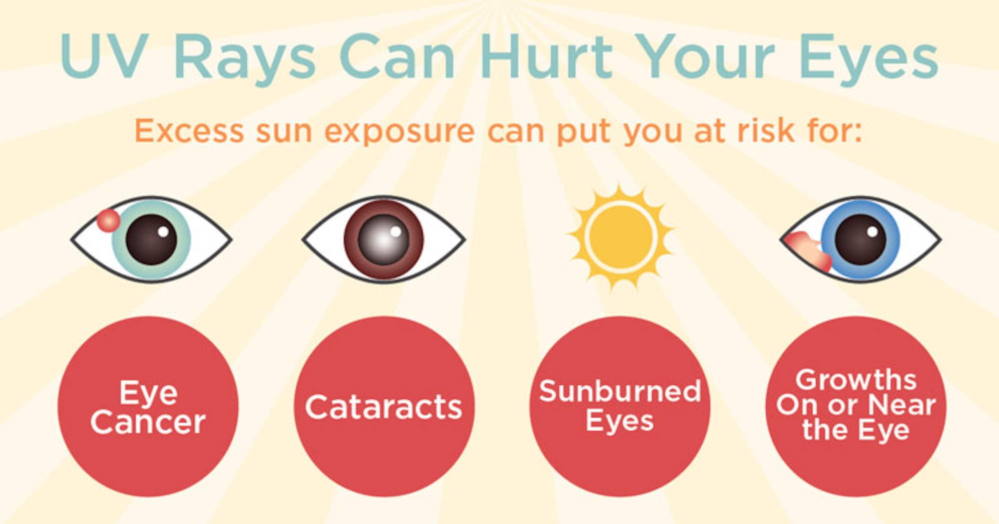


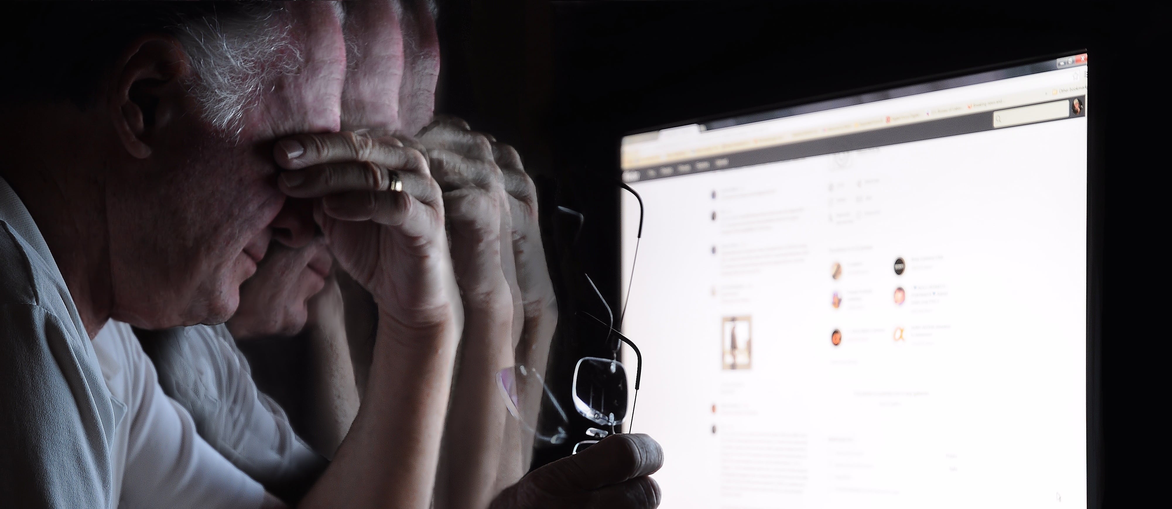

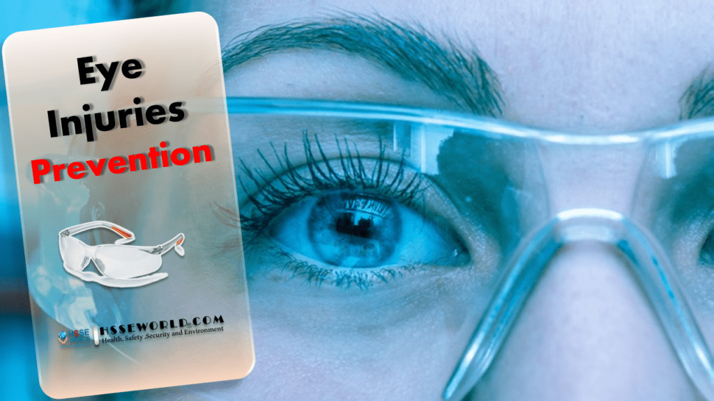


 This right lateral sclera image shows melanin pigment, the brownish coloured 'splotch' you can see in the sclera. This sign is common as a pigmentation spot in a person with darker skin and brown/hazel eyes, representing a genetic predisposition to liver dysfunction. However it does not present the same pathological circumstance as when the melanin sign appears in a blue eyed, fairer skinned person, which is more important/consequential.When the melanin is located close to the iris, it shows mild to moderate liver hardening with associated bacterial infection. When located in an area/s away from the iris, it represents more severe hardening of liver tissue, with sugar system involvement. A substantial diet and lifestyle change, with ongoing liver cleansing and support, is necessary in this circumstance to avoid the condition and sign worsening over time.


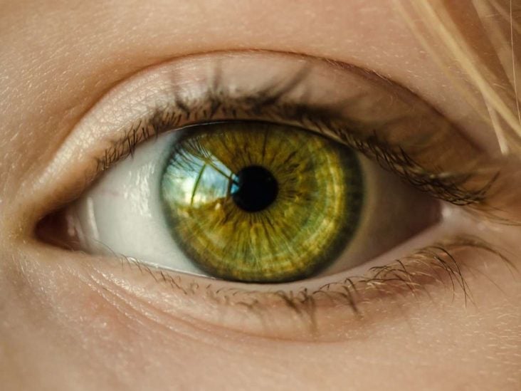
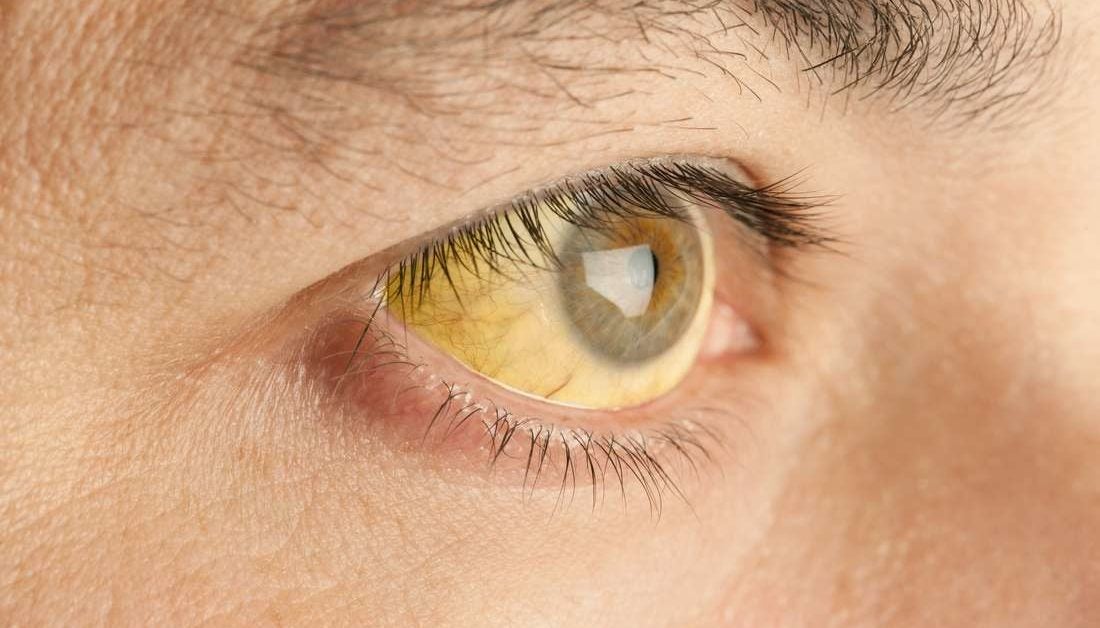
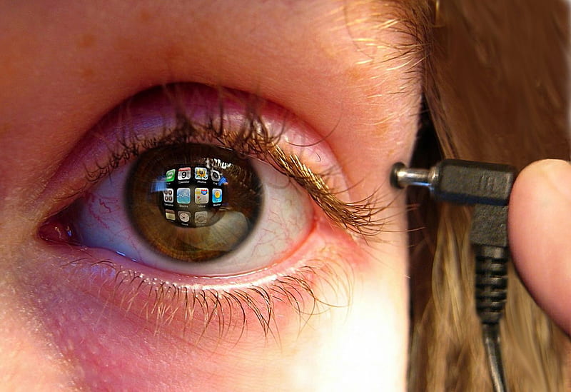


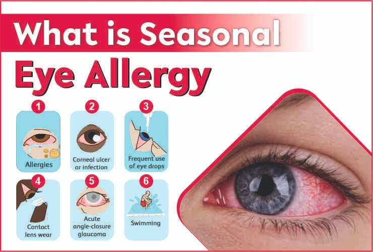


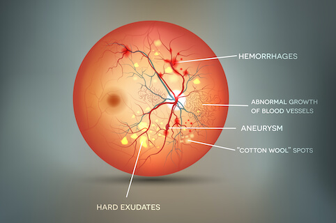

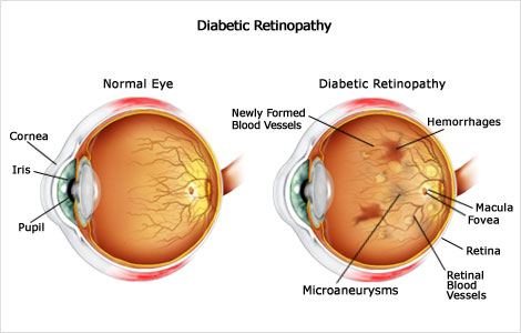

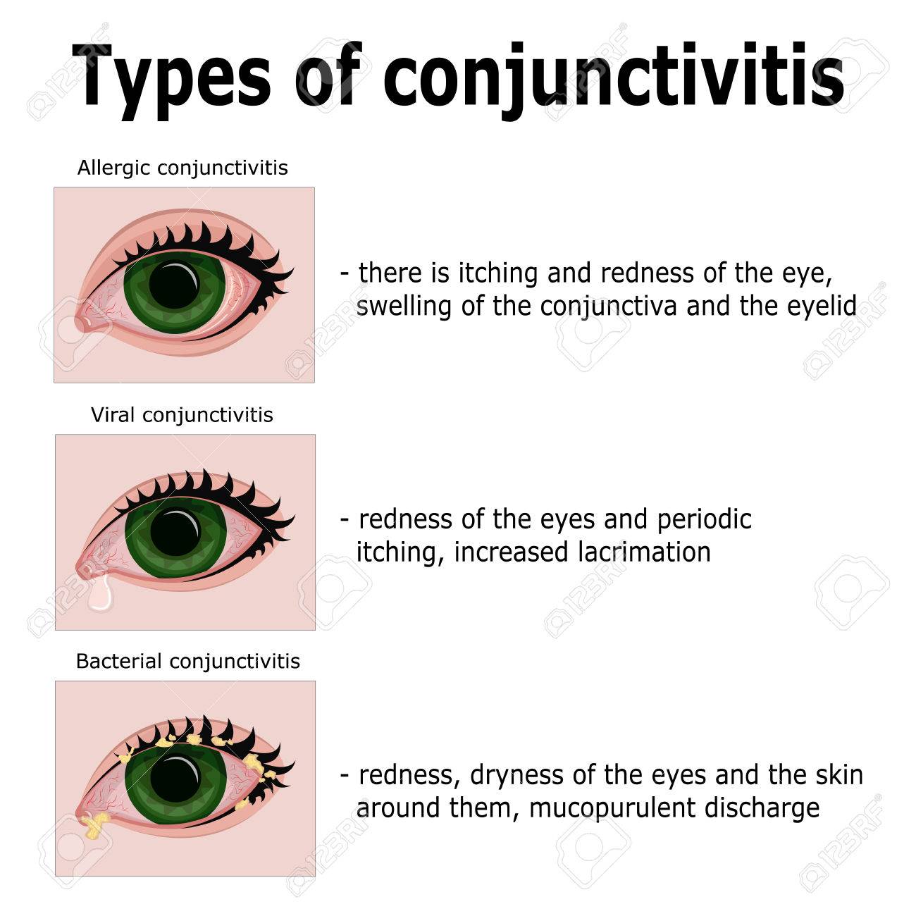


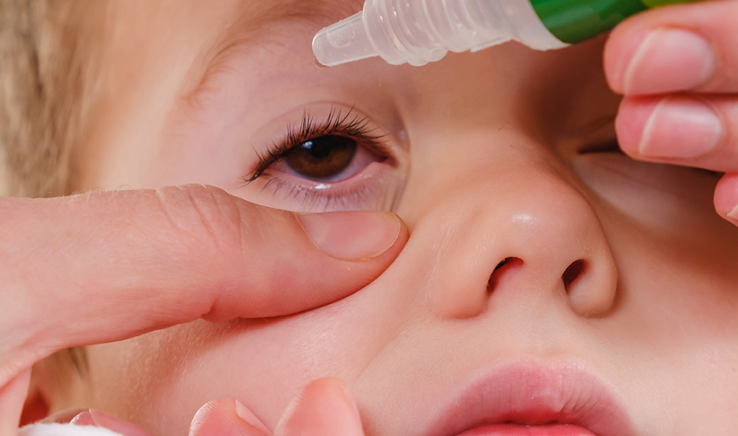


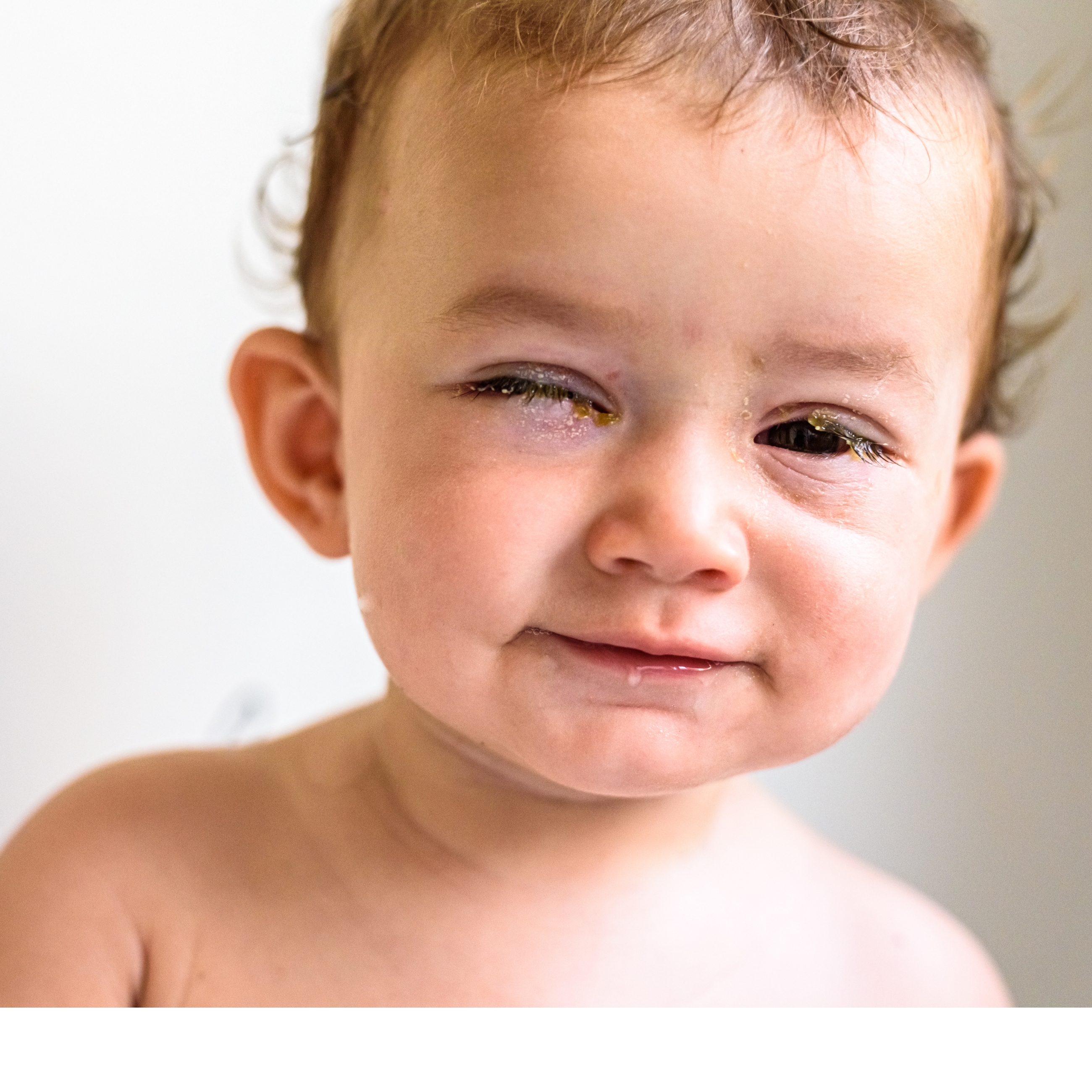
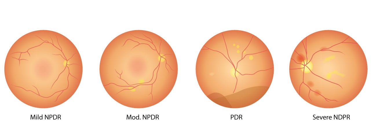


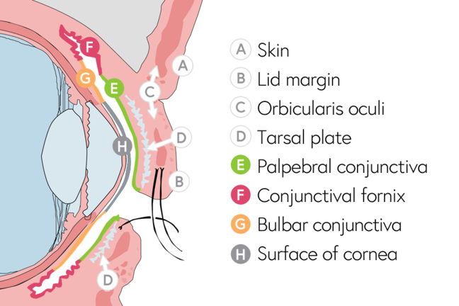

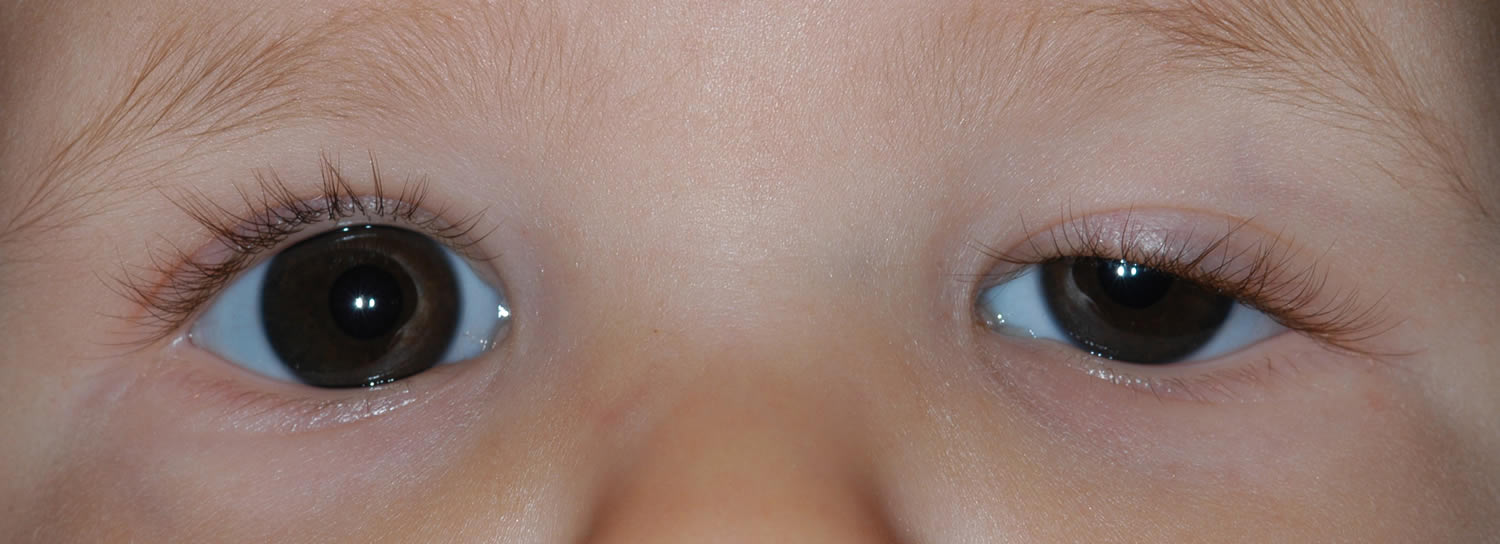
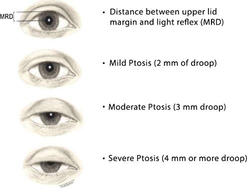


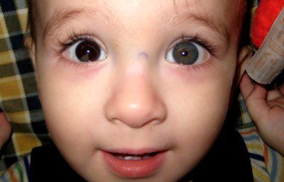


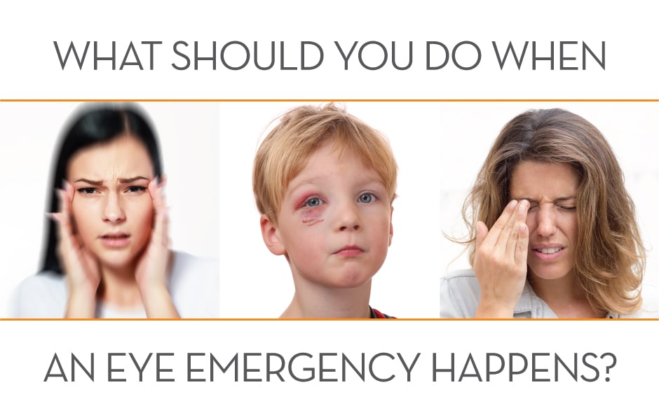
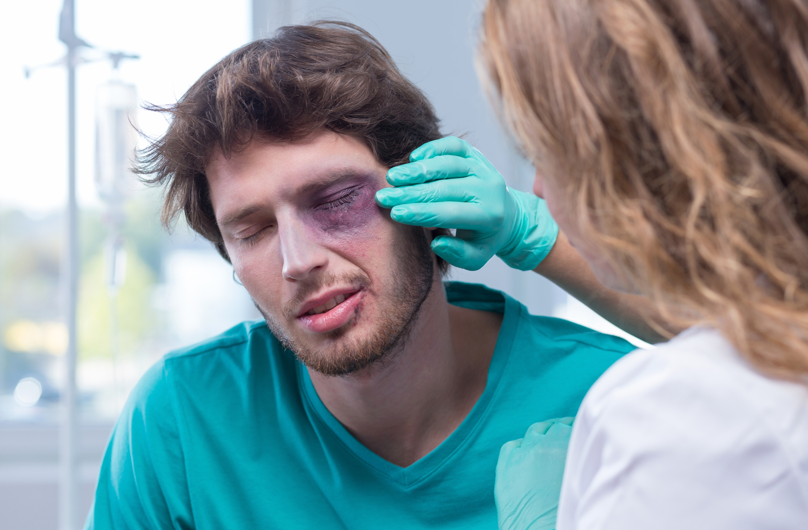
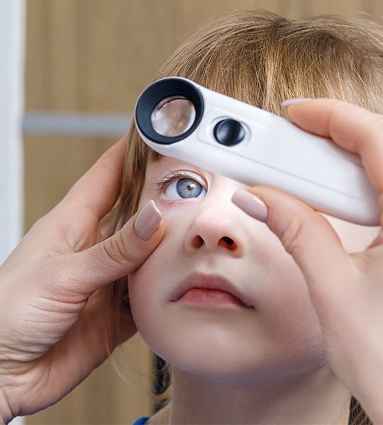
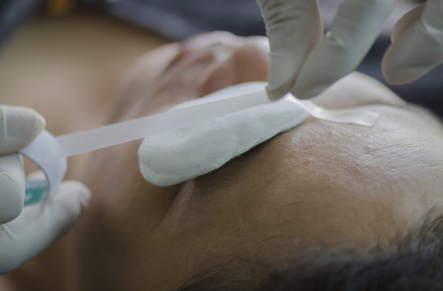

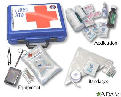

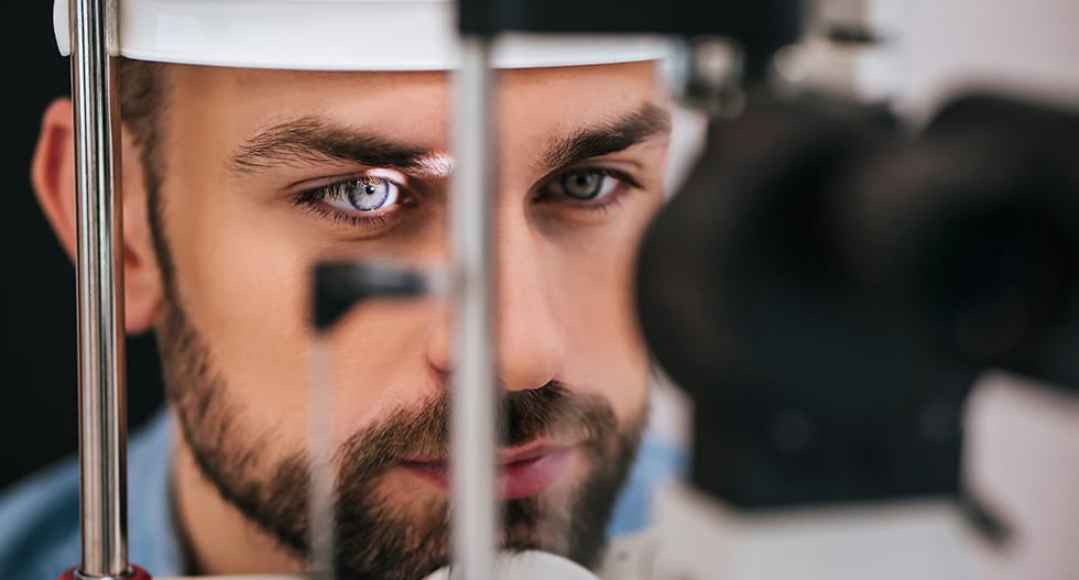


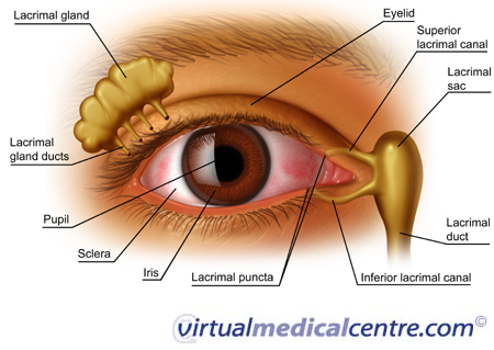


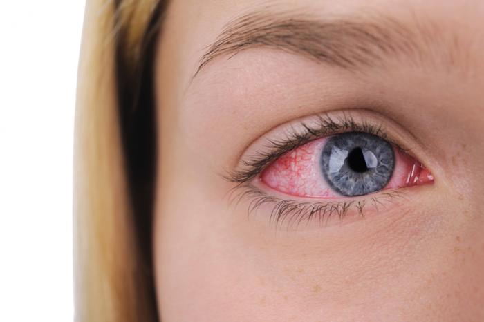
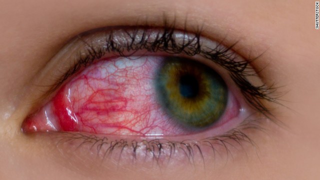
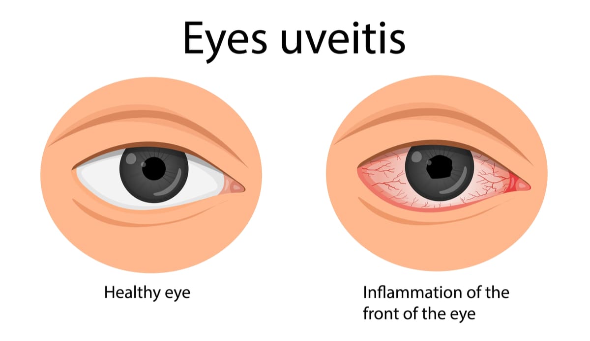
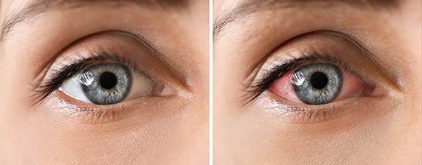
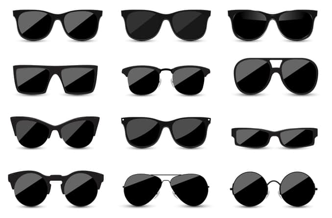
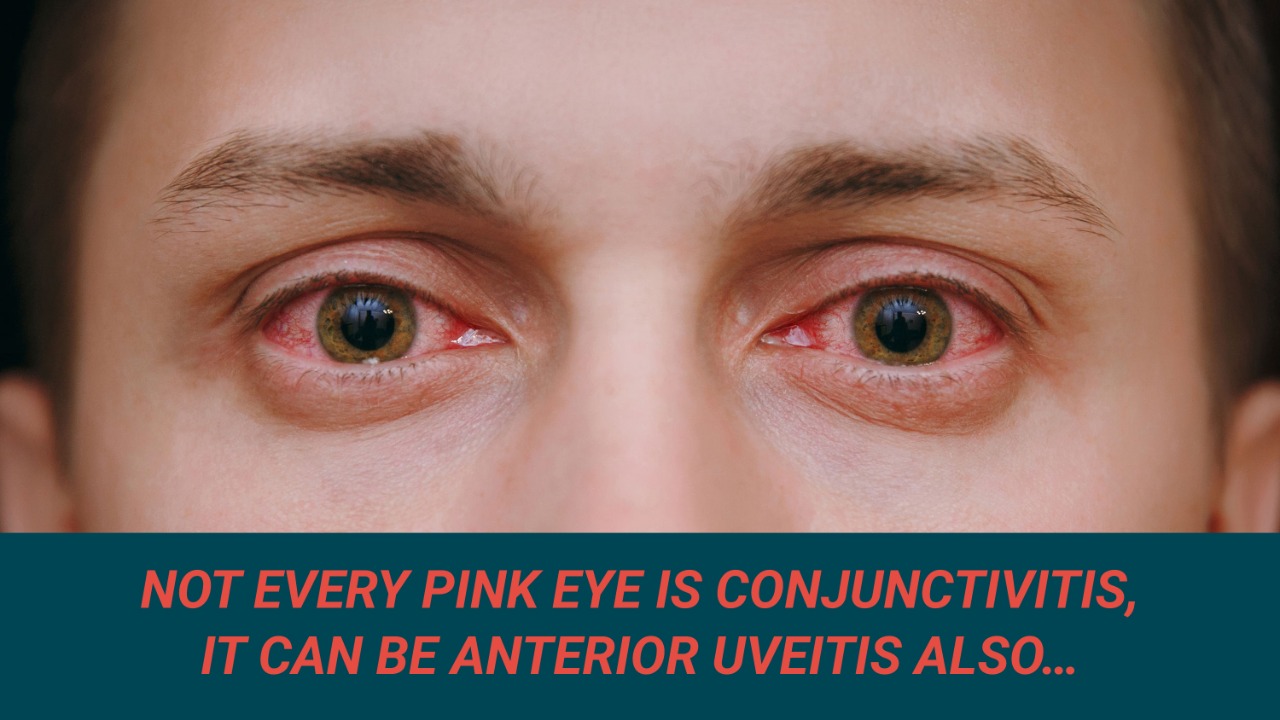


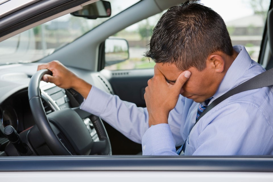

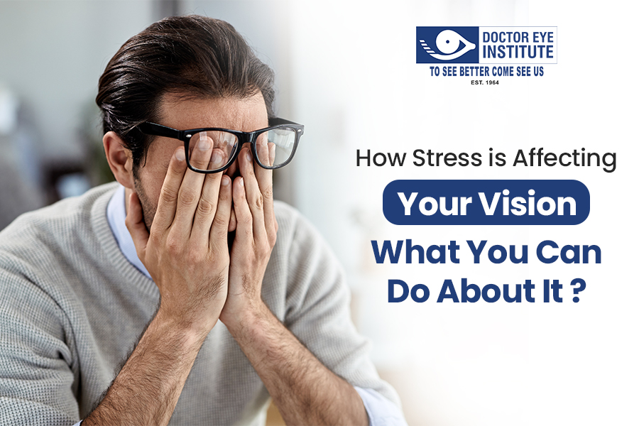


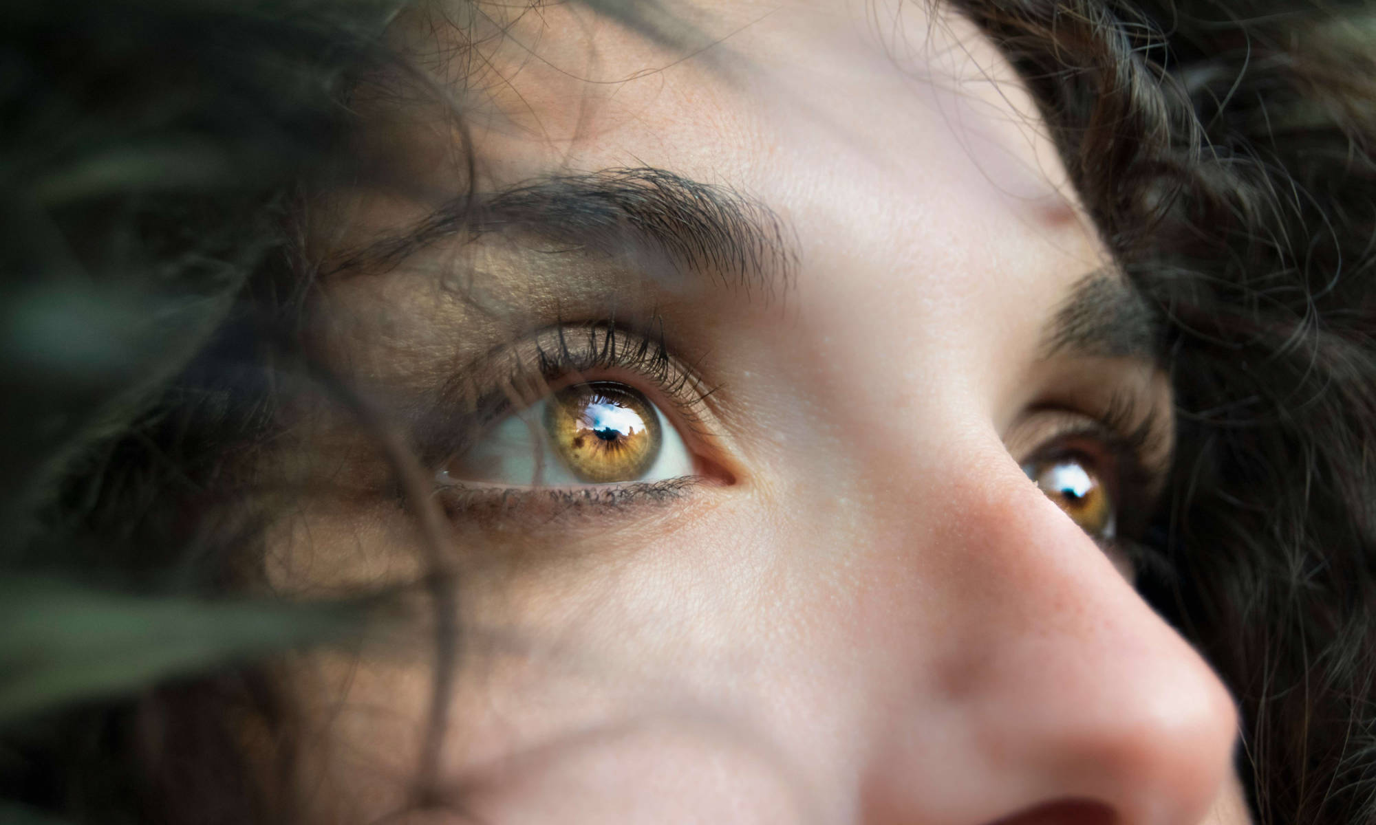


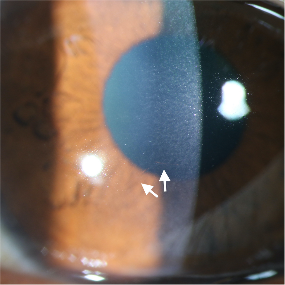
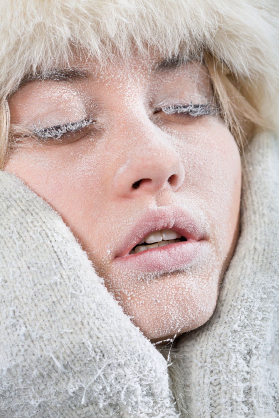
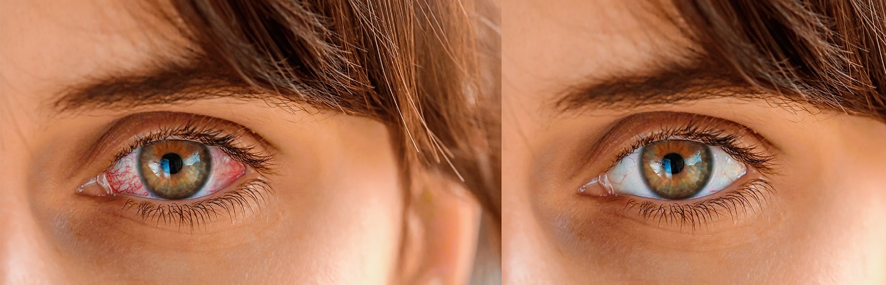
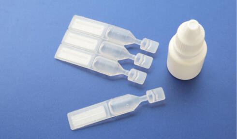


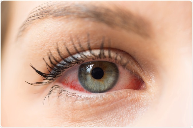
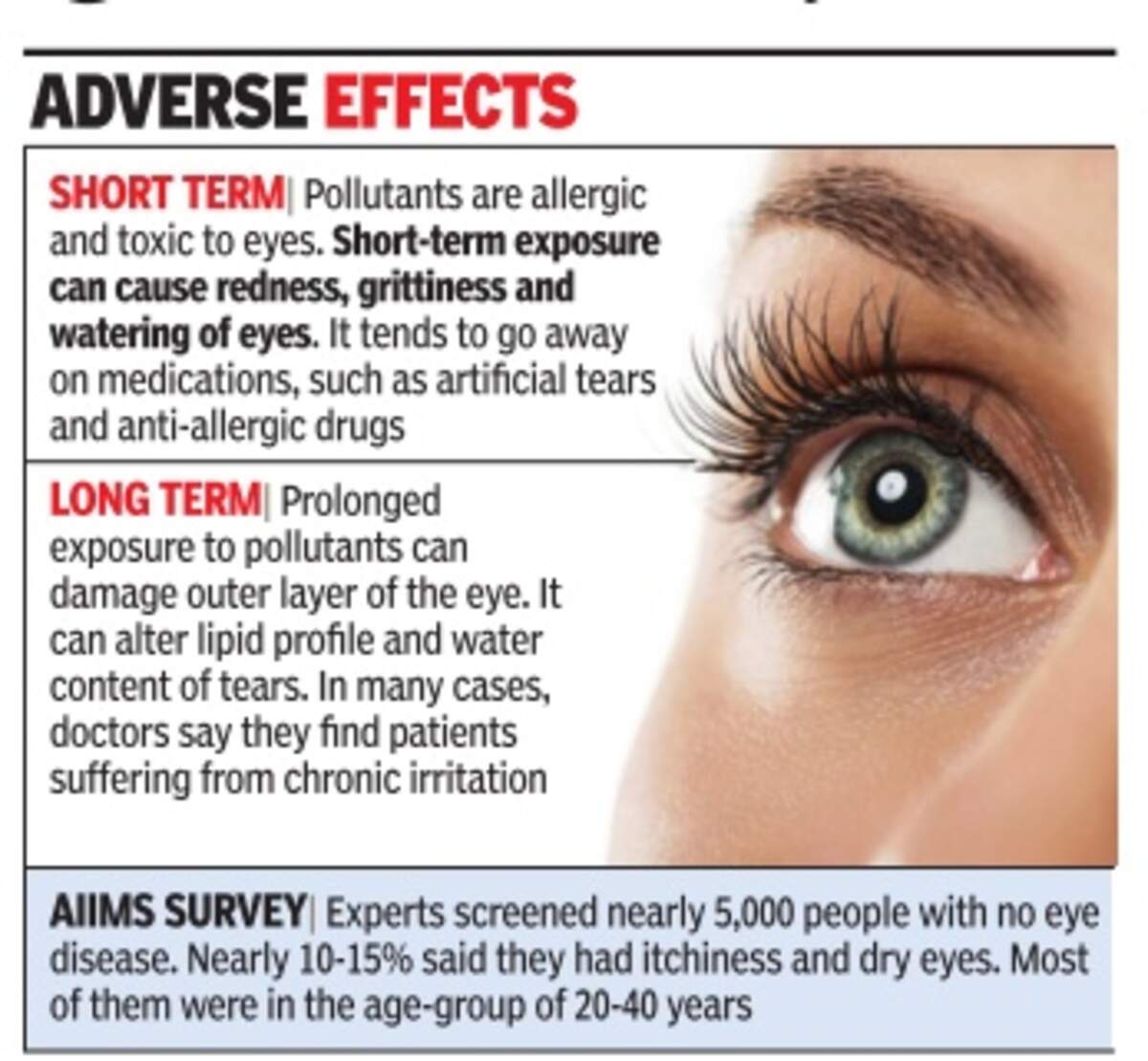


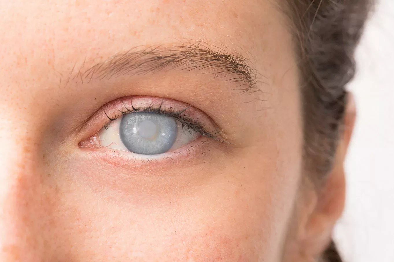

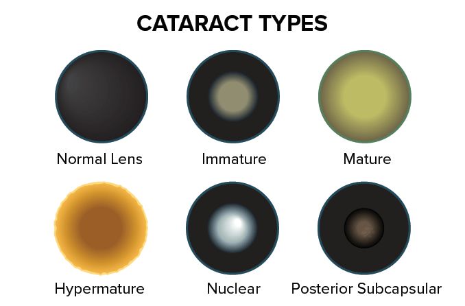
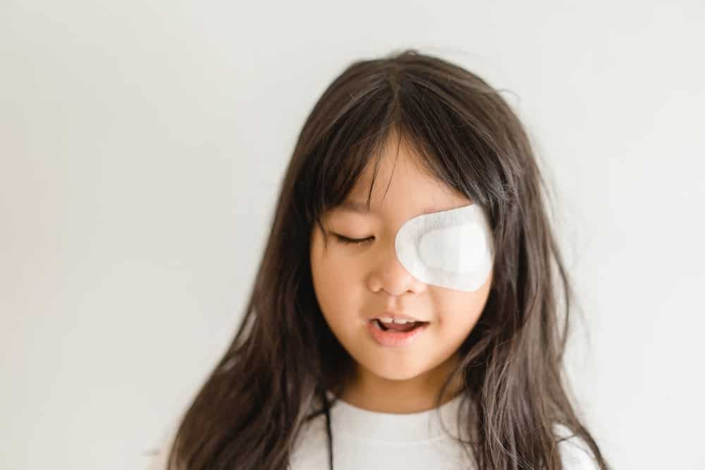


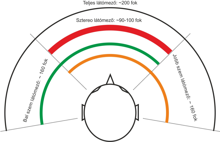
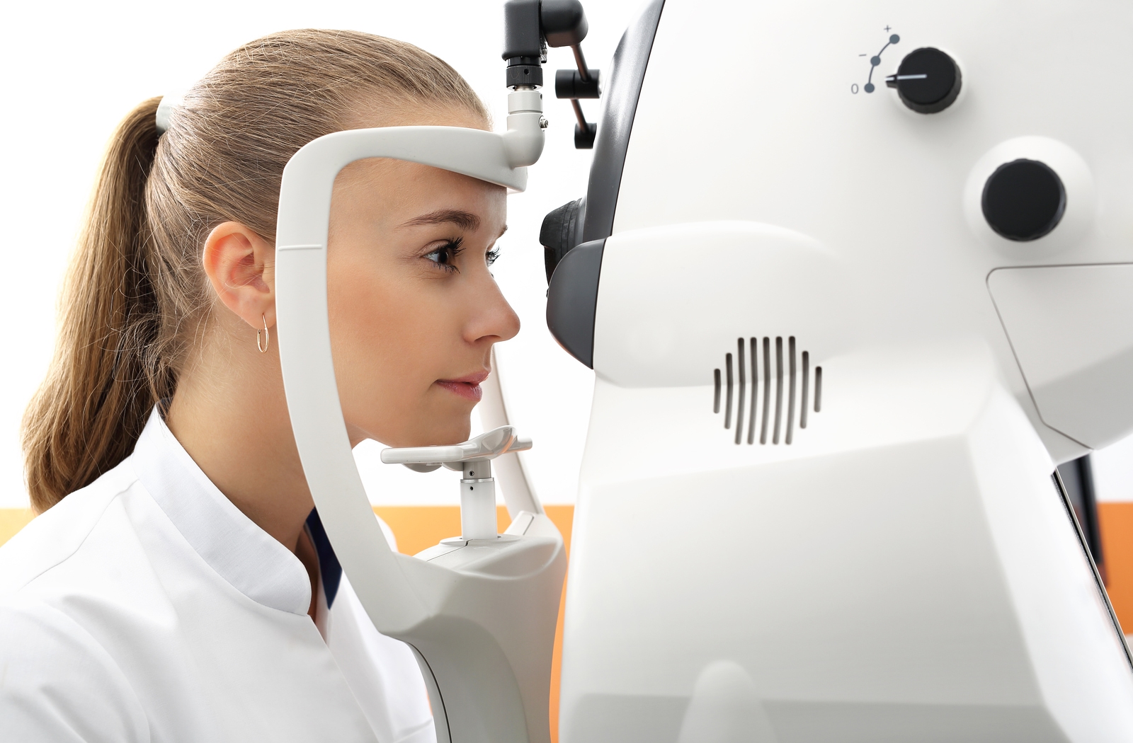


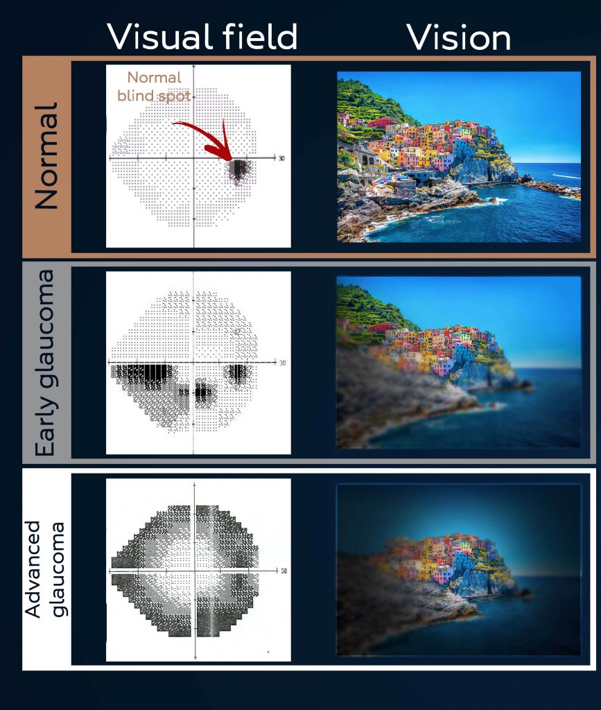


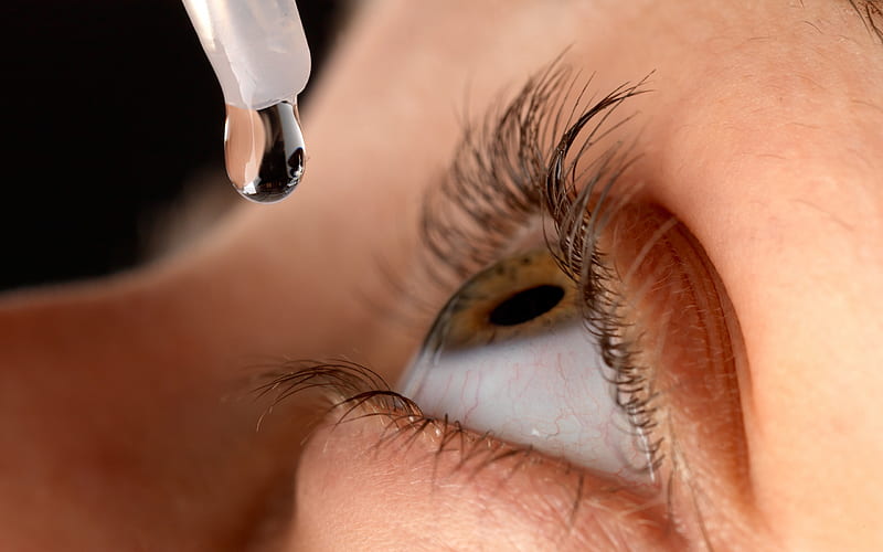

 وبلاگ تخصصی عینک شامل مجموعه مطالب پزشکی است که اطلاعات مفیدی در رابطه با عینک , چشم، لنز، سلامتی چشم و راه های پیشگیری از بیماریهای چشمی، کنترل و درمان آن را در اختیار شما کاربر محترم می گزارد.