ابزار وبمستر

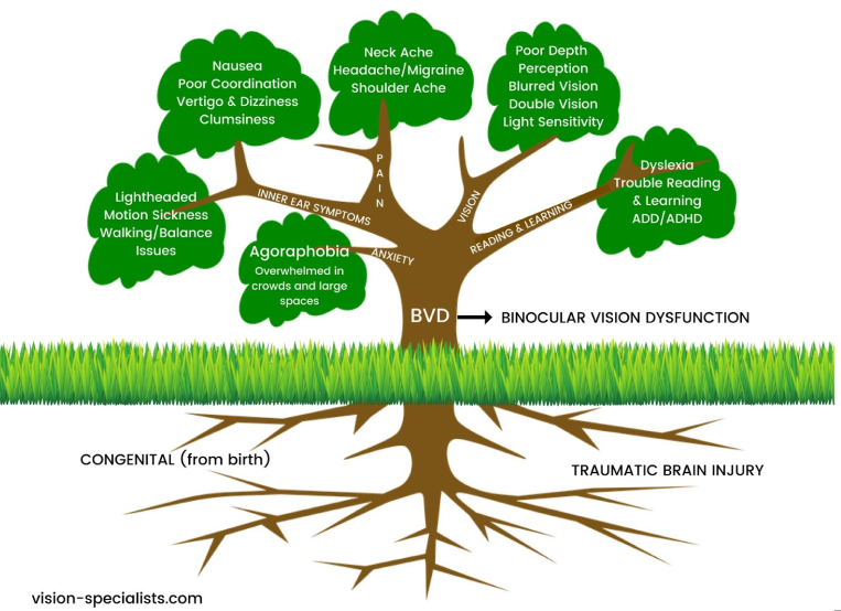
BVD(Binocular vision dysfunction) and other binocular vision issues can have a huge impact on your life, both at work and at home, which is why it’s so important to understand what BVD is and the signs and symptoms to watch for.

In order for the eyes to work together as a team, they must be in perfect alignment. When they’re not, a number of unpleasant and sometimes painful physical symptoms can occur. Headaches, dizziness and balance issues are some of the most common indicators that BVD is present.

Other signs include:

  • Reading problems (losing your place frequently, skipping lines), as well difficulty comprehending what was read.
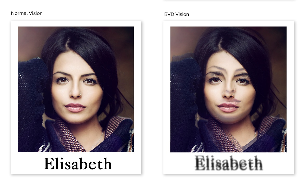
  • Severe light sensitivity and blurred/shadowed/doubled vision.
  • Anxiety and apprehension when in large, open indoor spaces with tall ceilings.

Treatment can include any of the following:

  • Custom micro-prism lenses that help realign the eyes, thus greatly reducing or even eliminating the symptoms of BVD.
  • Prism contact lenses that treat BVD, as well as contact lenses for astigmatism.[8]

Binocular vision dysfunction (BVD) is a visual condition where the line of sight from one eye tends to be slightly out of alignment with the line of sight from the other eye (usually vertical) and this puts heavy strain on the eye muscles as they are constantly trying to correct the alignment to achieve single focus vision.

The cause can be secondary to: normal facial asymmetry, acquired facial asymmetry from aging or head trauma from sports or injury damaging the nerves to your eye muscles causing the imbalance.

Binocular vision dysfunction means you see two images that compete in the middle where their fields of view overlap.

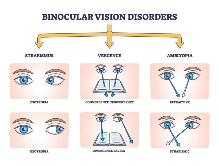
There are three forms of BVD:

1. Vertical Heterophoria
2. Superior Oblique Palsy
3. Horizontal misalignment

Symptoms of BVD

Those who suffer from Vertical Heterophoria or Superior Oblique Palsy tend to have a small amount of vertical eye misalignment, which the brain corrects by directing the eye muscles to properly reposition the eyes. However, using the eye muscles in this manner overworks them and they become strained and fatigued, causing the many symptoms of Vertical Heterophoria and Superior Oblique Palsy:

- Anxiety in crowds or large open spaces
- Overly sensitive to light and glare
- Double vision
- Shadowed, overlapping or blurred vision
- Skip lines or lose your place while reading.
- Quickly fatigue while reading and difficulty with comprehension.
- Closing or covering one eye to make it easier to see.
- Headaches
- Dizziness
- Lightheadedness
- Nausea
- Anxiety
- Motion sickness
- Poor depth perception
- Lack of good balance and drifting while walking
- Poor coordination and Clumsiness
- Aching eyes, especially with eye movement
- Neck, upper back or shoulder pain
- Head tilting [2]

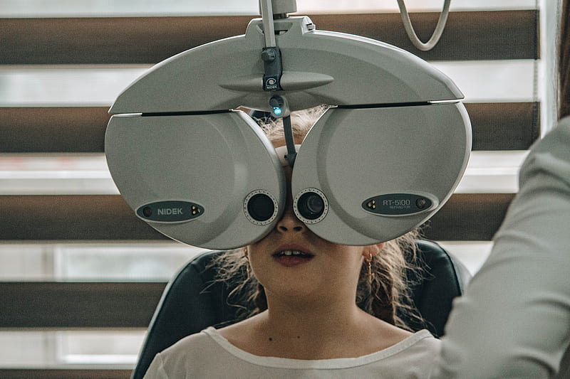
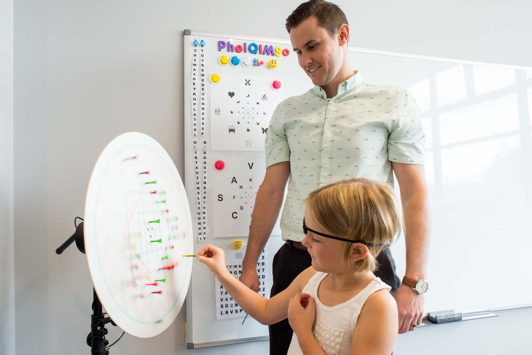
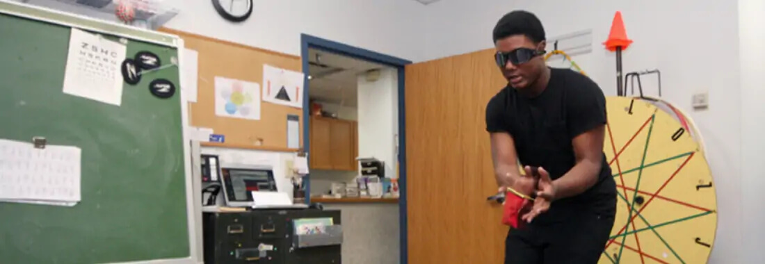
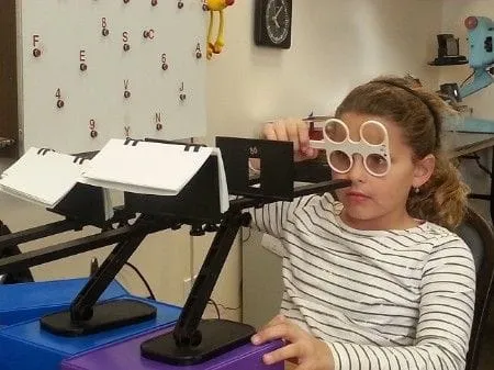
There are a number of tests the doctor may perform to assess any difficulties with vision, including:

Developmental Eye Movement (DEM): Reading eye movements and assessing their accuracy.
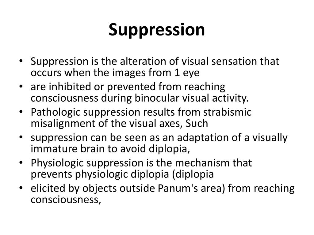
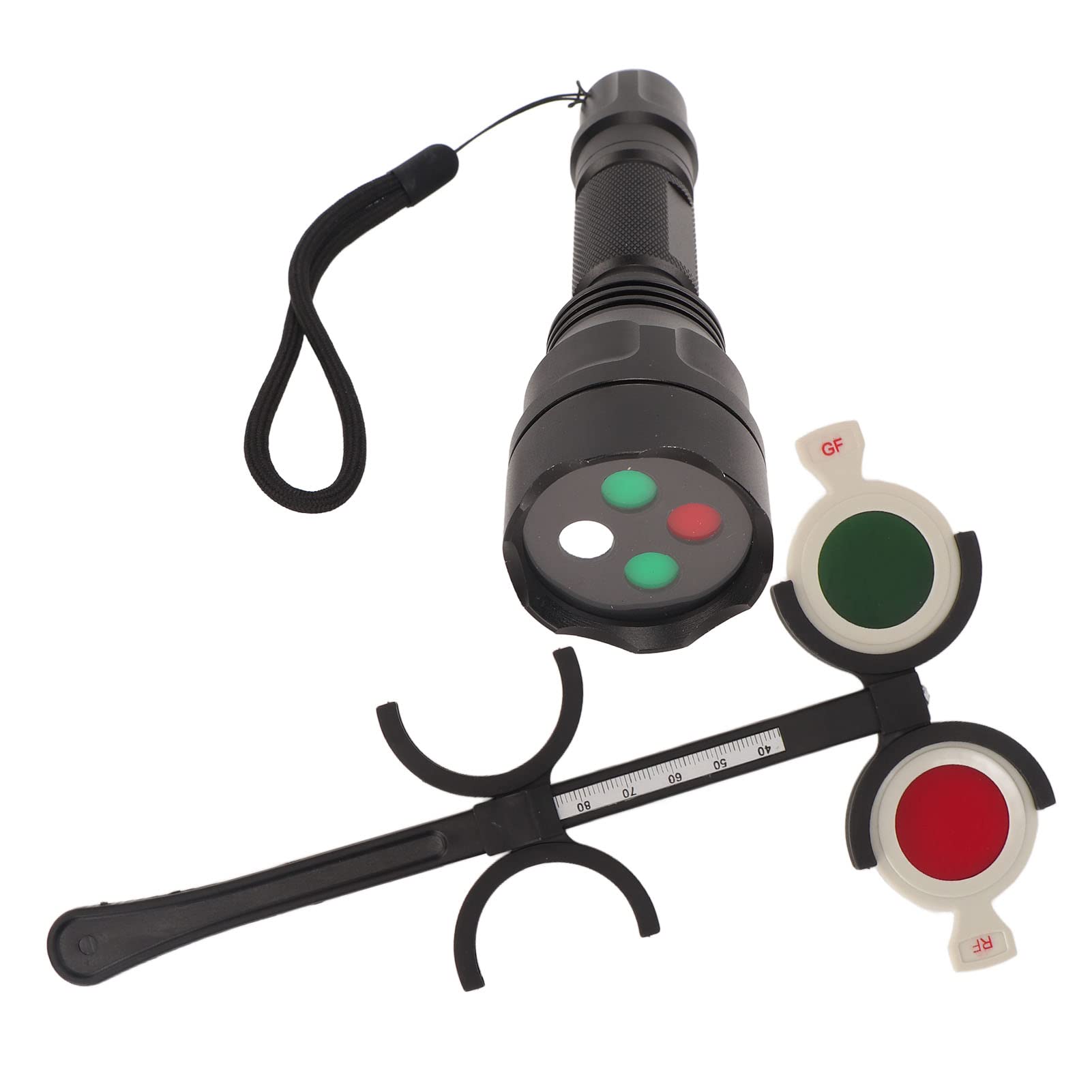
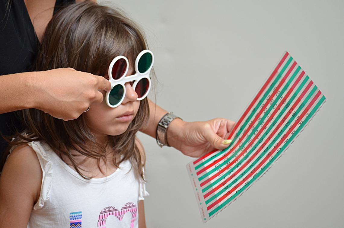
Sensory Fusion Assessment: This is a series of four separate examinations to discover if suppression, which can be part of an overall binocular vision disorder, is present.
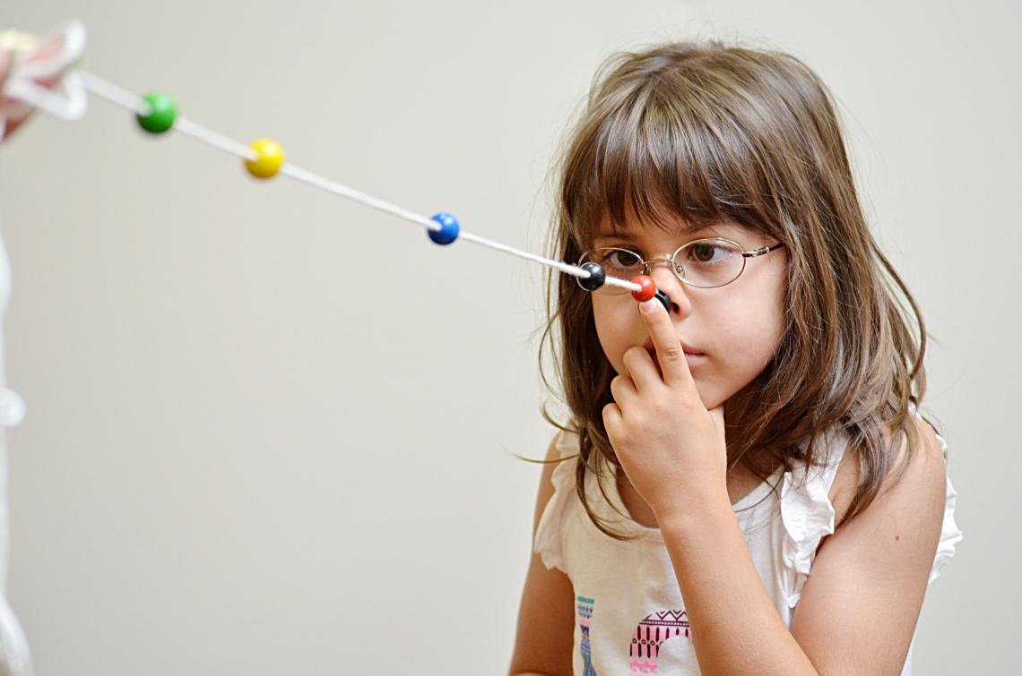
Near Point of Convergence (NPC): The test will find out if convergence and divergence dysfunctions are causing problems.
Accommodative Convergence/Accommodation (AC/A): Any evidence of accommodation which exists is discovered by the results of this test. [4]

Binocular vision is vision in which creatures having two eyes use them together. The word binocular comes from two Latin roots, bini for double, and oculus for eye. According to Fahle (1987), having two eyes confers six advantages over having one.

  1. It gives a creature a spare eye in case one is damaged.
  2. It gives a wider field of view. For example, humans have a maximum horizontal field of view of approximately 190 degrees with two eyes, approximately 120 degrees of which makes up the binocular field of view (seen by both eyes) flanked by two uniocular fields (seen by only one eye) of approximately 40 degrees.
  3. It can give stereopsis in which binocular disparity (or parallax) provided by the two eyes' different positions on the head gives precise depth perception. This also allows a creature to break the camouflage of another creature.
  4. It allows the angles of the eyes' lines of sight, relative to each other (vergence), and those lines relative to a particular object (gaze angle) to be determined from the images in the two eyes.These properties are necessary for the third advantage.
  5. It allows a creature to see more of, or all of, an object behind an obstacle. This advantage was pointed out by Leonardo da Vinci, who noted that a vertical column closer to the eyes than an object at which a creature is looking might block some of the object from the left eye but that part of the object might be visible to the right eye.
  6. It gives binocular summation in which the ability to detect faint objects is enhanced.


Once the fields of view overlap, there is a potential for confusion between the left and right eye's image of the same object.

This can be dealt with in two ways:

  • one image can be suppressed, so that only the other is seen,
  • or the two images can be fused.

If two images of a single object are seen, this is known as double vision or diplopia.

Fusion of images (commonly referred to as 'binocular fusion') occurs only in a small volume of visual space around where the eyes are fixating. Running through the fixation point in the horizontal plane is a curved line for which objects there fall on corresponding retinal points in the two eyes. This line is called the empirical horizontal horopter. There is also an empirical vertical horopter, which is effectively tilted away from the eyes above the fixation point and towards the eyes below the fixation point. The horizontal and vertical horopters mark the centre of the volume of singleness of vision. Within this thin, curved volume, objects nearer and farther than the horopters are seen as single. The volume is known as Panum's fusional area (it's presumably called an area because it was measured by Panum only in the horizontal plane). Outside of Panum's fusional area (volume), double vision occurs. eResearch by Navid Ajamin -- spring 2016

When very different images are shown to the same retinal regions of the two eyes, perception settles on one for a few moments, then the other, then the first, and so on, for as long as one cares to look.

This alternation of perception between the images of the two eyes is called binocular rivalry.

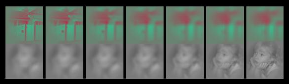
When different images are shown to the two eyes, awareness can alternate such that each is intermittently suppressed and only one image is seen at a time. For instance, a picture of a girl can be shown to the left eye and a picture of a house to the right. Perception can then alternate - swapping between seeing the girl and the house. This phenomenon is called Binocular Rivalry.

Binocular Rivalry has generated broad interest as it permits an opportunity to explore the relationship between changes in conscious vision and brain activity in the absence of changes to sensory input. However, the function of binocular suppression remains a point of contention.[7]

Binocular rivalry is a phenomenon of visual perception in which perception alternates between different images presented to each eye.

Humans have limited capacity to process an image fully at one time. That is why the binocular rivalry occurs. Several factors can influence the duration of gaze on one of the two images. These factors include context, increasing of contrast, motion, spatial frequency, and inverted images. Recent studies have even shown that facial expressions can cause longer attention to a particular image. When an emotional facial expression is presented to one eye, and a neutral expression is presented to the other eye, the emotional face dominates the neutral face and even causes the neutral face to not been seen.

How do you fix an eye misalignment

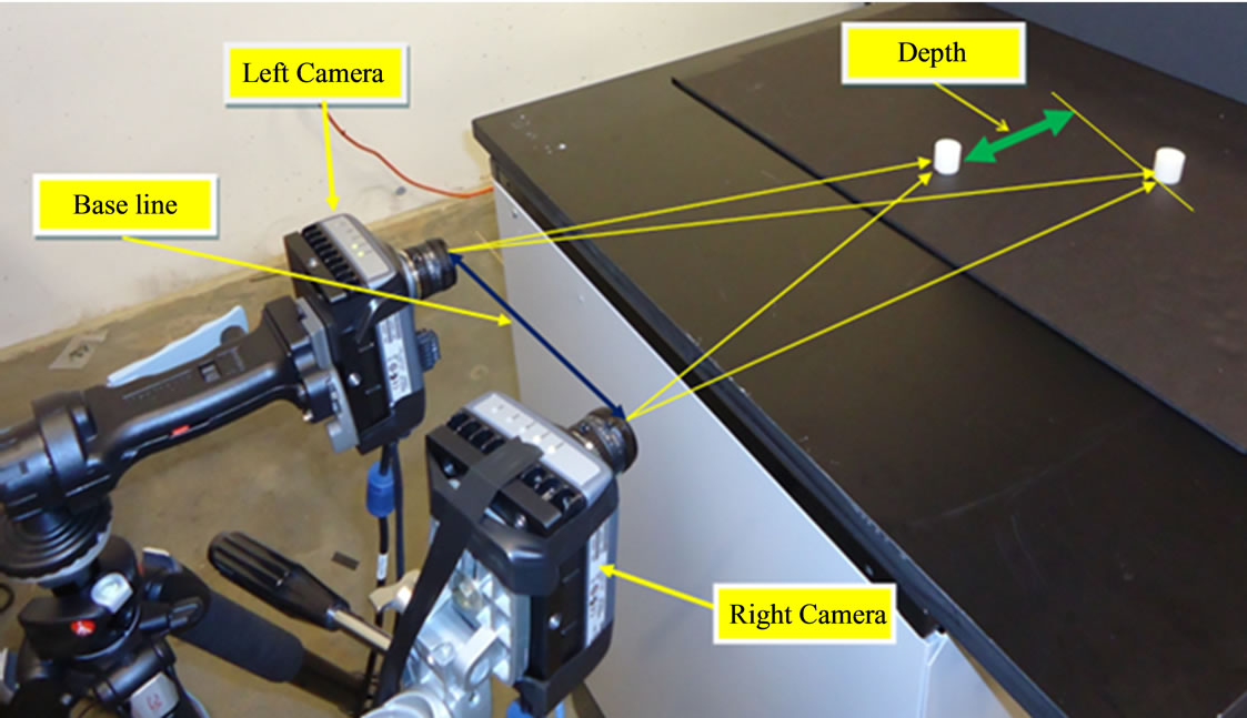
Binocular depth perception arises as a consequence of the slightly displaced point of view of the two eyes. The horizontal displacement of image features in the two eyes (i.e. binocular disparities) makes it possible to reconstruct the depth relationships in the visual world.

The term depth perception refers to our ability to determine distances between objects and see the world in three dimensions. To do this accurately, one must have binocular stereoscopic vision, or stereopsis.

Depth perception is the ability to judge depth and distance. Depth perception requires binocular vision, but it may be assisted by monocular cues such as motion parallax, or how objects move in relation to the movement of the head; interposition, or object overlap; and color and contrast cues that suggest distance.

What causes depth perception problems?

There is not one answer, but in fact several conditions that can contribute to poor depth perception:
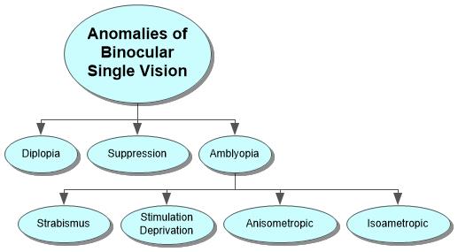
  • Strabismus This is a condition where both of the eyes cannot be aligned simultaneously. One or both eyes may turn outwards, inwards, downwards, or upwards. This is commonly referred to as being cross-eyed.
  • Blurred vision This is when one’s vision is not as sharp as normal and it makes it incredibly difficult to spot detail.
  • Amblyopia This is a condition where one eye cannot focus as well as the other and is often called a “lazy eye.”
  • Eye trauma Eye trauma is anything that disturbs or harms the eye. This prevents the eye or eyes from working as well as they should and can harm one’s vision.

Not everyone sees optimally. People suffering from amblyopia, optic nerve hypoplasia and strabismus often have reduced depth perception. A person with an injury to one eye, or a person missing one eye, may not be able to tell where objects are in relation to others. Visual therapy may help improve these problems.

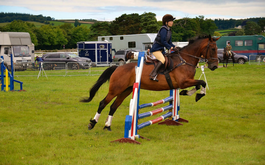
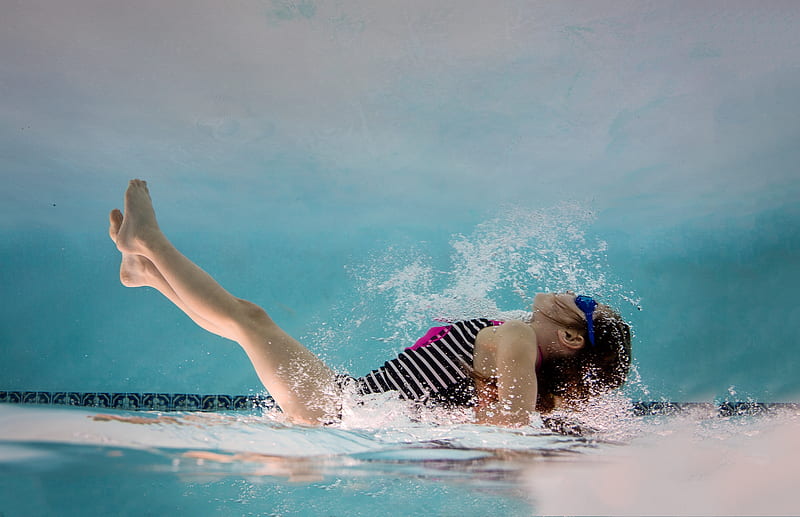
Depth perception plays an important part in many activities. Driving, sewing, threading a needle, watching 3D movies and even walking on uneven ground all require certain levels of depth acuity. People without functioning stereoscopic vision may not be able to perform these activities or may struggle with them.

Two-eyed, or binocular vision, allows each eye to see from different angles. The brain processes the information coming from each eye and forms it into one image—a process called convergence. If binocular vision is working as it should be, the brain can interpret the information, which is called stereopsis. Those that have vision in only one eye usually have to rely on other cues to aid their depth perception.

Binocular matching of local features in the retinal images may be used to obtain estimates of the absolute disparity (and distance) of objects or surfaces, as well as the relative disparity (or relative distances) between different objects.

Other phenomena of binocular vision include:

  • utrocular discrimination (the ability to tell which of two eyes has been stimulated by light),
  • eye dominance (the habit of using one eye when aiming something, even if both eyes are open),
  • allelotropia (the averaging of the visual direction of objects viewed by each eye when both eyes are open),
  • binocular fusion or singleness of vision (seeing one object with both eyes despite each eye's having its own image of the object),and
  • binocular rivalry (seeing one eye's image alternating randomly with the other when each eye views images that are so different they cannot be fused).

When different images are presented to the two eyes, they compete for perceptual dominance, such that one image is visible while the other is suppressed. This binocular rivalry is thought to reflect competition between monocular neurons within the primary visual cortex. However, neurons whose activity correlates with perception during rivalry are found mainly in higher cortical areas, and respond to input from both eyes. Thus rivalry may involve competition between alternative perceptual interpretations at a higher level of analysis. To investigate this, we tested the effect of rapidly alternating the rival stimuli between the two eyes. Under these conditions, the perceptual alternations exhibit the same temporal dynamics as with static patterns, and a single phase of perceptual dominance can span multiple alternations of the stimuli. Thus neural representations of the two stimuli compete for visual awareness independently of the eye through which they reach the higher visual areas. This finding places binocular rivalry in the general category of multistable phenomena, such as ambiguous figures, and provides a new way to study the neural cause and resolution of perceptual ambiguities.

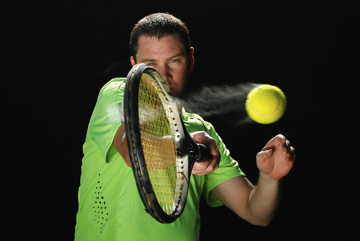
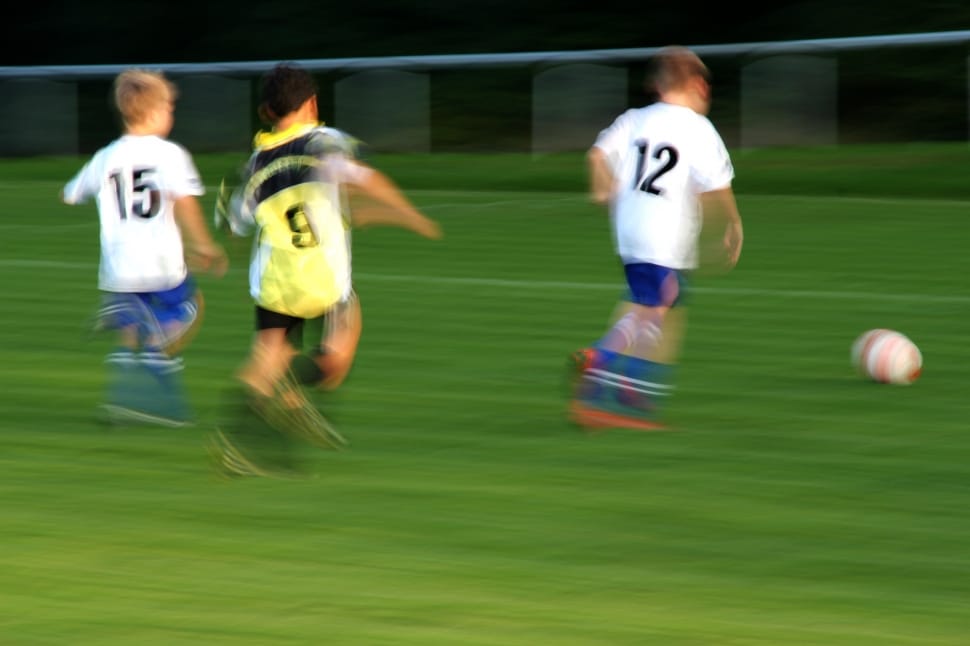
Binocular vision helps with performance skills such as catching, grasping, and locomotion.It also allows humans to walk over and around obstacles at greater speed and with more assurance.Orthoptists are eyecare professionals who fix binocular vision problems.[1]

Strabismus occurs when there are neurological or anatomical problems that interfere with the control and function of the extraocular muscles. The problem may originate in the muscles themselves, or in the nerves or vision centers in the brain that control binocular vision.

Grades of binocular vision

There are grades and methods of assessing binocular vision. The grades are the different steps in the development of stereopsis during the visual maturation. Testing of the grades is done by a synaptophore and graded as - no binocular single vision grade zero, simultaneous perception grade 1, fusion grade 2 and stereopsis grade 3. Limited form of testing can be done with worth four-dot test or Bagolini’s glasses.

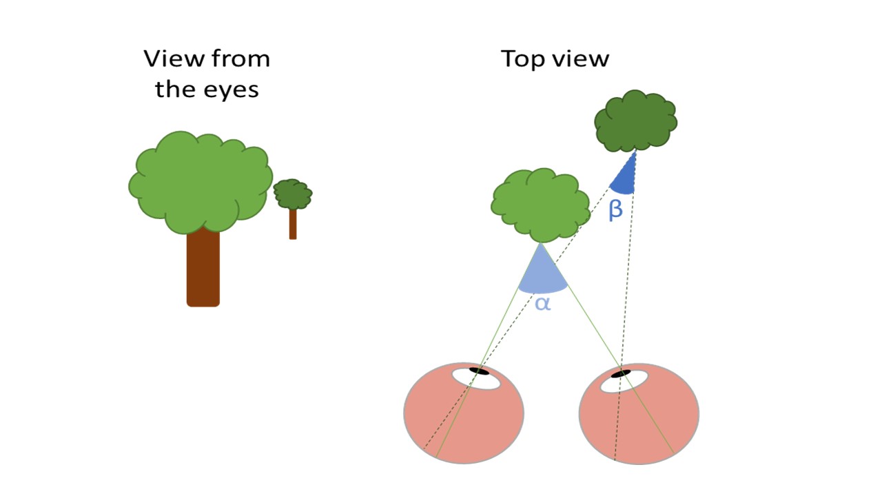
The drawing at the left shows the view of two trees from the perspective of the eyes. The light green tree stands in front of the dark green tree. The right drawing shows a top view of the scene. When the eyes are focusing on the light tree, the image is projected on the fovea of the left and right eye. The angle between both projections is angle α. The images of the dark tree are projected on different positions of the peripheral retina in the left and right eye with angle ß. Because angle ß is smaller than angle α our brain interprets the dark tree as further away than the light tree. The size of the difference between α and ß represents the disparity. Large differences in angle indicate large differences in depth

Stereopsis is not present at birth but develops in the first months of life. That full-term and pre-term children develop stereopsis at the same age post-birth shows that the development depends on visual experience rather than biological maturation of the system.In the early months of life, we develop coarse stereopsis, which operates on high contrast lines and edges and enables us to align our eyes.

Four basic types of Da Vinci stereopsis cues [8].

Four basic types of Da Vinci stereopsis cues

Alignment then permits the development of fusion and fine stereopsis. Fine stereopsis works over a much shorter range of disparities but enables us to make very fine depth judgments even in densely textured surfaces, such as grass or tree bark, where there are few or no depth cues monocularly.

important binocular visual skills:

- Tracking: the ability to move the eyes across a sheet of paper
- Fusion: the ability to use both eyes together at the same time
- Stereopis: binocular depth perception
- Convergence: the ability of the eyes to move and work as a team
- Visual Motor Integration: the ability to transform images from a vertical to a horizontal plane
[3]

Reference:

  1. en.wikipedia.org/wiki/Binocular_vision
  2. city-optometry.com
  3. children-special-needs.org
  4. rebuildyourvision.com
  5. intechopen.com/books/visual-cortex-current-status-and-perspectives/...
  6. deskarati.com/2014/03/10/binocular-rivalry
  7. psy.uq.edu.au/~uqdarnol/Rivalry.htm
  8. vision-specialists.com/articles/the-causes-symptoms-of-binocular-vision-dysfunction-your-questions-answered
  9. shopfelixgray.com/blog/understanding-depth-perception
  10. nearsay.com/c/670447/515154/a-guide-to-depth-perception
  11. eyehealthweb.com/depth-perception
  12. wisegeek.com/what-is-depth-perception.htm
  13. ncbi.nlm.nih.gov/m/pubmed/8602261
  14. eyewiki.aao.org/Stereopsis_and_tests_for_stereopsis
  15. opticianonline.net/cet-archive/4723
  16. allaboutvision.com/conditions/strabismus.htm

See also:

  • BINOCULAR VISION webeye.ophth.uiowa.edu Department of Ophthalmology & Visual Sciences
  • Binocular Vision Dysfunction (BVD) -- dizziness-and-balance.com

​According to the American Optometric Association (AOA), 25 percent of all children, or 1 in 4 students, have a vision problem significant enough to impact their learning.

Suppression of an eye is when the brain ignores the visual signals provided by one eye. Essentially, suppression is a method of compensating or adapting for a specific vision condition, like amblyopia (lazy eye) or strabismus (cross eyed). With normal vision, the eyes are aligned and have an equally clear image.

Suppression is a visual deficit that is associated with other strabismic and non-strabismic anomalies. Successful and long- lasting treatment of suppression is dependent upon the effectiveness of the treatment of the associated visual conditions.

A child may pass a visual acuity eye exam and still have an undetected vision problem— mainly because 20/20 eyesight does not indicate “perfect” functioning of the visual skills. In other words, visual problems do not only stem from blurry vision, but can be caused by reduced visual skills as well.

Vision problems do not go away on their own and will persist into adulthood if not treated. In many cases, adults are not even aware that their difficulties are caused by a vision problem— leading to years of avoidance of difficult activities.

Therefore, it is essential to obtain an accurate diagnosis of a vision problem, and appropriate treatment as early as possible.

Vision problems can impact:

  • Reading
  • Spelling
  • Handwriting
  • Reading comprehension
  • Homework completion
  • Classroom performance
  • Concentration and attention
  • Visual arts

Depiction of predictions for refoveating Müller's stimulus with eyes moving independently or eyes following Hering's law of equal innervation.

Any condition that hampers vision or the processing of vision may result in problems learning. Vision disorders may interfere with reading and learning. The eye is literally an extension of the brain. It is estimated that over 60% of the brain has some duties associated with vision input.

  1. Suppression is the act of one of the eye’s shutting down visual input being sent to the brain. This can happen intermittently or be constant. Suppression could also be isolated to one eye or alternate between both. Usually this happens when binocular vision cannot be achieved so in order to compensate an eye would suppress the visual information.
  2. Suppression may also occur when there is a difference in refractive error in each eye. This condition is called anisometropia. Deprivation of visual stimulus caused by a congenital cataract may also cause suppression.
  3. Suppression of an eye is a subconscious adaptation by a person's brain to eliminate the symptoms of disorders of binocular vision such as strabismus, convergence insufficiency and aniseikonia. The brain can eliminate double vision by ignoring all or part of the image of one of the eyes. The area of a person's visual field that is suppressed is called the suppression scotoma (with a scotoma meaning, more generally, an area of partial alteration in the visual field). Suppression can lead to amblyopia.
  4. Suppression essentially refers to the brain ignoring the visual signal provided by one eye. Let's start with a normal pair of eyes. When the eyes are aligned and pointing towards the same object in space and have an equally clear image, the brain uses small cues to determine the depth, size, shape, etc. of the object and combines the images of each eye together to see.

If one eye has a very blurry image (amblyopia, specifically refractive amblyopia due to an unequal glasses prescription) the brain struggles to combine the images of each eye together into a single image. To compensate, the brain suppresses or ignores visual input from the blurry eye. This same concept occurs if one eye is turned (strabismus or squint). In this example, the brain sees multiple images, which would be confusing. Again, the brain suppresses (ignores) visual input from the deviating eye and utilizes input from the fixating eye.

At its core, suppression is the visual system's method of compensating or adapting for a defect. To avoid the confusion of a blurry image superimposed on a clear image, or seeing two of one image in space, the brain simply ignores the abnormal image. Suppression is rather easily "learned" by the brain at an early age when the visual system has a higher degree of neuroplasticity. In contrast, a patient that suddenly acquires a strabismus or squint later in life may have trouble suppressing and constantly see double.

For amblyopic patients, a critical issue stems from long-term suppression. Suppression can be difficult to "unlearn". One of the key components of visual rehabilitation for patients that have amblyopia or strabismus is to teach the brain to use both eyes together. Tests of suppression check for the presence of suppression and, in some cases, quantify the amount of suppression the visual system is experiencing.

suppression amblyopia -- medical-dictionary.thefreedictionary.com eResearch by Navid Ajamin -- summer 2013

suppression of the central vision in one eye when the images from the two eyes are so different that they cannot be fused into one.

This may be due to:

  1. faulty image formation (sensory amblyopia);
  2. a large difference in refraction between the two eyes (anisometropic amblyopia);
  3. the two eyes, pointing in different directions (strabismic amblyopia).

Most suppression amblyopia can be reversed if appropriately treated before age 6 years.

a partial loss of vision, usually in one eye, caused by cortical suppression of central vision to prevent diplopia. It occurs commonly in strabismus in the eye that deviates and does not fixate. Early recognition of strabismus and amblyopia is essential because occlusive therapy that forces use of the bad eye may dramatically improve the child's vision if begun early. It becomes progressively less effective with increasing age but may improve vision even up to 9 years of age. Without therapy, near-blindness in the affected eye may result, but common acuity loss is 20/40 to 20/400.

Binocular interaction
Apart from binocular summation, the two eyes can influence each other in at least three ways.

Pupillary diameter. Light falling in one eye affects the diameter of the pupils in both eyes. One can easily see this by looking at a friend's eye while he or she closes the other: when the other eye is open, the pupil of the first eye is small; when the other eye is closed, the pupil of the first eye is large.

Eye Suppression 4 Dot Light Test

Accommodation and vergence. Accommodation is the state of focus of the eye. If one eye is open and the other closed, and one focuses on something close, the accommodation of the closed eye will become the same as that of the open eye. Moreover, the closed eye will tend to converge to point at the object. Accommodation and convergence are linked by a reflex, so that one evokes the other.
Interocular transfer. The state of adaptation of one eye can have a small effect on the state of light adaptation of the other. Aftereffects induced through one eye can be measured through the other.

Grating acuity, interocular suppression and binocular interaction were evaluated at different positions in the visual field of strabismic humans with alternating fixation. In all cases, the suppression of the deviated eye was most intense in a region corresponding to the fovea of the fixating eye, but reduced or absent in the periphery. In addition, suppression was found in part of the peripheral visual field of the fixating eye. For most subjects, the monocular properties of both eyes were normal. However, for one subject with a strong preference for one of the eyes, visual acuity and luminance detection were selectively impaired in the chronically suppressed regions of the non-preferred eye. Regardless of which eye was currently used for fixation, the visual information available to the subject came from both eyes. In parts of the visual field, the two eyes tended to replace each other. In the far periphery, and especially in the upper and lower hemifield, signals from the two eyes were often combined in a binocular perception of depth.

Related image Suppression may treated with vision therapy, though there is a wide range of opinions on long-term effectiveness between eye care professionals, with little scientific evidence of long-term improvement of suppression, if the underlying cause is not addressed (strabismus, amblyopia, etc.).


  • Schober's test. If suppression of the amblyopic eye is weak, the cross may be seen "blinking".
  • Schober's test often helps in the diagnostic assessment of children with anisometropic amblyopia.
  • The test picture contains two green rings and a red cross. When viewed through red - green lenses a binocular person sees both the rings and the red cross.

Sometimes the child tells that (s)he sees the cross "blinking", i.e the cross appears for a short time and then disappears again for a short time. In such a case suppression is weak and can usually be broken in a short time. Since we usually do not have follow-up visits sooner than a month, it is not possible to know how soon the function became normal. If the child has intensively trained the amblyopic eye, visual acuity may become normal within a month. Training must be both fun and demanding; nowadays many children like to play different electronic games, the smaller, the better.

ANTI SUPRESSION

Patients with unstable visual systems often learn a neurological adaptation called suppression, in which their brains block the visual input coming in from one eye. People unconsciously learn to suppress at very young ages to keep from seeing double. Suppression is never normal and is always a sign of an unstable visual system.

Vision therapy trains the eyes to work together but to do so both eyes have to be “on.” Therapists work with patients using special red-green lenses and a target with red-green letters. One eye can only see the red letters, and the other the green. If the visual system shuts off an eye, the patient won’t be able to see all of the letters. Anti suppression activities such as these make the patient aware of when she is suppressing and allow her to gain control and learn to leave both eyes on simultaneously. This is especially important for patients with wandering eyes, crossed eyes, and lazy eyes.

Reference:

  • strabismusworld.com
  • en.wikipedia.org/wiki/Binocular_vision
  • en.wikipedia.org/wiki/Suppression_(eye)
  • medical-dictionary.thefreedictionary.com
  • lea-test.fi/en/assessme/stpeter/index.html
  • amplifyeye.care/article/what-is-suppression
  • icarevision.com/eye-tests/are-you-using-both-eyes
  • sciencedirect.com/science/article/pii/0042698982900256
  • seevividly.com/info/Binocular_Vision/Vision_Tests/Suppression_Tests
  • allagesvisioncare.com/vision-therapy-treatment-devices-and-methods
  • aoa.org/healthy-eyes/eye-and-vision-conditions/vision-related-learning-problems
  • focusvisiontherapycenter.com/vision-therapy-glossary-a-z/suppression-of-binocular-vision
  • optometrists.org/vision-therapy/guide-vision-and-learning-difficulties/vision-and-learning-difficulties

See Also:

​Convergence insufficiency occurs when your eyes don't turn inward properly while you're focusing on a nearby object. When you read or look at a close object, your eyes should converge — turn inward together to focus — so that they provide binocular vision and you see a single image. But if you have convergence insufficiency, you won't be able to move your eyes inward to focus normally.

Convergence insufficiency is caused by complications coronating eye movements and muscles. Instead of the eyes coming together (converging) to focus on objects close by, one or both eyes point outward. Because the brain controls all eye movement, damage to the brain is the leading cause of convergence insufficiency. However, the exact cause of this condition remains a mystery. The working theory among researchers is that neurogenerative disease such as Parkinson’s disease, myasthenia gravis and Alzheimer’s disease in some way cause CI.

Convergence insufficiency (CI) is a common eye condition that affects the ability of the eyes to work together. This condition occurs when the eyes are unable to converge or move inward effectively, making it difficult to focus on objects that are close up. This can cause a variety of symptoms, including eye strain, headaches, blurred vision, and difficulty reading.

Symptoms

Not everyone with convergence insufficiency experiences symptoms. Signs and symptoms occur while you're reading or doing other close work and may include:

  1. Tired, sore or uncomfortable eyes (eyestrain)
  2. Headaches
  3. Blurred vision
  4. Difficulty reading — words seem to float on the page, you lose your place or you read slowly
  5. Double vision
  6. Difficulty concentrating
  7. A "pulling" feeling around your eyes
  8. Sleepiness
  9. Squinting, rubbing or closing one eye
  10. Trouble concentrating. It can be difficult to focus and pay attention. In school, children may do work slowly or avoid reading, which can affect learning.

If you or your child experiences symptoms of convergence insufficiency or has problems reading, consult an eye care professional, such as an ophthalmologist or an optometrist. A technician called an orthoptist may assist the eye care professional in evaluating and treating convergence insufficiency.

Convergence insufficiency results from misalignment of the eyes when focusing on nearby objects. The exact cause isn't known, but the misalignment involves the muscles that move the eye. Typically, one eye drifts outward when you're focusing on a word or object at close range.

Complications

Difficulties with reading and concentrating can adversely affect a child's learning. Convergence insufficiency typically isn't detected in routine eye exams or school-based vision screenings. A child with the condition may be evaluated for learning disabilities because of his or her reading troubles.

Tests and diagnosis

People with convergence insufficiency may have otherwise normal or "20-20" vision, and the condition may not be detected during a routine eye exam. To diagnose convergence insufficiency, your eye doctor may do the following, including special eye-focusing tests:

Treatments and drugs

If convergence insufficiency isn't causing symptoms, you generally don't need treatment. But for people with symptoms, treatment with eye-focusing exercises can increase the eyes' convergence ability. Treatment may take place in the office of a trained therapist or at your home.

Treatments may include:

A study sponsored by the National Eye Institute of the National Institutes of Health compared home-based treatment with doctor office-based treatment for convergence insufficiency in children ages 9 to 17. Study results showed that the most effective therapy was a weekly hourlong session of in-office vision therapy with at-home reinforcement exercises. Other studies have also found that office-based treatment is effective about 75 percent of the time.

Home-based treatment with pencil pushups or computer programs hasn't been shown to be as effective — in some studies, it works only about one-third of the time. But home treatment costs less and is more convenient. Only a small percentage of eye care providers offer in-office therapy for convergence insufficiency. Many people who can't find or can't afford in-office therapy opt for home-based treatment.

If you choose home treatment, many experts recommend using computer software programs along with pencil pushups. The combined approach may be more effective, and the computer therapy is more engaging for children.

Treatment for convergence insufficiency may take three months or longer, though you'll likely start to see improvement in your symptoms after four weeks. After your convergence ability has improved, you can help maintain your improved vision by continuing to read and do other near tasks. Treatment can permanently cure convergence insufficiency, but symptoms may come back after an illness, lack of sleep or when you're doing a lot of reading or other close work. In rare cases, eye-focusing exercises don't work and your doctor may recommend surgery.

eResearch by Navid Ajamin -- spring 2013

Take a medical history. This may include questions about problems you have with focusing, blurred or double vision, headaches, and other signs and symptoms.

Vision Therapy for Convergence Insufficiency

Measure the near point of convergence (NPC). This test measures the distance from your eyes to where both eyes can focus without double vision. For this simple test, the examiner holds a small target, such as a glass ball, printed card or penlight, in front of you and slowly moves it closer to you until either you experience double vision or the examiner recognizes that your eyes can no longer focus together.

Assess positive fusional vergence (PFV). During this test, you're asked to read letters on an eye chart while looking through prism lenses. The examiner will note when you begin to have double vision.

Perform a routine eye exam. If you have any other vision problems, such as nearsightedness, your ophthalmologist or optometrist may conduct tests to assess the degree of the problem.

Pencil pushups. In this simple exercise, you focus on a small letter on the side of a pencil as you move it closer to the bridge of your nose, stopping the movement if you have double vision. The exercise is often done for 15 minutes a day, five or more days a week.

Computer vision therapy. Eye-focusing exercises are done on a computer using special software designed to improve convergence. You may print out the results to share with your eye doctor.

Reading glasses. Glasses with built-in prisms force your eyes to work harder to align and are sometimes used for people who need help with their reading vision. But they can be tiring to your eyes and generally haven't proved effective.

Your brain controls all your eye movements. When you look at a nearby object, your eyes move inward to focus on it. This coordinated movement is called convergence. It helps you do close work like reading or using a phone.

Convergence insufficiency is a problem with this movement. The condition causes one or both eyes to drift outward when you look at something close by.

Doctors don’t know what causes convergence insufficiency. However, it’s associated with conditions that affect the brain.

These may include:

  • traumatic brain injury
  • concussion
  • Parkinson’s disease
  • Alzheimer’s disease
  • Graves’ disease
  • myasthenia gravis

Convergence insufficiency appears to run in families. If you have a relative with convergence insufficiency, you’re more likely to have it, too.

Your risk is also higher if you use the computer for long periods of time.
Diagnosing convergence insufficiency

It’s common for convergence insufficiency to go undiagnosed. That’s because you can have normal vision with the condition, so you can pass a normal eye chart exam. Plus, school-based eye exams aren’t enough to diagnose convergence insufficiency in children.

You’ll need a comprehensive eye exam instead. An ophthalmologist, optometrist, or orthoptist can diagnose convergence insufficiency.

Visit one of these doctors if you are experiencing reading or visual problems. Your child should also see an eye doctor if they’re struggling with schoolwork.

At your appointment, your doctor will do different tests.

They might:

Ask about your medical history. This helps your doctor understand your symptoms.
Perform a full eye exam. Your doctor will check how your eyes move separately and together.
Measure near point of convergence. Near point convergence is the distance you can use both eyes without seeing double. To measure it, your doctor will slowly move a penlight or printed card toward your nose until you see double or an eye moves outward.
Determine positive fusional vergence. You’ll look through a prism lens and read letters on a chart. Your doctor will note when you see double.

Vision Exams

Following symptom analysis, a comprehensive vision exam is vital. These exams are not just about checking visual acuity; they involve a series of tests specifically designed to evaluate the eyes’ ability to converge when focusing on close objects. Key tests include:

  • Cover Test: Determines how the eyes move and work together.
  • Near Point of Convergence (NPC): Measures the closest point at which the eyes can focus together without double vision.
  • Positive Fusional Vergence (PFV) at Near: Assesses the ability to sustain focus on a close target without experiencing double vision or discomfort.

Reference:

  • mayoclinic.com
  • eyefitness.fit/what-is-convergence-insufficiency
  • nvisioncenters.com/conditions/convergence-insufficiency
  • dizzinessandheadache.com/blog/convergence-insufficiency.html
  • healthline.com/health/eye-health/convergence-insufficiency#diagnosis
  • grandvisioninstitute.com/eye-care-services/vision-therapy-optometrist/vision-therapy-for-convergence-insufficiency

دو تصور غلط در مورد معاينه چشم وجود دارد. يكي اين است كه اگر خوب مي بينيد نيازي به معاينه چشم نداريد و ديگري اينكه "تست بينايي" كه حدت بينايي را با استفاده از تابلو هاي مخصوص اندازه گيري مي كند (مشابه تست هاي بينايي كه در هنگام اخذ گواهينامه رانندگي انجام مي شود) همان معاينه چشمي است با اسمي ديگر. اما بايد توجه داشت كه چشم پزشك در واقع علاوه بر تست بينايي، چشم شما را از نظر بيماري هاي ديگري نيز كه ممكن است علائم زودرس نداشته باشند ولي نياز به درمان زودرس دارند معاينه مي كند. بنابراين معاينه كامل چشمي بسيار بيشتر از يك تست بينايي است.

چه كساني بايد مورد معاينه چشمي قرار گيرند؟

صرف نظر از سن و سلامت جسماني، هر شخصي بايد بصورت دوره اي و منظم مورد معاينه چشمي قرار گيرد. در بزرگسالان معاينه چشمي از جهت درست بودن شماره عينك و تشخيص زوردس بيماري ها اهميت دارد. در كودكان، معاينه چشمي نقش بسيار مهمي در تكامل بينايي كودك دارد.

از آنجاييكه بينايي نقش مهمي در فرايند يادگيري كودكان دارد، اهميت معاينات دوره اي در كودكان دو چندان است. مشكل بينايي كودك گاهي خود را بصورت افت تحصيلي و مشكل در انجام تكاليف مدرسه نشان مي دهد. در بسياري موارد، كودكان به اين دليل كه نمي دانند ديد "طبيعي" چگونه بايد باشد شكايتي از ديد خود ندارند. اگر كودك شما از نظر درسي در مدرسه مشكل دارد و يا در خواندن و يادگيري دچار مشكل است حتماً بايد جهت اطمينان از عدم مشكلات چشمي معاينه شود.

در معاينه چشم چه مشكلاتي مورد توجه قرار مي گيرند؟

مشكلاتي كه چشم پزشك در معاينه چشمي بدانها توجه مي كند

عبارتند از:

  • عيوب انكساري: شامل دوربيني، نزديك بيني، آستيگماتيسم، ...

  • تنبلي چشم (آمبليوپي): اين مشكل در موارد استرابيسم و يا اختلاف زياد بينايي دو چشم رخ مي دهد. در اين وضعيت مغز تصوير گرفته شده از چشم مشكل دار را ارسال نمي كند. آمبليوپي در صورت عدم درمان ممكن است باعث اختلال در تكامل بينايي شده و اختلال دائمي بينايي را بهمراه داشته باشد. اين اختلال معمولاً با بستن چشم بدون مشكل براي مدتي مشخص درمان مي شود.

  • استرابيسم (انحراف چشم): چشم پزشك چشم بيمار را از نظر هماهنگي حركتي و وضعيت قرار گيري نسبت به يكديگر بررسي مي كند. استرابيسم مي تواند سبب اختلال در درك عمق و آمبليوپي شود.

  • بيماريهاي چشمي: بسياري از بيماريهاي چشمي نظير گلوكوم و مشكلات ناشي از ديابت در مراحل اوليه علائم واضحي ندارند. چشم پزشك در معاينه چشمي به اين بيماري ها توجه كرده و در صورت برخورد با اين علائم درمان هاي اوليه را آغاز مي كند. در بسياري موارد تشخيص و درمان زودرس بيماريها سبب كاهش عوارض و از دست دادن دائمي ديد مي شود.

  • بيماريهاي ديگر: معاينه عروق ته چشم، پرده شبكيه و ديگر قسمت هاي چشم مي تواند ابتلا بيمار به بيماري هاي غير چشمي نظير فشار خون، ديابت، چربي بالا و بعضي بيماري هاي ديگر را نشان دهد.

معاينه چشمي به چه فواصلي بايد انجام شود؟ How Often Should You Get an Eye Exam

چشم پزشكان توصيه مي كنند كه هر فردي بسته به داشتن ريسك فاكتور و سلامت جسماني هر 1 تا 3 سال يكبار معاينه كامل چشمي شود.

كودكان: اين زمان در كودكان متفاوت است. تخمين زده مي شود كه از هر 20 كودك پيش دبستاني و هر 4 كودك دبستاني 1 كودك مشكل چشمي دارد كه در صورت عدم درمان مي تواند سبب كاهش دائمي بينايي شود. كودكاني كه علائمي نداشته و ريسك پاييني دارند بايد در 6 ماهگي، 3 سالگي و قبل از ورود به مدرسه معاينه كامل چشمي شوند. اين كودكان پس از آن بايد هر 2 سال مورد معاينه قرار گيرند.

اما كودكان داراي ريسك فاكتور مشكلات بينايي، نياز به معاينات بيشتري دارند.

بعضي از اين ريسك فاكتورها عبارتند از:

  • سابقه خانوادگي بيماري هاي چشمي family history of ocular diseases

  • سابقه صدمات چشمي the history of eye injuries

  • تأخير در تكامل developmental delay in children

  • انحراف چشم unusual ocular deviation

  • تولد زودرس premature birth

كودكاني كه از عينك يا كنتاكت لنز استفاده مي كنند اغلب نياز به معاينات سالانه دارند تا در صورت تغيير در شماره چشم، عينك آنها اصلاح شود.

Image result for sight test for kids

بزرگسالان: بطور كلي، بسته به ميزان تغييرات بينايي و سلامت جسماني، بزرگسالان بايد تا سن 40 سالگي هر 2 تا 3 سال تحت معاينه كامل چشمي قرار گيرند. در بيمارني كه به بيماري هايي نظير ديابت و فشار خون مبتلا هستند معاينات بيشتري توصيه مي شود زيرا اين بيماري ها تاثير سويي بر بينايي دارند.در افراد بالاي 40 سال بهتر است معاينه چشمي هر 1 تا 2 سال صورت گيرد. زيرا بعضي بيماري هاي نظير پير چشمي، كاتاراكت و دژنراسيون ماكولا با افزايش سن اتفاق مي افتند.از آنجا كه ريسك بيماري هاي چشم با افزايش سن بالا مي رود افراد بالاي 60 سال باز هر سال معاينه شوند.

Optometrists(OD) and ophthalmologists(eye MD) use a wide variety of tests and procedures to examine your eyes. These tests range from simple ones, like having you read an eye chart, to complex tests, such as using a high-powered lens to visualize the tiny structures inside of your eyes.

What is the normal eyesight of a child

An eye examination is a series of tests performed by an ophthalmologist (medical doctor), optometrist, or orthoptist, optician (UK), assessing vision and ability to focus on and discern objects, as well as other tests and examinations pertaining to the eyes.

eye and vision tests that you are likely to encounter during a comprehensive eye exam:

  • Ocular Motility (Eye Movements) Testing ; Stereopsis (Depth Perception) Test
  • Autorefractors And Aberrometers ; Peripheral Visual Field Test
  • Applanation Tonometry ; Contrast sensitivity Test
  • Non-Contact Tonometry ; Fluorescein Angiogram
  • Contact Lens Fittings ; Color Blindness Test
  • Retinal Tomography ; The Glaucoma Test
  • Visual Acuity Tests ; Keratometry Test
  • Visual Field Test ; Slit Lamp Exam
  • Pupil Dilation ; Retinoscopy
  • Ultrasound ; Cover Test
  • Refraction

Visual Ability

The basic mechanical skills of the visual system are: eye movements, which are important for following a moving object or looking from one object to another, as well as the ability to look at a single object, whether still or moving, for as long as is necessary; eye teaming, which refers to both eyes pointing at the same thing at the same time; without this ability there can be confusion and disorientation in processing visual information for meaning and response; and focusing, which is the ability to see clearly at any distance for any period of time with minimal effort. Other very important aspects of visual function are peripheral visual awareness and eye/hand coordination.

Eye health evaluation

A wide variety of microscopes, lense, and digital technology will be used to assess the health of all the structures of the eye and the surrounding tissues. Dilating eye drops are often used to temporarily widen the pupil for better views of the structures inside the eye. In addition to measuring the pressure inside of the eye, this also is part of the eye exam where a doctor of optometry can detect otherwise unknown eye and systemic diseases.

Supplemental testing

Additional testing may be needed based on the results of the previous tests to confirm or rule out possible problems, to clarify uncertain findings, or to provide a more in-depth assessment.

At the completion of the examination, the doctor will assess and evaluate the results of the testing to determine a diagnosis and develop a treatment plan. He or she will discuss with you the nature of any visual or eye health problems found and explain available treatment options. In some cases, referral for consultation with, or treatment by, another doctor of optometry or other health care provider may be indicated. If you have questions about any diagnosed eye or vision conditions, or treatment recommendations, don't hesitate to ask your doctor for additional information or explanation. eResearch by Navid Ajamin -- spring 2012

The 8-Point Eye Exam

The key to any examination is to be systematic and always perform each element.

1. Visual acuity In the clinic, visual acuity is typically measured at distance. Otherwise, in a consult setting outside of the clinic, it’s measured at near. Don’t forget to have a near card with you. Make sure the patient is wearing his or her correction. Always have a pair of +3.00 readers with you, as many people in the emergency room won’t have their glasses with them. A pinhole occluder will also reduce the impact of uncorrected refractive error. If the patient is unable to see the biggest optotype on the card, the progression (from better to worse) is counting fingers (CF), hand motions (HM), light perception (LP) with projection, LP without projection and no light perception (NLP). For children who are too young to use Allen pictures, employ the “central, steady, maintain (CSM)” approach. Central: Is the corneal light reflex in the center of the pupil? Steady: Can the patient continue fixating when the light is slowly moved around? Maintain: Can the patient maintain fixation with the viewing eye when the previously covered eye is uncovered?

2. Pupils Look for anisocoria. If present, carefully check the pupil size in both well-lit and dark conditions. Check the reactivity of each pupil with a penlight or Finoff transilluminator. Use the swinging flashlight test to look for a relative afferent pupillary defect.

MYOPIA CONTROL: YOUR CHILD’S VISION

3. Extraocular motility and alignment Have the patient look in the six cardinal positions of gaze. Test with both eyes open to assess versions — repeat monocularly to test ductions. Use the cover/uncover test to assess for heterotropias. Use the alternate cover test to assess for the total amount of deviation. This amount minus any heterotropia is the amount of heterophoria.

4. Intraocular pressure Goldmann applanation tonometry is the gold standard and should be used in the clinic whenever possible. Outside of the clinic, Tono-Pen tonometry is much more practical. If you suspect a ruptured globe, skip this part of the exam.

5. Confrontation visual fields Assess each quadrant monocularly by having the patient count the number of fingers that you hold up. If acuity is particularly poor, have the patient note the presence of a light. Use the colored lid of an eyedrop bottle to define the position of a scotoma more accurately.

6. External examination Look for any ptosis by measuring the margin-to-reflex distance, which is the distance from the corneal light reflex to the margin of the upper lid. Look for lagophthalmos. Note any unusual growths or lesions that may require a biopsy. Palpate lymph nodes and the temporal artery if indicated by the history or exam. Measure proptosis or enophthalmos with an exophthalmometer. Perform a full cranial nerve exam for patients with diplopia or other neurologic symptoms.

7. Slit-lamp examination Lids/lashes/lacrimal system: Normal anatomy and contours? Any lesions? Conjunctiva/sclera: White and quiet? Injection? Lesions? Cornea: Clear? Epithelial disruptions? Stromal opacities? Endothelial lesions? Anterior chamber: Deep? Cell or flare? Iris: Round pupil? Transillumination defects? Nodules? Lens: Clear? Nuclear, cortical or subcapsular cataract? Anterior vitreous: Inflammation? Hemorrhage? Pigmented cells?

8. Fundoscopic examination Optic nerve: Cup-to-disc ratio? Focal thinning? Pallor? Symmetric? Macula: Foveal light reflex? Drusen, edema or exudates? Vessels: Contour and size? Intraretinal hemorrhage? Periphery: Tears or holes? Lesions? Pigmentary changes?

Understanding your vision and pinpointing the problems can be somewhat of a challenge at times.

What can sometimes be even more confusing is knowing what kind of eye exam to get: a comprehensive one or a regular one?

Knowing the difference between a comprehensive eye exam and a regular or routine eye exam is crucial in keeping your eyes healthy. It’s important to know which one to get done when it comes time to check your eyes.

The Difference Between a Comprehensive Eye Exam and a Regular One

A comprehensive eye exam normally takes about half an hour to an hour to complete, depending on how many exams you need to take. A comprehensive eye exam is a collection of a bunch of different tests used to diagnose disease and vision impairments.

Refraction

In physics, "refraction" is the mechanism that bends the path of light through the eye. In an eye exam, the term refraction is the determination of the ideal correction of refractive error. Refractive error is an optical abnormality in which the shape of the eye fails to bring light into sharp focus on the retina, resulting in blurred or distorted vision. Examples of refractive error are myopia, hyperopia, and astigmatism.

A refraction procedure consists of two parts: objective and subjective.

Objective refraction

An objective refraction is a refraction obtained without receiving any feedback from the patient, using a retinoscope or auto-refractor.

To perform a retinoscopy, the doctor projects a streak of light into a pupil. A series of lenses are flashed in front of the eye. By looking through the retinoscope, the doctor can study the light reflex of the pupil. Based on the movement and orientation of this retinal reflection, the refractive state of the eye is measured.

An auto-refractor is a computerized instrument that shines light into an eye. The light travels through the front of the eye, to the back and then forward through the front again. The information bounced back to the instrument gives an objective measurement of refractive error without asking the patients any questions.

Subjective refraction

A subjective refraction requires responses from the patient. Typically, the patient will sit behind a phoropter or wear a trial frame and look at an eye chart. The eye care professional will change lenses and other settings while asking the patient for feedback on which set of lenses give the best vision.

Cycloplegic refraction

Sometimes, eye care professionals prefer to obtain a cycloplegic refraction, especially when trying to obtain an accurate refraction in young children who may skew refraction measurements by adjusting their eyes with accommodation. Cycloplegic eye drops are applied to the eye to temporarily paralyze the ciliary muscle of the eye.

Retinal examination

  1. Direct exam. Your eye doctor uses an ophthalmoscope to shine a beam of light through your pupil to see the back of the eye. Sometimes eyedrops aren't necessary to dilate your eyes before this exam.
  2. Indirect exam. During this exam, you might lie down, recline in a chair or sit up.

What Else Can Your Eye Exam Include?

Your ophthalmologist may suggest other tests to further examine your eye. This can include specialized imaging techniques such as:

  • topography
  • fundus photos
  • fluorescein angiography (FA)
  • optical coherence tomography (OCT)

Each part of the comprehensive eye exam provides important information about the health of your eyes. Make sure that you get a complete examination as part of your commitment to your overall health.

These tests can be crucial. They help your ophthalmologist detect problems in the back of the eye, on the eye's surface or inside the eye to diagnose diseases early.

Eye testing for infants

Babies should be able to see as well as adults in terms of focusing ability, color vision and depth perception by 6 months of age.

A doctor examining a child's eye

To assess whether your baby's eyes are developing normally, the doctor typically will use the following tests:

  • Tests of pupil responses evaluate whether the eye's pupil opens and closes properly in the presence or absence of light.
  • "Fixate and follow" testing determines whether your baby's eyes are able to fixate on and follow an object such as a light as it moves. (Infants should be able to fixate on an object soon after birth and follow an object by the time they are 3 months old.)
  • Preferential looking involves using cards that are blank on one side with stripes on the other side to attract the gaze of an infant to the stripes. In this way, vision capabilities can be assessed without the use of a typical eye chart.

Recommended examination frequency for the pediatric patient

At-risk

Asymptomatic / low risk

Patient age (years)
At 6 to 12 months of age or as recommendedAt 6 to 12 months of ageBirth through 2
At least once between 3 and 5 years of age or as recommendedAt least once between 3 and 53 through 5
Before first grade and annually, or as recommended thereafterBefore first grade and annually thereafter6 through 17

The extent to which a child is at risk for the development of eye and vision problems determines the appropriate re-evaluation schedule. Children with ocular signs and symptoms require a prompt, comprehensive examination. Furthermore, the presence of certain risk factors may necessitate more frequent examinations based on professional judgment.

Factors placing an infant, toddler or child at significant risk for eye and vision problems include:

  • Prematurity, low birth weight, prolonged supplemental oxygen at birth.
  • Family history of myopia, amblyopia, strabismus, retinoblastoma, congenital cataracts, metabolic or genetic disease.
  • Infection of mother during pregnancy (e.g., rubella, toxoplasmosis, venereal disease, herpes, cytomegalovirus or human immunodeficiency virus).
  • Maternal smoking, use of alcohol or illicit drug use during pregnancy.
  • Cortical visual impairment.
  • Difficult or assisted labor, which may be associated with fetal distress.
  • High or progressive refractive error.
  • Strabismus.
  • Anisometropia.
  • Academic performance problems.
  • Known or suspected neurodevelopmental disorders.
  • Systemic health conditions with potential ocular manifestations.
  • Wearing contact lenses.
  • Functional vision in only one eye.
  • Eye surgery or previous eye injury.
  • Taking prescription or nonprescription drugs (e.g., over the counter medications, supplements, herbal remedies) with potential ocular side effects.
Eye exams aren’t just about vision. They’re about your health

Don’t Do Anything Visually Stressful

It’s important that you don’t overexert your eyes in the hours before your eye exam. Using digital devices, reading, driving for prolonged periods, etc can all place considerable strain on your eyes, and this means that you are more likely to suffer from eye fatigue following your eye exam. For similar reasons, you should also try and get a good amount of sleep before your eye exam. Try and schedule your appointment for the morning to make sure that your eyes are as rested as possible.

Don’t Drink Coffee

Many people start the day with a cup of coffee, but what you might not realize is that drinking caffeine can affect your blood pressure, and the more you drink, the more significant this change is likely to be. This might not seem that important, but as part of your eye exam, your eye doctor will look at the blood vessels that are found at the back of the eye. These can reflect high blood pressure and potentially cause your eye doctor to be unnecessarily concerned.

Similarly, patients should also avoid drinking alcohol 24 hours before their appointment if possible. Alcohol also affects your blood pressure, as well as potentially making your eyes feel dry and irritated. And this could make your tests less comfortable.

Reference:

  • aao.org
  • allaboutvision.com
  • mayoclinic.org
  • en.wikipedia.org
  • webmd.com
  • precision-vision.com
  • optometrists.org/vision-therapy
  • urban-optics.com/blog/what-should-you-not-do-before-an-eye-exam.html

See also:

  • Why do eye prescriptions differ between optometrists and ophthalmologists?
  • Surprising Health Problems an Eye Exam Can Catch
  • The Benefits Of An Enhanced Eye Examination
  • What Are the Different Types of Eye Exams?
  • What to expect from a DMV vision test
  • 4 serious age-related eye problems
  • Interesting Facts on Eye Chart
  • Eyesight standards

What we see is the result of signals sent from the eyes to the brain. Usually the brain receives signals from both (bi) eyes (ocular) at the same time. The information contained in the signal from each eye is slightly different and with well-functioning binocular vision, the brain is able to use these differences to judge distances and coordinate eye movements.

Binocular vision anomalies are among the most common visual disorders. They are usually associated with symptoms such as headaches, eye strain, eye pain, blurred vision, and occasionally double vision. There are many reasons binocular vision might become reduced or lost altogether, including: Reduced vision in one eye, Loss of coordination of movement between the two eyes (strabismus) and Problems with the brain comparing images from both eyes.[12]


In Binocular Vision Dysfunction(BVD), the line of sight from one eye tends to be slightly out of alignment with the line of sight from the other eye. This misalignment can be vertical, horizontal or both and puts heavy strain on the eye muscles as they are constantly trying to correct the misalignment to achieve single focus. This heavy strain on the eye muscles results in dizziness, headaches, disorientation, neck aches and reading difficulties. These common symptoms are not traditionally thought to be to be associated with your vision but they are.

Symptoms of Binocular Dysfunction may include: (the inability to coordinate the eyes together effectively)

  • Only being able to read for short periods
  • Difficulty maintaining body control
  • Bumping into walls or objects
  • Occasionally seeing double
  • Poor depth perception
  • Frequent eye rubbing
  • Poor handwriting
  • Motion sickness
  • Double vision
  • Visual fatigue
  • Headaches

Who should receive a Binocular Vision Assessment?

The short answer is that anyone with two eyes has the potential to need a Binocular Vision Assessment.

The main groups of people that we see for Binocular Vision Assessments include:

Amblyopia – Amblyopia, sometimes called lazy eye, is a vision condition where one or both eyes fails to see 20/20 because of barriers to visual development. Amblyopia often results in glasses wear and patching, but should also include a Binocular Vision Assessment. If one eye is weaker than the other, an assessment of eye teaming should be performed so further treatment can be initiated. Treatment prescribed at a Binocular Vision Assessment helps many patients with amblyopia achieve 20/20 vision even when their previous doctor told them nothing more could be done.

Strabismus – This is a general term for eye turn. An eye turn is a form of binocular vision dysfunction (makes sense right, the eyes have such a hard time working together that one turns away). There are several treatment options for strabismus and a Binocular Vision Assessment allows the doctor to determine the best treatment course for the specific case.

Double Vision – Double vision results when the two eyes do not work together. People who see double, even occasionally, should have a Binocular Vision Assessment to determine

1) what is causing the double vision 2) how to resolve the double vision.

Headaches – Not every headache is caused by vision. But if you get headaches after reading, working on the computer, or at the end of a school or work day it is important to rule out vision as a contributing factor.

Eyestrain – This one seems obvious, and an eye exam with the right doctor can solve many cases of eyestrain. Many doctors, however, do not test the necessary visual skills that result in eyestrain, which is why a Binocular Vision Assessment is needed.


Struggling Students – 80% or more of what we learn in class occurs through the visual system. This creates a situation where many struggling students have undiagnosed vision conditions (1 in 4 students actually have a vision condition significant enough to impact learning). Step number one with a struggling student: make sure they can see and hear. This means more than can they see 20/20 and can they hear the tone. A Binocular Vision Assessment evaluates the visual skills necessary for success in the classroom.

Acquired Brain Injury Patients – Head injuries result in a number of visual deficits, often times affecting the visual skills evaluated in a Binocular Vision Assessment. A Neuro-Optometric Assessment, performed by an optometrist with residency-training in neuro-optometry, is tailored to the needs of acquired brain injury patients. Rehabilitation cannot be successful if visual barriers are not addressed early in the process. You must see to improve.


Patients in Occupational, Speech or Balance Therapy – Vision plays a central role with most everything we do. If a person is receiving therapy for deficits in one area, vision should be evaluated prior to therapy to know if additional visual barriers are present. Handwriting is a visual-motor task. Language requires sight-sound connections. Balance relies of visual-vestibular input.

Athletes – Competitive sports require extremely high levels of visual function. An Athlete Vision Assessment is tailored to the needs of athletes and the specific visual demands of their sport.

Along with the aforementioned symptoms of BVD, additional problems associated with the condition include:

Vertical Heterophoria (VH). This is a condition in which there’s a very slight, often imperceptible difference in the height of the eyes. The right eye may be marginally higher than the left eye or vice versa. If not detected and treated, VH can cause pain and discomfort throughout the patient’s life.

Post Concussive Syndrome. This condition can develop following a blow to the head sustained during a sporting event, motor vehicle accident, military action or other trauma. When a patient has post concussive syndrome, the headaches and dizziness that commonly characterize the condition can last for weeks or even months.


There are three forms of Binocular Vision Dysfunction:

  • Vertical heterophoria
  • Superior oblique palsy
  • horizontal misalignment
Vision Therapy


Vertical heterophoria can be present at birth, but symptoms can only occur later in life after prolonged strain on the muscles surrounding the eye. The eyes will try to overcompensate for the small height difference and move up or down straining the eye muscles continuously so images can be seen clearly together, instead of resulting in double vision. However, after a certain time, prolonged eye muscle strain can lead to vertical heterophoria. The muscles simply give out. This is when symptoms of dizziness, headaches, and blurred vision appear.[10]

Superior oblique palsy is an eye disorder involving a weak or paralyzed superior oblique muscle, responsible for rotation. It can be congenital, or acquired through an injury.[9]

Sensory strabismus is strabismus due to vision loss or impairment, leading to horizontal, vertical or torsional misalignment or to a combination thereof, with the eye with poorer vision drifting slightly over time. Most often, the outcome is horizontal misalignment.[11]

The corrective measures taken by the eye muscles in order to keep the lines of sight aligned, i.e. vision that is not blurred or double, results in overuse of the eye muscles making them strained and fatigued which results in many of the symptoms of BVD. Head tilt is known to occur in Vertical Heterophoria and Superior Oblique Palsy to minimize misalignment and avoid double vision. In order to achieve single focused vision, the brain takes a corrective measure by tilting the head slightly resulting in neck pain and other BVD symptoms.[8]


Our visual system has evolved to keep track of head and body movements

Most people get along just fine without true binocular vision. Some do have some difficulty with certain tasks under certain situations. Driving a motor vehicle, especially if the left eye is blurred or otherwise unused, can sometimes be troublesome. Threading a needle is chore. Some sports need good binocular vision as does viewing holographs.

A young child who is delayed in learning to walk or, later, bumps into things (more than normal) should be examined by an eye doctor, preferably an optometrist or someone who understands and can test binocular function. Sometimes there is a fairly straightforward diagnosis and management plan. There are a number of vision system causes for loss of binocular function. It is possible, although much more rare, for higher level neurological dysfunction to be the culprit. These would be problems within the brain or the connections between the eyes and the visual processing center in the brain. eResearch by Navid Ajamin -- Spring 2011

What are the causes for loss of binocular vision?
There are number of causes for the lose of binocular vision. The two primary issues are amblyopia and strabismus.[1]

What Is a Binocular Vision Assessment


What is Binocular Vision?
Usually the brain gets images from both (bi) eyes (ocular) at the same time. The brain combines the two images into one, to make vision. The images that the brain gets from the eyes are however slightly different from each other. The brain uses these small differences to work out how far away an object is. This is called depth perception. It can also help to work out how quickly an object is moving towards or away from a person. This is a type of movement perception.

What causes loss of binocular vision?
There are lots of reasons why binocular vision might become reduced or lost altogether.

Reasons include:

  • Reduced vision in one eye
  • Loss of coordination of movement between the two eyes (squint)
  • Problems with the brain comparing images from both eyes [2]

Binocular Vision Problems
Headaches, eyestrain, fatigue, blurred and double vision are common symptoms for someone with a binocular vision problem. A perfectly healthy eye with 20/20 vision can still have a disorder of the focusing system or the extra-ocular muscles. Binocular vision problems can be a major problem for young students and can impact reading and learning.

Vision therapy can effectively treat and relieve the symptoms of most binocular vision problems. All children should have a professional eye exam before 30 months, to rule out any possible binocular vision problems.[3]


Why Binocular Vision Dysfunction in Children Is Frequently Mistaken for Something Else

Binocular interaction
Apart from binocular summation, the two eyes can influence each other in at least three ways.

Pupillary diameter. Light falling in one eye affects the diameter of the pupils in both eyes. One can easily see this by looking at a friend's eye while he or she closes the other: when the other eye is open, the pupil of the first eye is small; when the other eye is closed, the pupil of the first eye is large.


Accommodation and vergence. Accommodation is the state of focus of the eye. If one eye is open and the other closed, and one focuses on something close, the accommodation of the closed eye will become the same as that of the open eye. Moreover, the closed eye will tend to converge to point at the object. Accommodation and convergence are linked by a reflex, so that one evokes the other.

Interocular transfer. The state of adaptation of one eye can have a small effect on the state of light adaptation of the other. After effects induced through one eye can be measured through the other.[4]

With stereo vision you see an object as solid in three spatial dimensions--width, height and depth--or x, y and z. It is the added perception of the depth dimension that makes stereo vision so rich and special.

Stereopsis (from the Greek στερεο- stereo- meaning "solid", and ὄψις opsis, "appearance, sight") is a term that is most often used to refer to the perception of depth and 3-dimensional structure obtained on the basis of visual information deriving from two eyes by individuals with normally developed binocular vision. Because the eyes of humans, and many animals, are located at different lateral positions on the head, binocular vision results in two slightly different images projected to the retinas of the eyes. The differences are mainly in the relative horizontal position of objects in the two images. These positional differences are referred to as horizontal disparities or, more generally, binocular disparities. Disparities are processed in the visual cortex of the brain to yield depth perception. While binocular disparities are naturally present when viewing a real 3-dimensional scene with two eyes, they can also be simulated by artificially presenting two different images separately to each eye using a method called stereoscopy. The perception of depth in such cases is also referred to as "stereoscopic depth".[7]


There are two aspects of stereopsis:
the nature of the stimulus information specifying stereopsis, and the nature of the brain processes responsible for registering that information.

The distance between the two eyes on an adult is almost always 6.5 cm and that is the same distance in shift of an image when viewing with only one eye. Retinal disparity is the separation between objects as seen by the left eye and the right eye and helps to provide depth perception. Retinal disparity provides relative depth between two objects, but not exact or absolute depth. The closer objects are to each other, the retinal disparity will be small. If the objects are farther away from each other, then the retinal disparity will be larger. When objects are at equal distances, the two eyes view the objects as the same and there is zero disparity.[4]

Retinal disparity, sometimes called binocular disparity, is part of the process in visual perception that generates the depth and dimensionality. In the sequence of perception, this would occur at the surface/object stage. Specifically, retinal disparity is the space between the eyes that allows binocular vision to create depth perception.

The diagram below indicates a left and right eye. Both eyes converge on a box but due to retinal disparity, the angle of viewing is slightly different for each eye. The brain combines the two images to create the perception of a three-dimensional object.[13]

Retinal disparity is usually thought of as a 2D vector representing the deviation from retinal correspondence. It's assumed to decompose naturally into two orthogonal components, called horizontal and vertical disparity. Extensive literature has shown these components to be processed in fundamentally different ways. But when eye movements and non-identical correspondence patterns are taken into account, the simple definition of retinal disparity breaks down. In general, neither horizontal, nor vertical disparity, nor, indeed, the disparity vector itself, are well defined entities. Retinally, a binocular target is represented by one 2D position vector for each eye, or four dimensions. If disparity is assumed to be the difference between these projection vectors and a retinal correspondence pattern, the resulting entity has eight degrees of freedom - four more than a retinally located 2D disparity vector would have. Only when empirical retinal correspondence obeys certain constraints can disparity be reduced to such a vector. But even then it can not be simply split into retinal horizontal and vertical components, because moving eyes change the relationship between retinal locations and epipolar projection geometry. A practical consequence of these theoretical issues is demonstrated using the induced effect as an example.[14]


Stereoscopy creates the illusion of three-dimensional depth from given two-dimensional images. Human vision, including the perception of depth, is a complex process, which only begins with the acquisition of visual information taken in through the eyes; much processing ensues within the brain, as it strives to make sense of the raw information. One of the functions that occur within the brain as it interprets what the eyes see is assessing the relative distances of objects from the viewer, and the depth dimension of those objects.[6]

Stereo Vision Has Many Advantages

Stereo vision--or stereoscopic vision --probably evolved as a means of survival. With stereo vision, we can see WHERE objects are in relation to our own bodies with much greater precision--especially when those objects are moving toward or away from us in the depth dimension. We can see a little bit around solid objects without moving our heads and we can even perceive and measure "empty" space with our eyes and brains.[5]

Two Eyes = Three Dimensions (3D)
Each eye captures its own view and the two separate images are sent on to the brain for processing. When the two images arrive simultaneously in the back of the brain, they are united into one picture. The mind combines the two images by matching up the similarities and adding in the small differences. The small differences between the two images add up to a big difference in the final picture! The combined image is more than the sum of its parts. It is a three-dimensional stereo picture.[5]

Stereoblindness (also stereo blindness) is the inability to see in 3D using stereopsis, or stereo vision, resulting in an inability to perceive stereoscopic depth by combining and comparing images from the two eyes.


Reference:

  1. eyecarecontacts.com/binocular_vision_report.html
  2. ssc.education.ed.ac.uk/resources/vi&multi/eyeconds/binoc.html
  3. drsmilburn.com/eyenews.html
  4. en.wikipedia.org/wiki/Binocular_vision
  5. vision3d.com/stereo.html
  6. en.wikipedia.org/wiki/Stereoscopy
  7. en.wikipedia.org/wiki/Stereopsis
  8. ithacaeyecare.com/binocular-vision-dysfunction-
  9. northportwellnesscenter.com/blog
  10. innerharbouroptometry.com/optometry-blog/vertical-heterophoria
  11. en.wikipedia.org/wiki/Strabismus
  12. opto.ca/health-library/binocular-vision
  13. artnet.nmu.edu/foundations/doku.php?id=retinal_disparity
  14. jov.arvojournals.org/article.aspx?articleid=2133633
  15. artisanoptics.com/artisan/the_eye_wire___artisan_optics_blog
  16. nvcofny.com/eye-care/what-are-the-symptoms-of-binocular-vision-dysfunction
  17. optometrists.org/childrens-vision/guide-to-childrens-eye-exams/what-is-a-binocular-vision-assessment

Vision therapy is has been shown over decades to successfully play a role in the management of several conditions, including;

  • Lazy eye
  • Eye turns (strabismus)
  • Convergence Insufficiency
  • Dyslexia
  • ADHD
  • Learning difficulties

Who benefits from vision therapy?

Many people can benefit from vision therapy. Some of the most common groups of people who may benefit from vision therapy include:

  • Children with visual problems such as amblyopia (lazy eye), strabismus (crossed eyes), and visual perception disorders like dyslexia.
  • Adults with visual problems such as binocular vision disorders (double vision, convergence insufficiency), acquired brain injuries, and visual problems caused by trauma.
  • Athletes of all ages who want to improve their visual skills, such as hand-eye coordination, reaction time, and visual tracking, in order to enhance their sports performance.
  • People who have suffered from a stroke or have been diagnosed with neurological conditions such as multiple sclerosis, traumatic brain injury, or cerebral palsy.
  • Individuals who experience visual symptoms such as headaches or eye strain caused by visual problems, or those who have been diagnosed with a specific visual disorder such as myopia, hyperopia, or astigmatism.
  • People who have had cataracts or other eye surgery and need to rehabilitate their vision.
  • Individuals who have reading difficulties, difficulty with eye-hand coordination, or those who are experiencing difficulties in school or at work due to visual problems.

It’s worth noting that vision therapy is not a one-size-fits-all solution and not everyone will benefit from it. An eye examination and consultation with a vision therapist are necessary to determine if vision therapy is appropriate and beneficial.

Also Read: What is Vision Therapy? How is it helpful in treating Vision Problems?

What are the principles of vision therapy?

Vision therapy works by targeting specific areas of the visual system, including the eyes, the brain, and the nerves that connect them. The therapy includes a variety of exercises and activities that focus on improving visual skills such as eye coordination, focusing, and tracking.

What is vision therapy?

Vision therapy is a program that aims to improve a person’s visual abilities. It uses a variety of ways – such as eye exercises, testing, occlusion (patching) lenses and prisms – to treat a range of visual problems.

Vision therapy may be used to treat problems such as:

  • amblyopia (lazy eye)
  • eye alignment and coordination problems (including turned eyes or squints)

Each program is designed to suit the specific needs of the individual.

How vision works

Vision is the process of deriving meaning from what is seen. It is more than simply the ability to distinguish fine details (visual acuity). Vision also involves:

  • accommodation (focusing)
  • convergence (eye aiming)
  • binocularity (eye coordination)
  • fixation and eye movement abilities
  • eye-hand coordination
  • visual form perception.

Vision continues to develop after birth and is influenced by the visual environment and someone's experience.

Vision problems can exist even if you have healthy eyes and can see clearly. Difficulties may occur in your eye muscle control and coordination.

If you have vision problems, you may experience visual discomfort when performing visually demanding activities.

How does vision therapy treat eye problems?

According to the Australian Bureau of Statistics

, approximately half the Australian population has some vision problem that requires treatment.

While most people have refractive errors (such as short-sightedness, long-sightedness, astigmatism and presbyopia), in some, their eye problems can be improved by vision therapy.

Although vision therapy is available to people of all ages, it is more effective in children and young adults.

Vision therapy for children and young people

One common problem in children is they may have difficulty coordinating their eyes. To see something clearly, both eyes must be aimed correctly and focused at the right distance.

Any problems with aiming or focusing the eyes can cause a variety of symptoms, including:

  • intermittent double vision
  • blurred vision
  • headaches
  • eyestrain.

Children with these problems often do not complain about them, but may simply avoid tasks (such as reading), which are difficult or cause discomfort.

An optometrist may suggest a program of vision therapy to improve eye coordination and focusing.

What does a vision therapy program involve?

A vision therapy program (also known as visual training) is designed by an optometrist to meet individual needs. So, your program may differ from someone else’s.

Vision therapy is typically used to improve the coordination and control of eye movements and a program may include:

  • diagnostic tests
  • training procedures
  • exercises
  • use of lenses and prisms – these may be integral to the successful treatment of your vision problem.

The frequency of optometrist visits, amount of home training and duration of the therapy will depend on the nature and severity of your eye problem.

Sometimes you will work with the optometrist in their office and then other times, you may have work to do at home (such as activities and exercises).

Regular practice is important to achieve the best results. Therapy will teach you how to have better eye control to improve your understanding of what you are seeing and reading. You may find that therapy gives you greater confidence and improves your performance in daily activities – such as at school, university, or work.

Reference:

  • optometrists.org/vision-therapy/vision-therapy-for-children/what-is-the-youngest-age-to-start-vision-therapy
  • betterhealth.vic.gov.au/health/conditionsandtreatments/vision-therapy
  • bynocs.com/top-10-ways-vision-therapy-can-benefit-your-visual-health

See Also:

  • What Are Visual Skills?
  • What is a Behavioral Optometrist?
  • Vision Therapists at 4D Vision Gym

A thorough eye exam can test all of the eight vision skills needed to read. If your child is having trouble in school, it could be eyesight-related. Learn more about vision changes in school-age children. Good vision is vital to reading well. And although vision may not be the only cause of reading difficulties, it is one cause that is sometimes overlooked.

Needed to read eight vision skills
Reading requires the integration of eight different vision skills. The typical school eye chart test checks only one. Quick eye examinations may cover only one or two. Since a comprehensive eye examination will cover the eight vision skills, it is a must for anyone who is having trouble reading .

The eight skills include:

  1. Visual acuity,or the ability to see objects clearly at a distance. Visual acuity is sometimes measured in a school vision screening. Normal visual acuity is referred to as 20/20 vision (or 6/6 vision in the metric system) — a measure of what can normally be seen at a distance of 20 feet, or 6 meters. If a problem is discovered in the screening, a thorough optometric examination should follow.
  2. Visual fixation,or the ability to aim the eyes accurately. One type of fixation, called direct fixation, has to do with the ability to focus on a stationary object or to read a line of print. The other type, called pursuit fixation, is the ability to follow a moving object with the eyes.
  3. Accommodation,or the ability to adjust the focus of the eyes as the distance between the individual and the object being observed changes. Children frequently use this skill in the classroom as they shift focus between books and blackboards.
  4. Binocular fusion,or the brain's ability to gather information received from each eye separately and form a single, unified image. Eyes must be precisely aligned physically or double vision may result. If it does, the brain often subconsciously suppresses or inhibits the vision in one eye to avoid confusion. That eye may then develop poorer visual acuity (amblyopia or lazy eye).
  5. Stereopsis,a function of proper binocular fusion enhancing the perception of depth, or the relative distances of objects from the observer.
  6. Convergence,or the ability to turn the two eyes toward each other to look at a close object. Any close work, such as desk work, requires this vision skill.
  7. Field of vision,or the area over which vision is possible. It is important to be aware of objects on the periphery (left and right sides and up and down) as well as in the center of the field of vision.
  8. Perception,the total process of receiving and recognizing visual stimuli. Form perception is the ability to organize and recognize visual images as specific shapes. A reader remembers the shapes of words, which are defined and recalled as reading skills are developed.


Most people don’t realize that you need 17 visual skills
to succeed in reading, learning, sports, and in life!

1. Eye Movement Control
The ability to move both eyes together to point at an intended target or follow along a path, like a line of text

2. Simultaneous Focus at Far
Forming a clear image of something in the distance

3. Sustaining Focus at Far
Keeping an image of something in the distance clear

Eye chart with sports figures

4. Simultaneous Focus at Near
Forming a clear image of something close to the eyes

5. Sustaining Focus at Near
Keeping a clear image of something close to the eyes

6. Simultaneous Alignment at Far
Lining up both eyes at the same point the distance

7. Sustaining Alignment at Far
Holding both eyes lined up at the same point in the distance

8. Simultaneous Alignment at Near
Lining up both eyes at the same point up close

9. Sustaining Alignment at Near
Holding both eyes lined up at the same point up close

10. Central Vision (Visual Acuity)
This is where "20/20" vision comes in!

11. Peripheral Vision
Being able to see what's on either side of you while your eyes are pointed forward

12. Depth Awareness
Being able to tell that things are further away or closer up than each other (also know as depth perception)

13. Color Perception
Being able to tell different colors apart (if you are not color-blind)

14. Gross Visual-Motor
Moving yourself through space without bumping into things by using information from your vision

15. Fine Visual-Motor
Writing, sewing, texting, and doing other small and close-up activities with accuracy by using information from your vision

16. Visual Perception
Being aware of your environment and what is going on around you in your visual field (the area you can see)

17. Visual Integration
Bringing together your vision and your other senses to accomplish complex tasks, like reading while walking a balance beam


Important Vision Skills for Sports

Dynamic Visual Acuity

If you are playing a sport like racquetball, tennis, soccer or hockey, you need to be able to clearly see objects while you and/or the objects are moving fast. Without good dynamic visual acuity, you will have a difficult time in sports like these.

Visual Concentration

Image result for Visualization sports

When you commit an error on an easy ground ball or miss a short putt, you might be distracted by things that are happening around you. Our eyes normally react to anything that happens in our field of vision-spectators, other participants or even rustling leaves on an overhanging branch. Visual concentration is the ability to screen out these distractions and stay focused on the object or the target.

Eye Tracking

When you are playing any sport with a ball or a fast-moving opponent, you need to be able to follow objects without much head motion. Eye tracking helps you maintain better balance and quickly react to the situation.

Eye-Hand-Body Coordination

Eye-hand-body coordination is how your hands, feet and body and other muscles respond to the information gathered through your eyes. It is an important part of most sports because it affects both timing and body control.

Visual Memory

When you are pushing a fast break up the basketball court, leading a rush up the ice in hockey or catching the big wave amid a crowd of surfers, you need to process and remember a fast-moving, complex picture of people and things. This is called visual memory. The athlete with good visual memory always seems to be in the right place at the right time.

Visualization

Picture yourself hitting a perfect drive ... long and right down the middle of the fairway. Believe it or not, picturing yourself doing it can actually help you do it. Through visualization, you see yourself performing well in your "mind's eye" while your eyes are concentrating on something else, usually the ball. Using scanning techniques, researchers have found that the same areas of the brain that light up during performance also are at work when you visualize the performance.

Peripheral Vision

Image result for soccer

When a soccer player sees her teammate out of the corner of her eye, she is using her peripheral vision. Much of what happens in sports does not happen directly in front of you. Therefore, increasing your ability to see action to the side without having to turn your head is important.

Visual Reaction Time

The pitcher releases the ball and you swing ... a little late and you hit a weak foul down the line, or worse, you miss the ball completely. Or maybe you can't quite return that tennis serve. You need to improve your visual reaction time, or the speed with which your brain interprets and reacts to your opponent's action.

Depth Perception

In racket sports, depth perception enables you to quickly and accurately judge the distance between yourself, the ball, your opponents, teammates, boundary lines and other objects. If you consistently over- or underestimate the distance to your target, poor depth perception may be the reason.

Is your child myopic (or nearsighted)?

Which type of myopia does your child have?

Myopia is a condition in which one can see clearly up close but unable to see clearly far away. In a myopic person, the eye is longer than it should be and so light coming into the eye from far away is focused in front of the retina rather than right on it, producing a blurred image. If the myopia starts in a child at a young age, chances are that it will worsen rapidly as the child grows.

There is actually more than one type of myopia, depending on its cause:

  • Infantile myopia: the child is born with high myopia, often associated with abnormal pregnancy or poor nutrition during gestation.
  • Stress­-induced myopia: the child begins reading at a very young age and tends to be very detail­-oriented. This is myopia caused by excessive near ­work at an age when the child ought to be spending time playing outdoors.
  • Myopia induced by binocular dysfunction: the child’s binocular system did not develop properly, therefore reading creates excess stress on the focusing system which in turn, induces elongation of the eye resulting in worsening eyesight.

Here are some of the many visual skills that are improved through Sports Vision Training.

  1. Visual Acuity at All Distances (Adaptable Clear Vision)
    Do the two eyes maintain clear vision at varying distances at all times? Clear vision at all distances is important to sports success.
  2. Eye Focusing Skills - Shifting and Sustaining Sharp Vision: Do the two eyes shift and sustain focusing power quickly and easily? Accurate eye focusing skills are a big boon for an athlete in any game with moving objects and/or players, especially in a
    Vision Therapy and Sports Vision Training
    fast-paced game. Sports Vision Training can enhance focusing power, agility, and speed.
  3. Eye Tracking, Eye Teaming, and Eye Movement Skills: Do the two eyes aim, move, and work as an effective coordinated team? Fast, fluid, binocular vision with coordinated eye movements and smooth eye tracking skills are essential to success in sports. Sports Vision Training conditions all of these visual skills simultaneously.
  4. Depth Perception: Good binocular depth perception enables accurate assessment of relative distances, such as where the athlete's body is in relation to other objects or people. Sports Vision Training can increase and stabilize binocular depth perception.
  5. Peripheral Vision and Awareness: Widening your peripheral field of vision and peripheral awareness allows a better perception of action, motion, space, and objects to the sides of your eyes and body without having to move your eyes in that direction. In other words, you can perceive what is happening around you without having to move your eyes away from a single focal point or visual target.Good or heightened peripheral vision and awareness add greatly to sports performance, such as in team sports. In addition, good peripheral vision contributes greatly to a well functioning vestibular system (good balance).

Of these three types of myopia, myopia induced by binocular dysfunction is the easiest to treat with vision therapy. When a child’s eyes do not work together properly as a team, he may respond by having a performance issue in reading, or he may learn to work his focusing system extra hard in an attempt to overcome the binocular dysfunction. In such cases, treating the underlying binocular dysfunction is key to eliminating the excess strain which induced the worsening myopia to begin with.

Because the binocular dysfunction varies from person to person, we do not prescribe the same vision therapy exercises for myopia control to all patients. There are many programs for natural vision improvement out there, but we don't typically recommend them because they are not customized to address each individual patient's binocular problems.

? What are Visual Skills

For stress­-induced myopia, vision therapy may be helpful but it requires constant vigilance and continual therapy. Patients will need to learn drills to reduce the stress on their eyes, learn proper posture and improve visual hygiene. In addition, plus lenses may be prescribed to further decrease the stress on their eyes when reading. For such patients, orthokeratology, an overnight contact lens which reshapes the eye while sleeping, tends to work better in arresting the worsening of their eyesight.

There are several key visual skills that are enhanced through sports vision programs for athletes that aim to achieve their optimal sports performance, these include:
Dynamic visual acuity: this refers to the patient’s ability to see objects clearly while in motion. This is exceptionally important as hand-eye coordination and reflex reactions are essential for success in most sporting activities.
Contrast sensitivity: good contrast sensitivity is needed to determine the difference between an object and its surroundings. Contrast sensitivity is particularly important in situations where there may be low light, fog or glare that could diminish the natural contrast between objects and backgrounds.
Eye tracking: this refers to the ability to follow a fast-moving object, such as a ball or puck.
Switching eye focus: athletes need to be able to change their focus quickly and accurately from one distance to another.
Binocular vision skills: also known as eye teaming skills, these skills determine how well your eyes work with one another to produce a single, clear image.

Processing speed: visual processing speed is defined as the amount of time it takes to make a correct judgement about a visual stimulus – for example, how fast a ball is travelling towards them.
Peripheral awareness: athletes also need to be able to be aware of what is happening at the edges of their vision while also concentrating on a fixed object in front of them.

Sports vision testing can enable your eye doctor to spot any weaknesses that you may have in any of these key visual skills. By identifying them, it is possible for you to undergo treatment to overcome theses issues and meet your specific goals that will ultimately enhance your overall athletic performance. This is known as sports vision training.

Reference:

  • hope.vision/child
  • novartisophthalmics.com
  • covd.org/page/visual_skills
  • maranaeyecare.com/sports-vision.html
  • sjvisiontherapy.com/specialties/myopia
  • drslotnick.com/vision-services/optometric-vision-therapy
  • visiondevelopmentinstitute.com/sports-vision-training-specialist
  • drbasueyehospital.com/snellen-chart-the-eye-test-chart-for-accurate-vision-measurement
  • enchroma.com/blogs/beyond-color/why-sports-are-better-with-enchroma-color-blind-glasses
  • aoa.org/patients-and-public/caring-for-your-vision/sports-and-vision/important-vision-skills-for-sports
nicetoview.blogfa.com
عینک eyewear وبلاگ تخصصی عینک شامل مجموعه مطالب پزشکی است که اطلاعات مفیدی در رابطه با عینک , چشم، لنز، سلامتی چشم و راه های پیشگیری از بیماریهای چشمی، کنترل و درمان آن را در اختیار شما کاربر محترم می گزارد.

Always Be Healthy
همیشه سلامت باشید

Navid Ajamin نوید عجمین
eMail: navid.aj@outlook.com
موضوعات وب
پیوندها

unicef for every child

شمارنده