ابزار وبمستر

Senile scleral plaques are benign, age-related changes in the sclera (the white part of the eye) that are common in older individuals. These plaques typically appear as well-defined, greyish, or whitish areas, often located just in front of the insertion points of the horizontal rectus muscles. While usually asymptomatic, they can sometimes become calcified and appear more opaque.

Key characteristics of senile scleral plaques:

Age-related: Primarily found in older individuals, with prevalence increasing with age.

Location: Commonly situated just in front of the medial and lateral rectus muscle insertions.

Appearance: Greyish, well-defined, and sometimes rectangular or ovoid areas of increased scleral translucency.

Calcification: May undergo calcification, becoming more opaque and white.

Symptom: Generally asymptomatic, but can be mistaken for foreign bodies on imaging.

Prevalence: Increases with age, with some studies indicating a prevalence of 25% in those aged 80 and over, according to Radiopaedia.org.

Etiology: Believed to be caused by a combination of factors including actinic damage (sun damage) and tractional forces from eye muscles, according to the National Institutes of Health (NIH).

Reference:

  • aao.org/education/image/senile-calcific-plaque

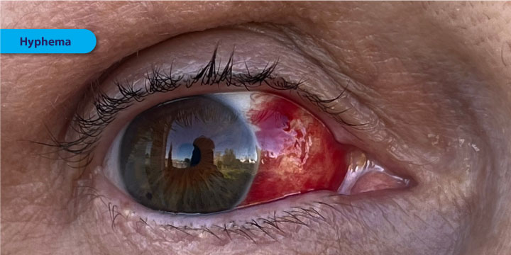
Hyphema is the collection of blood in the anterior chamber of the eye. The most common cause of hyphema is blunt trauma, though spontaneous hyphemas can occur in the setting of sickle cell disease or other increased bleeding states. Hyphemas are graded based on the degree of blood obscuring the cornea.

Hyphema is the presence of blood in the anterior chamber of the eye, the space between the cornea and the iris. It's typically caused by trauma to the eye, but can also occur after surgery or, rarely, spontaneously.

Symptoms include blurred vision, eye pain, light sensitivity, and visible blood in the eye. Immediate medical attention is crucial to prevent potential complications like glaucoma or vision loss.

Hyphema is defined as accumulated red blood cells (RBC) in the anterior chamber of the eye. Blood must be grossly visible, either on direct inspection or slit-lamp examination. Blood accumulates from disruption of the vessels of the iris or ciliary body, usually due to trauma or underlying medical conditions. The anterior chamber is the area bounded by the cornea anteriorly, the angle laterally, and the lens and iris posteriorly. This space normally contains clear, aqueous humor, which is produced by the ciliary body and drained through the Canal of Schlemm. The angle, an important anatomic location, is where the trabecular network and the Canal of Schlemm are located. Blockage of this location inhibits aqueous drainage leading to an increase in intraocular pressure.

Hyphema Causes and Risk Factors

In rare cases, certain medical problems like juvenile xanthogranuloma and cancer can cause a hyphema.

About 70% of hyphemas happen in children, especially in males ages 10 to 20. They’re usually caused by blunt injuries from activities like:

  • Sports
  • Industrial accidents
  • Falls
  • Fights
  • Shooting BB and airsoft guns

Less common causes include:

  • Eye surgery
  • Unusual blood vessels on your iris
  • Eye infections from a herpes virus
  • Blood clotting problems
  • Eye cancers

Some things that affect your blood can make you more likely to have a hyphema, such as:

  • Leukemia
  • Hemophilia
  • Sickle cell disease
  • Von Willebrand disease
  • Blood-thinning (anticoagulant) drugs

Hyphema Diagnosis

Your doctor will ask whether you’ve ever had an eye injury and what happened before the hyphema. It’s important for them to know if, for example, you were hit in the eye with a baseball or you ran into a tree branch.

The doctor will do an eye exam. This involves:

  • A visual acuity test to check how well you can see. They’ll also check the pressure inside your eye (called intraocular pressure).
  • Looking inside your eye with a special microscope called a slit lamp. A hyphema looks like a clot or layered blood in the front of your eye. If the anterior chamber is filled with blood, it’s called a total, black, or eight-ball hyphema. The doctor can also see if you have a microhyphema, which looks like a haze of red blood cells.
  • A CT scan to look at your eye sockets and other parts of your face, if the injury is severe
  • Screening for sickle cell disease or thalassemia in people of African descent

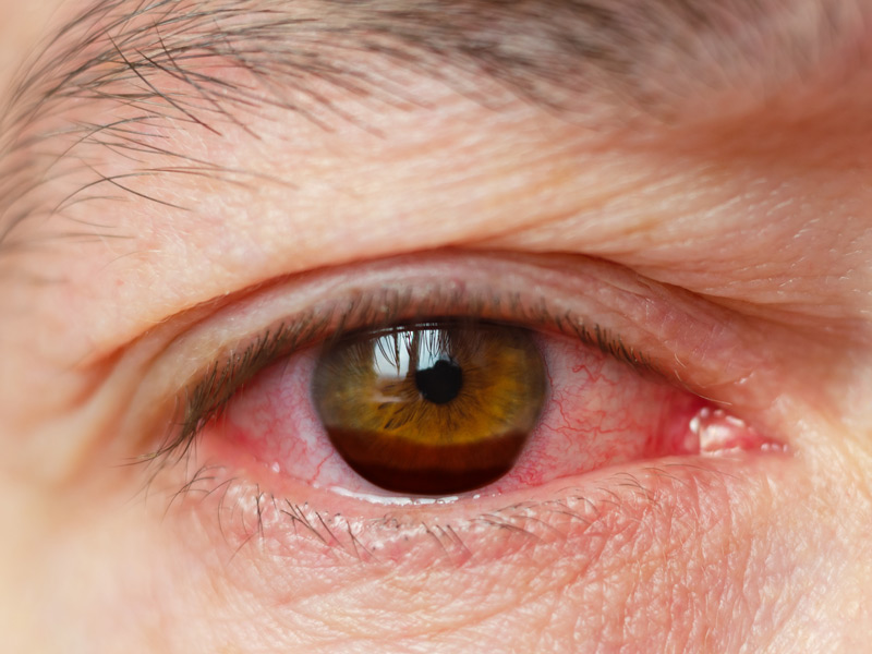
What are the symptoms of hyphema?

Let’s focus on the signs and symptoms that indicate hyphema:

Hyphema results in visual disturbance and vision changes- of course the degree of hyphema matters to a great extent in determining visual disturbance. If you have pupillary obstruction, you will be only able to detect some amount of light or identify hand movements.

In hyphema, the aqueous humour is replaced with blood- which eventually leads to bloody eyes. Make sure to book an online appointment in the initial stage- the eye specialists will let you know about the sizable hyphema.

In hyphema, blood in your anterior chamber causes pressure and irritation. It gives rise to unnecessary eye pain after a certain point in time.

Also referred to as photophobia, light sensitivity might end up hurting your eyes and causing headaches. If too much light exposure triggers your eye health, contact a doctor without any further ado.

  • Visual Disturbance
  • Bloody Eye
  • Eye Pain
  • Light Sensitivity

Severity and grading of hyphemas

Hyphemas are often graded based on the amount of blood present within the anterior chamber of the eye. This grading system helps healthcare professionals assess the severity of the condition and guide treatment decisions. The most commonly used grading system categorizes hyphemas into four grades: eResearch by Navid Ajamin -- winter 2024

Grade 0 (Microhyphema):

In this grade, there is a small amount of blood that is not easily visible to the naked eye. The blood may be detected using specialized medical equipment.

Grade 1:

In a Grade 1 hyphema, approximately one-third of the anterior chamber is filled with blood. This is when the blood is clearly visible and noticeable to the patient and healthcare provider.

Grade 2:

A Grade 2 hyphema involves blood filling about one-half of the anterior chamber. This level of hyphema is more extensive and can significantly affect vision.

Grade 3:

The most severe grade, Grade 3, occurs when the anterior chamber is filled with blood to the extent that it completely obstructs the view of the iris and pupil. This can lead to a significant decrease in vision and other complications.

The grading of hyphema helps in determining the appropriate course of treatment and monitoring.

Lower-grade hyphemas may be managed with conservative approaches, including rest, elevation, and eye drops to control inflammation and intraocular pressure.

Higher-grade hyphemas, especially Grade 3, are more likely to require closer monitoring and potential medical or surgical intervention to prevent complications such as elevated intraocular pressure or rebleeding.

It’s important to note that the grading system may vary slightly among medical professionals, but the general principles of assessing the amount of blood in the anterior chamber remains consistent.

If you suspect a hyphema, seek immediate medical attention to receive a proper diagnosis and appropriate treatment based on the severity of your condition.

HOW IS A HYPHEMA TREATED?

When treating a hyphema, the goals are to help clear the blood out, make sure the eye pressure stays normal, and prevent more bleeding. It is important for a person with a hyphema to rest and limit their activity for several days. The head should be kept higher than the feet, even while sleeping, and the eye needs to be protected with a shield. Doctors may prescribe steroid eye drops to help with inflammation and dilating drops (to make the pupil bigger) to help with pain. With a hyphema, it is important not to take aspirin or ibuprofen as they can make the bleeding worse.

Sometimes, the blood from a hyphema can clog normal eye drainage and cause high eye pressure. If the eye pressure stays too high for too long this can cause glaucoma and permanent eye damage. This can be more common in people with sickle cell anemia.

IS SURGERY EVER INDICATED FOR A HYPHEMA?

If the blood does not clear with rest and medication or if the eye pressure is too high for too long, then surgery may be needed to remove the blood.

ARE THERE ANY LONG-TERM EFFECTS FROM A HYPHEMA?

While many people heal well from a hyphema and get their vision back, there can be complications. If the eye bleeds again while healing or other parts of the eye are injured, there may be permanent vision problems. Sometimes the eye’s drainage canals are damaged due to the injury, causing glaucoma (vision loss from high eye pressure). An ophthalmologist can check for damage to the drainage canals with special tools and testing and then decide if ongoing monitoring is needed. It is important for parents, schools, and communities to encourage use of eye protection during sports and play to prevent eye injuries, including hyphema.

More details about hyphema:

Hyphema is essentially blood collecting in the front part of the eye, specifically within the anterior chamber.

Causes:

While trauma is the most common cause, other causes include eye surgery, certain medical conditions (like uveitis or blood disorders), and even spontaneous occurrences.

Symptoms:

Besides visible blood, patients with hyphema may experience pain, blurry or distorted vision, sensitivity to light, and headaches.

Treatment:

Treatment usually involves eye protection (like an eye shield), rest, and pain management. In some cases, more intensive treatments like surgery or hospitalization may be necessary.

Complications:

Untreated hyphema can lead to serious complications such as glaucoma, vision loss, and damage to the eye's structures.

Importance of seeking medical care:

It is crucial to seek immediate medical attention from an ophthalmologist if you suspect a hyphema, as prompt diagnosis and treatment are essential to prevent long-term vision problems.

Hyphema Treatment

Depending on the severity of a hyphema and associated risk factors, your eye doctor may recommend a combination of the following precautions and treatments:

  • Limited physical activity
  • Head elevation (including when sleeping)
  • Wearing an eye shield
  • Frequent follow-up visits for a few weeks or months
  • Pain medicine
  • Anti-inflammatory medicine (topical or oral)
  • Other medications

In the case of a severe hyphema, surgery may be required.

Do not use over-the-counter pain medications that contain aspirin or nonsteroidal anti-inflammatory drugs (NSAIDs) if you have a hyphema, as these medications can increase the risk of rebleeding in the eye.

Even if your eye feels fine and you don't notice vision problems, see an eye doctor immediately if you have eye trauma that could cause a hyphema. Make sure to attend all follow-up visits your doctor recommends.

Also, routine eye exams are very important after having a hyphema, as your risk of elevated eye pressure and glaucoma may be higher even years later.

How Can I Prevent A Hyphema?

The best way to avoid a traumatic hyphema is to wear safety glasses or other protective eyewear whenever you are involved in potentially hazardous activities.

Protective sports glasses should be worn when playing baseball, softball, racquetball, basketball, hockey or other sports that pose a risk of trauma to the eyes.

Also, be aware that sports like boxing significantly increase your risk of a traumatic hyphema. And if you participate in paintball games, wear protective headgear that includes a clear, impact-resistant shield that fully protects your face and eyes.

Reference:

  1. aapos.org/glossary/hyphema
  2. bceye.com/what-is-a-hyphema
  3. specscart.co.uk/blog/hyphema
  4. ncbi.nlm.nih.gov/books/NBK507802
  5. webmd.com/eye-health/hyphema-eye-internal-bleeding
  6. my.clevelandclinic.org/health/diseases/22586-hyphema
  7. allaboutvision.com/conditions/hyphema

The sclera is the medical term for the white of the eye. In general, sclerology refers to an alternative medicine practice that relies on examining the sclera for clues to overall health. These clues can involve the overall shape or color of the sclera, as well as the presence and pattern of red lines in the sclera.

This practice has apparently been around for thousands of years. Very old Chinese drawings of the sclera suggest that this method was used in ancient China. In addition, this method is also believed to have been practiced by certain Native American tribes, but this is based on word of mouth and not on actual writings or drawings.The basic idea behind sclerology is that subtle emotional or physical changes happening in the body will first show up in the sclera of the eye. For this reason, a main claim is that this method can detect these subtle problems before they begin to manifest a full range of symptoms. Some practitioners believe that a person’s innate weaknesses or predispositions for certain diseases can be seen in the sclera many years before they would be detected by Western science methods. By recognizing these weaknesses or predispositions, a person can then begin to alter his or her lifestyle to try to prevent, or at least delay, the onset of symptoms.

SCLEROLOGY BASICS COURSE – Grand Medicine Eyology | Iridology

Many sclerologists either are or work closely with herbalists or naturopaths and will suggest certain supplements or nutritional aids to deal with any problems seen in the sclera. Others may suggest relaxation or other stress-reducing techniques for a person whose sclera suggests that he or she is dealing with emotional problems. Another common goal of this practice is to pinpoint the true cause of a patient’s problematic symptom. For example, a patient struggling with being overweight despite exercising and healthy eating habits may see a sclerologist to help determine the true cause of his or her weight problem. A weight problem may result from thyroid problems, hormonal imbalances, or a wide range of other metabolic problems. Sclerology claims that the true cause of these types of issues will often be visible in the sclera.

Sclerology also claims that it can help monitor treatments. As a treatment changes the body, it will show in the sclera. Sclerology can therefore be used to monitor Western medical treatments or alternative treatment plans.

Although the majority of medical professionals do not believe that sclerology is a valid science, there are no known dangers to this practice. The only concern regarding this practice voiced by most doctors is that patients with serious health issues should not rely on sclerology and postpone Western medical treatment. Another drawback is that sclerology sessions tend to be expensive but insurance does not cover them.

Iridology / Sclerology

Eyes have long been referred to as the “Windows of the Soul.” But few people are aware of just how true this observation is. Accurate analysis of iris structure and pigmentation provides information about you or your clients that is hard to find through other methods. This information is so valuable that Iridology deserves to become a widely practiced assessment tool in both the physical and psychological health fields. There are a multitude of factors that influence our health and personality, and many of these factors are reflected in the iris. Look closely at your eyes in a mirror, and then at the irises of those around you. You will see many different patterns of iris fibres and colours. Like fingerprints or faces, no two are exactly alike, and neither are we! The iris structure is so unique; it is now being used for security identification at ATM machines and airports.

Iridology involves examination of the iris – the colour portion of the eye, and sclerology exams the red lines in the whites of the eyes. These analyses are a science and an art and are not diagnostic tools. It is a gentle non-invasive way of identifying inherited strengths and deficiencies in the body. Observing youreyes, enables me to get a broad overview of your health and genetic make-up.

Reference:

  • optimalhealthcentre.co.nz/iridology-sclerology
  • wise-geek.com/what-is-sclerology.htm

گاهی وقت ها سفیدی چشم به رنگ زرد درمی آید. این تغییر رنگ می تواند علل مختلفی داشته باشد، از جمله افزایش ماده ای به نام بیلی روبین در خون. بیلی‌روبین یکی از رنگدانه های صفراوی است که از تجزیه هموگلوبین حاصل می شود.زردي چشم ممكنست علل مختلفي داشته باشد. غير از علل عمومي آن در چشم ممكن است به دليل بيماري هاي پلك و يا ملتحمه ايجاد شده باشد كه با معاينه مشخص مي گردد. در غير اين موارد، تابش طولاني مدت آفتاب باعث ايجاد لكه هاي زرد رنگي در اطراف سياهي چشمي مي گردد و در بعضي از موارد بصورت ساختماني و بدون علت بيماري اين حالت ديده مي شود كه درمان خاصي براي آن وجود ندارد.

اگرچه زردی چشم زردی چشم همیشه نشانه ابتلا به هپاتیت نیست بلكه ممكن است فرد به سندرم «ژیلبرت» مبتلا شده باشد.

چند عامل شناخته شده که باعث ایجاد زردی چشم می‌شوند:

  1. التهاب حاد کبد Acute inflammation of the liver
  2. التهاب مجاری صفراوی Inflammation of the bile ducts
  3. گرفتگی مجرای صفرا Bile duct obstruction
  4. آنمی / کم خونی Anemia - وقتی تعداد زیادی از گلبول‌های خونی از بین بروند، میزان بیلی روبین تولیدی نیز افزایش می‌یابد) و یا کم خونی همولیتیک (افزایش سریع در سطح بیلی‌روبین به تغییر رنگ چشم منجر می‌شود. گاهی کل بدن بیمار تحت تاثیر کم خونی همولیتیک قرار می‌گیرد که این به ظاهر شدن لکه‌های زرد در تمام قسمت‌های بدن می‌انجامد.
  5. سندروم ژیلبرت- یک بیماری ارثی کبدی که در آن توانایی آنزیم‌ها برای انجام پروسه‌های آنزیمی کاهش می‌یابد
  6. بیماری کلستازیس - در این بیماری گردش صفرا در کبد منقطع می‌شود و به جای دفع در کبد باقی می‌ماند.
  7. تب هموراژیک - ملتحمه لایه‌ی پوستی بسیار ظریفی است و ممکن است بدون هیچ دلیل خاصی بترکد. این ترکیدن می‌تواند به تغییر رنگ چشم از سفید به قرمز روشن یا زرد منجر شود.بیماران مبتلا به هموراژیک ممکن است در خلال ترکیدن بافت هیچ درد یا اشکالی در بینایی احساس نکنند، اما، خارش و ورم چشم دو مورد از علائم رایج آن هستند.
  8. میخوارگی (اعتیاد به مصرف الکل) - همواره باید از نوشیدن الکل پرهیز کرد چون برای سلامتی بسیار خطرناک است و احتمال هپاتیت، مشکلات قلبی، سیروز، بی اشتهایی، عادات نادرست خواب، و سایر مشکلات و بیماری‌ها را افزایش می‌دهد.
  9. سیگار کشیدن
  10. تابش زیاد نور آفتاب - در واقع سفیدی چشم ها حالت مات و کدر پیدا می کند که این مسئله ارتباطی با بالا رفتن بیلی روبین خون ندارد.
  11. مصرف برخی داروها - مثل شیمی درمانی، هورمونی و یا مربوط به بدن سازی
    هپاتیت
  12. بیماری های گوارشی - این بیماری ها صرفا یک علامت ندارند و علامت های زیاد دیگری هم مشاهده می شود
  13. زردی ژنتیکی و نژادی - در این افراد پوست بدن و ملتحمه چشم و حتی زیر زبان به زردی می زند.این افراد هنگام گرسنگی و تشنگی دچار زردی پوست هم می شوند که معمولا گذراست.
    غلظت بیلی روبین (زردی خون) برخی از افراد به طور ژنتیكی بیش از دیگران است. این سندرم بیشتر در مردان به علت مسائل هورمونی پس از دوران بلوغ بروز می كند. در دوران بلوغ علایم این سندرم ظاهر می شوند البته آنان از بدو تولد به ژیلبرت مبتلا بوده اند.ممکن است زنان هم به ژیلبرت مبتلا باشند ولی چندان دچار زردی نمی شوند.این سندرم، بسیار شایع است به گونه ای كه ۱۰ درصد مردم جهان به آن مبتلا هستند.
  14. گرسنگی های طولانی مدت چند روزه، مصرف برخی از داروهای هورمونی و مربوط به بدنسازی باعث بروز بیشتر زردی چشم می شود. البته سایر آزمایش های افراد مبتلا به ژیلبرت، طبیعی است. مبتلایان به ژیلبرت در مصرف مواد غذایی، فعالیت بدنی و مصرف دارو محدودیتی ندارند و لازم نیست از داروی خاصی به علت ابتلا به این سندرم استفاده كنند.
  15. تب شالیزار (لپتوسپیروز) - رایج در افرادی است که در مناطق گرم‌تر زندگی می‌کنند. این عفونت معمولا به خاطر مصرف آب آشامیدنی ناسالم (آلوده به ادرار حیوانات) به وجود می‌آید. کسانی که به نوشیدن آب از حوضچه‌های راکد عادت دارند احتمال دارد به تب شالیزار مبتلا بشوند.

لکه زرد اطراف چشم ها
بیماری های قلبی می توانند یکی از دلایل به وجود آمدن این لکه ها باشند. این لکه های زردرنگ که اغلب در نزدیکی گوشه داخلی پلک به وجود می آیند، نرم، کوچک و بدون درد هستند و مشکلی در بینایی ایجاد نمی کنند. این مشکل اغلب با تصلب شرایین، دیس لیپیدمی و بیماری عروق کرونر در ارتباط است.

افرادی که دارای چشم حساس اند، بر اثر التهاب مزمن و جذب خون دچار زردی چشم می شوند که با گذشت زمان با استفاده از اشک مصنوعی رفع می شود. eResearch by Navid Ajamin -- spring 2013

افرادی که مبتلا به بیماری هپاتیت هستند نیز به عارضه زردی چشم دچار می شوند.بیماری هپاتیت، بیماری خطرناکی است که بلافاصله بعد از دیدن زردی چشم و رنگ زرد ادرار باید درمان شود.

افرادی که دارای چشم حساس اند به مرور زمان بر اثر التهاب مزمن با جذب خون و رفع قرمزی چشم به زردی چشم مبتلا می شوند این عارضه با گذشت زمان و تجویز اشک مصنوعی و ضدعفونی کننده ها توسط پزشک متخصص چشم رفع می شود.

زردی چشم در بعضی از افراد به صورت خال بروز می کند که از طریق عمل جراحی برطرف می شود.

زردی نوزادی

از مواردی كه در روزهای اول پس از تولد باید به دقت مورد توجه والدین قرار گیرد، تغییر رنگ پوست یا ملتحمه چشم نوزاد به زردی است.

بیش از 60 درصد نوزادان در روزهای اول پس از تولد دچار زردی می‌‌شوند. بروز این زردی در نوزادانی كه زودتر از موعد به دنیا آمده‌اند، بیشتر است.

علت بروز این زردی تجزیه بیشتر گلبول‌های قرمز و تولید ماده‌ای به نام بیلی روبین است.

نكته حائز اهمیت آن است كه در نوزادان این ماده به آسانی از سد مغزی - خونی عبور می کند و موجب آسیب سلول‌های مغزی می‌گردد كه در آینده خود را به صورت عقب‌ماندگی ذهنی و معلولیت‌های حركتی و ناشنوایی نشان می‌دهد. بنابراین زردی نوزاد از مواردی است كه باید آن را جدی تلقی کرد و حتما درمان نمود.

Image result for yellowish eyes

10 Home Remedies for Jaundice in Newborns -- parenting.firstcry.com

چنانچه میزان این زردی بالا نباشد، درمان با نور انجام می‌شود، اما در صورت زردی بالا، جهت جلوگیری از آسیب مغزی، تعویض خون باید صورت گیرد.

One type of discoloration of the front of the eye is conjunctival icterus,

which is the medical term for yellow eyes.(Sometimes, the term scleral icterus also is used to describe yellow eyes.)

The eyes usually start to turn yellow when a compound called bilirubin accumulates in the blood.

This type of yellowing is often referred to as jaundice.
Yellowing of the eyes and skin are almost always symptoms of a condition that requires medical treatment.
This can prevent serious complications, including organ damage.

Home remedies for yellow eyes include eating a healthful diet high in fiber and lean protein.
The best way to get rid of the yellowing is to treat the underlying cause and any other conditions present.

When jaundice is caused by an infection, such as hepatitis C or malaria, a person may need to take antibiotics, antifungals, or antivirals.

When jaundice is the result of alcohol or drug use, a person may need medical assistance to help with quitting or reducing consumption.

If dietary habits are behind jaundice, a person should eat more fruits, vegetables, whole grains, beans, legumes, and lean meats.

Jaundice can also result from organ damage, sometimes caused by:anemia, an injury , cirrhosis ,a blockage, cancer

Depending on the extent of damage and the organs affected, treatments may include surgery, radiation, chemotherapy, or blood transfusions.

Aside from a yellowing of the skin, one of the clearest signs of jaundice in an infant is the yellowing of the eyes.

Jaundice is very common in newborns, and only around 1 in 20 infants affected will require medical treatment.

Neonatal jaundice can usually be resolved by increasing breast-feeding sessions to 8–12 times daily.

The aim is to speed up digestion and bilirubin removal.

When treatment is necessary, a doctor may recommend phototherapy with fiber optic blankets.

Yellow eyes are only one symptom of newborn jaundice.

Jaundice is very common in newborn infants because the liver is still maturing.

New parents should also watch for the following symptoms:

yellow skin, lack of energy, irritability, fever, trouble with eating

Bilirubin often builds up faster than the immature liver of an infant can break it down, causing jaundice to occur frequently.

Some causes of newborn jaundice require further treatment. These include:

  • Blood incompatibility jaundice: When a mother and a fetus do not have compatible blood types, the mother's body may attack the red blood cells of the fetus while it is in the womb. As the mother's antibodies are already breaking down the infant's red blood cells before birth, this type of jaundice may occur as early as 1 day old.
  • Jaundice of prematurity: Premature babies are at the greatest risk of jaundice because their livers are highly underdeveloped. Premature babies may have more severe jaundice or jaundice alongside a number of other conditions.
  • Infections: Some bacterial infections, such as sepsis, can cause newborn jaundice.
  • Hemorrhage: Internal bleeding can cause jaundice. Premature infants face a particularly high risk of hemorrhages.

Melanin 
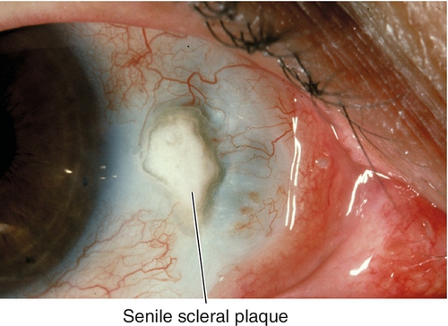
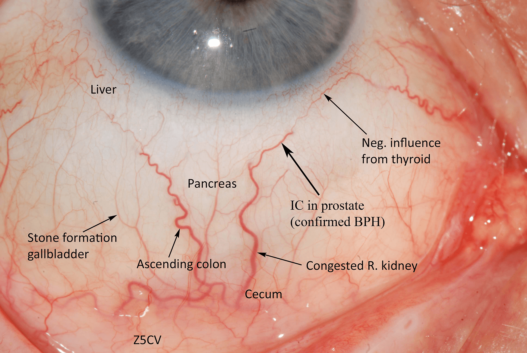
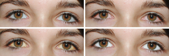
This right lateral sclera image shows melanin pigment, the brownish coloured 'splotch' you can see in the sclera. This sign is common as a pigmentation spot in a person with darker skin and brown/hazel eyes, representing a genetic predisposition to liver dysfunction. However it does not present the same pathological circumstance as when the melanin sign appears in a blue eyed, fairer skinned person, which is more important/consequential.When the melanin is located close to the iris, it shows mild to moderate liver hardening with associated bacterial infection. When located in an area/s away from the iris, it represents more severe hardening of liver tissue, with sugar system involvement. A substantial diet and lifestyle change, with ongoing liver cleansing and support, is necessary in this circumstance to avoid the condition and sign worsening over time.

Parasites & Worms The sclera image shows a very distinct parasite sign – a Protozoa to be specific. The line (in the left of the sclera) is very red and acidic and is shaped like a hook. Attached to this ‘hook’ are multiple enclosed spaces (circles or boxes) which is also an indicator that worms and parasites are present in the body. Parasites play a major role in the disease process, and can be detectable in many different signs within the sclera and the body. Many parasites, worms and viruses are polymorphic - which means they adapt very well to their environment. Some can play a beneficial role within the body when living in healthy, oxygenated tissue, but are also able to take on a negative form, attacking the body and increasing acidity and toxicity levels when their environment is mouldy or necrotic. With correct nutrition and a strict worm and parasite cleanse, these can be addressed and expelled from the body, with the lines eventually fading away.

 The Perpendicular The Perpendicular is a sign that shows trauma, from either cyst, tumour or physical injury, and is represented as one line joining another – usually from the in- or outside of a semi-circular line. This sign often appears in the uterine and testicular areas, usually present in clients suffering from neoplastic testicular/prostate involvement or cystic ovaries. Some people’s bodies just like to make cysts/tumours, more often than not though they are benign. It is important to look for additional signs that may explain the type of cyst/tumour and other possible related health problems to get a more accurate understanding of the body’s current state of health. To the right of the lower quadrant in the below eye is a Perpendicular located in the prostate. This Perpendicular line is also very obviously thickening toward the iris – which may represent a sign of active neoplasm. It is also important to note that there are worm/parasite pockets and bacteria present in this area, which are very common with this sign.

Liver Dysfunction The sclera image has a very strong, acidic line running into the liver area of the sclera (to the left of the iris). This shows acute congestion of the liver and is simply called the Liver Dysfunction sign. This may be caused from excessive intake of alcohol and drugs, an unhealthy diet high in processed foods or a profession in which airborne smoke-type toxins were regularly inhaled. This can be a common sign, with some sort of liver congestion evident in most sclera’s. With correct nutrition, gentle liver cleansing and ongoing liver support, this congestion can be reduced and the line will fade as a result of improvement in health.

Drug Imbedment The sclera image is of drug imbedment primarily in the colon – a straight line running parallel to a wavy line. This is a D1, early stages of drug imbedment of simple tissue. This can be reversed and the lines will, in turn, fade away with correct cleansing and nutrition. However, if the issue and general health of the individual is not addressed, it could lead to drug-induced lowered function, loss of function, tissue destruction and eventually drug-induced neoplasm.

Uneven Parallels: Fatty Buildup There are multiple uneven parallels in the medial quadrant of this right eye. This sign presents as it is named, as uneven parallels (one thicker line parallel to a thinner line), and indicates a degree of fatty buildup within the arterial walls. Although in this circumstance these lines here are are quite faint and thin, they are present throughout both of this client's eyes which may indicate a growing issue that should be monitored. If accompanied by fat metabolism dysfunction markings or liver congestion, it may indicate an issue with this clients ability to digest and metabolise fats, which may be causing the fatty buildup. Diet changes and supplementation should see these lines retreat.

Yellow Central heterochromia

Someone with central heterochromia has different colors within the same eye. Complete heterochromia is when they have two different colored eyes. Heterochromia of the eye is caused by variations in the concentration and distribution of melanin, the pigment that gives color to the skin, hair, and eyes.

What we do know about eye color determination is that it involves two pigments: melanin (brown pigment), and lipochrome (yellow pigment). It also depends on how the iris scatters light. When you see someone with light-blue eyes, it means there is an absence of melanin or brown pigmentation. Conversely, when you see someone with dark-brown eyes, they have an abundance of melanin.

زردی چشم ناشی از پینگوکولا (Pinguecula)

A pinguecula is a yellowish, slightly raised thickening of the conjunctiva on thewhite part of the eye (sclera), close to the edge of the cornea.

Pinguecula treatment depends on how severe the symptoms are. It's especially important for anyone with pingueculae to protect their eyes from the sun, since it's the sun's harmful UV rays that causes pingueculae to develop in the first place and encourages them to keep growing.

To help protect your eyes from pingueculae, shield your eyes from the sun whenever you are outdoors in daylight (even on overcast days because the sun's UV rays penetrate clouds).

Consider purchasing photochromic lenses, which darken automatically in sunlight and provide 100 percent UV protection. Photochromic lenses also shield your eyes from harmful high-energy blue light. Ask your eye care professional for details.

If a pinguecula is mild but accompanied by dry eye irritation or foreign body sensation, lubricating eye drops may be prescribed to relieve symptoms. Scleral contact lenses sometimes are prescribed to cover the growth, protecting it from some of the effects of dryness or potentially from further UV exposure.

Pingueculae also can lead to localized inflammation and swelling that is sometimes treated with steroid eye drops or non-steroidal anti-inflammatory drugs (NSAIDs). If dry eye is the cause of the pinguecula, eye drops formulated to treat dry eyes also may be prescribed.

Surgical removal of a pinguecula may be considered if it becomes especially uncomfortable, if it interferes with contact lens wear or blinking or if it is cosmetically bothersome.

26 possible conditions:- healthline.com/symptom/yellow-eyes

Image result for pinterest yellow eyes

  1. Jaundice occurs when there is excessive bilirubin in your system.
  2. Hepatitis refers to an inflammatory condition of the liver. It's commonly caused by a viral infection.
  3. A biliary obstruction blocks the bile ducts, which carry bile to the small intestine for digestion and waste removal.
  4. Damage to the liver from excessive drinking can lead to ARLD(Alcohol-Related Liver Disease).
  5. Cirrhosis is the severe scarring and poor function of the liver caused by long-term exposure to toxins such as alcohol or viral infections.
  6. Gallstones can block your bile duct and cause abdominal pain.
  7. Thalassemia is a blood disorder in which the body makes an abnormal form of hemoglobin.
  8. G6PD deficiency is a genetic condition caused by a lack of the G6PD enzyme in the blood.
  9. Acute pancreatitis is an inflammation in the pancreas, which causes pain and swelling in the upper left side of the abdomen, nausea, and burping.
  10. An ABO incompatibility reaction can occur if you receive the wrong type of blood during a blood transfusion.
  11. Newborn jaundice is a yellowing of a baby's skin and eyes.
  12. Red blood cells are normally shaped like discs, which allows them to travel through blood vessels. Sickle cell disease causes red blood cells to be sickle-shaped. (Sickle Cell Anemia)
  13. Drug-induced immune hemolytic anemia is a rare blood disorder.
  14. Liver Cancer
  15. Pancreatic cancer is one of the deadliest forms of cancer and is often difficult to detect.
  16. Breast milk jaundice is associated with breast-feeding.
  17. Yellow fever is a serious, potentially deadly flu-like disease spread by mosquitoes. It's characterized by a high fever and jaundice.
  18. Infectious mononucleosis, or mono, refers to a group of symptoms usually caused by the Epstein-Barr virus (EBV).
  19. Chlamydia is a sexually transmitted infection that may not present any noticeable symptoms. Although sometimes without symptoms.
  20. Calculus of gallbladder with acute cholecystitis occurs when a person has both gallstones and gallbladder inflammation.
  21. Hepatitis B is liver inflammation caused by the hepatitis B virus (HBV).
  22. The hepatitis E virus is transmitted via the intestinal tract and isn't caused by the hepatitis A virus.
  23. Hepatitis D, also known as the hepatitis delta virus, is an infection that causes the liver to become inflamed.
  24. the different types of hepatitis C.
  25. Hepatitis A is inflammation of the liver caused by the hepatitis A virus. This highly contagious form of hepatitis can be spread through contaminated food or water.
  26. Weil's disease is a severe form of the bacterial infection leptospirosis.

Treatment of yellow eyes focuses on the underlying medical condition.

Accompanying symptoms might include itchy skin, fullness in the stomach, fatigue, fever, pale stools, dark urine, loss of appetite, nausea and sudden weight loss.The best treatment of yellow eyes is determined by a number of tests, including one that measures the amount of bilirubin in the blood, a complete blood count and other liver tests.

The test results, along with a review of symptoms, medical history, a physical exam and possibly imaging tests, will help determine the proper diagnosis.If the underlying cause of yellow eyes is found to be an infection like hepatitis C or malaria, antibiotics, anti-fungal or anti-viral medications may be prescribed.

If alcohol or drug use are part of the diagnosis, giving up those substances will start the healing process.Diet also can play an important role. The liver processes and metabolizes most digested nutrients, and it works harder when foods are difficult to digest. This includes large amounts of refined sugars, salt and saturated fats.People with jaundice are advised to stay well-hydrated and to eat more liver-friendly foods — fruits and vegetables, whole grains, lean proteins, nuts and legumes.

As the liver begins to heal with treatment, the jaundice and yellow eyes will subside.In some cases, surgery may be necessary to correct a contributing factor like a blocked bile duct.

The following tips may help to reduce the yellowing of eyes:

  • Stay hydrated.
  • Consume enough dietary fiber, which can be found in whole fruits, vegetables, beans, legumes, and whole grains.
  • Eat lean protein, such as that from fish, nuts, and legumes
  • Avoid processed or packaged foods.
  • Avoid foods rich in saturated and trans fats.
  • Avoid refined carbohydrates, which can be found in sugary baked goods and candies.
  • Do not consume alcohol excessively.
  • Stop smoking or using tobacco products.
  • Refrain from using illegal drugs or abusing prescription medications.
  • Exercise regularly.

Reference:

  • prasadnetralaya.com/are-yellow-eyes-normal
  • owlcation.com/stem/rare-eye-colors
  • wikipedia.org/wiki/Gilbert's_syndrome
  • healthline.com/symptom/yellow-eyes
  • improveeyesightHQ.com/alcohol-and-eye.html
  • allaboutvision.com/conditions/yellow-eyes
  • allaboutvision.com/conditions/pinguecula.htm
  • medicalnewstoday.com/articles/321746.php
  • medicalnewstoday.com/articles/312403.php
  • medicalnewstoday.com/articles/312403.php#causes-in-children-and-adults
  • holisticeye.com.au/sclera-markings/?offset=1437965471821

See also:

  • How to whiten your eyes allaboutvision.com
  • What are Jaundice and Kernicterus? cdc.gov
  • Best Home Remedies For Yellow Eyes pickaremedy.com

The sclera (/ˈsklɪərə/) (from the Greek skleros, meaning hard), also known as the white of the eye, is the opaque, fibrous, protective, outer layer of the eye containing collagen and elastic fiber. In humans the whole sclera is white, contrasting with the coloured iris, but in other mammals the visible part of the sclera matches the colour of the iris, so the white part does not normally show. In the development of the embryo, the sclera is derived from the neural crest.

In children, it is thinner and shows some of the underlying pigment, appearing slightly blue. In the elderly, fatty deposits on the sclera can make it appear slightly yellow.

Red, Yellow, and Super-White Sclera

The sclera forms the posterior five-sixths of the connective tissue coat of the globe. It is continuous with the dura mater and the cornea, and maintains the shape of the globe, offering resistance to internal and external forces, and provides an attachment for the extraocular muscle insertions. The sclera is perforated by many nerves and vessels passing through the posterior scleral foramen, the hole that is formed by the optic nerve. At the optic disc the outer two-thirds of the sclera continues with the dura mater (outer coat of the brain) via the dural sheath of the optic nerve. The inner third joins with some choroidal tissue to form a plate (lamina cribrosa) across the optic nerve with perforations through which the optic fibers (fasciculi) pass.

The thickness of the sclera varies from 1mm at the posterior pole to 0.3 mm just behind the rectus muscle insertions. The sclera's blood vessels are mainly on the surface. Along with the vessels of the conjunctiva (which is a thin layer covering the sclera), those in the episclera render the inflamed eye bright red.

In many vertebrates, the sclera is reinforced with plates of cartilage or bone, together forming a circular structure called the scleral ring. The cornea and sclera make up the outer tunic of the eye. Each is a connective tissue containing collagen fibrils embedded in a proteoglycan-rich extrafibrillar matrix, but whereas the cornea is uniquely transparent, the sclera is totally opaque.

Related image

The eyes of all non-human primates are dark with small, barely visible sclera.

The sclera is made up of three divisions: the episclera, loose connective tissue, immediately beneath the conjunctiva; sclera proper, the dense white tissue that gives the area its color; and the lamina fusca, the innermost zone made up of elastic fibers.

Image result for sclera pinterest

The sclera is covered by the conjunctiva, a clear mucus membrane that helps lubricate the eye. It is thickest in the area surrounding the optic nerve.

The sclera is made up of three divisions:

  • the episclera, loose connective tissue, immediately beneath the conjunctiva
  • sclera proper, the dense white tissue that gives the area its color
  • the lamina fusca, the innermost zone made up of elastic fibers.

Image result for sclera structure

There are a number of abnormalities associated with the sclera. eResearch by Navid Ajamin -- spring 2012

Some are genetic and include:

  • Melanosis: excess deposits of melanin (pigment) on the surface of the sclera, which can become inflamed and uncomfortable
  • Scleral Coloboma: missing tissue that results in notching and bulging of the sclera (lesions)
  • Ectasia: a thinning and bulging of the sclera

What does a healthy sclera look like?

A healthy sclera is white. But what does it mean when the sclera takes on a different hue? If your whites become yellow or otherwise discolored, consult with your ophthalmologist. In some cases, this could signal an underlying health condition.

Reference:

  • eaglet-eye.com
  • en.wikipedia.org/wiki/Sclera
  • healthline.com/human-body-maps/sclera#1
  • link.springer.com/chapter/10.1007/978-0-387-73906-9_13
  • morancore.utah.edu/section-04-ophthalmic-pathology/sclera
  • aao.org/eye-health/tips-prevention/discolored-sclera-whites-of-my-eyes-turn-yellow
nicetoview.blogfa.com
عینک eyewear وبلاگ تخصصی عینک شامل مجموعه مطالب پزشکی است که اطلاعات مفیدی در رابطه با عینک , چشم، لنز، سلامتی چشم و راه های پیشگیری از بیماریهای چشمی، کنترل و درمان آن را در اختیار شما کاربر محترم می گزارد.

Always Be Healthy
همیشه سلامت باشید

Navid Ajamin نوید عجمین
eMail: navid.aj@outlook.com
موضوعات وب
پیوندها

unicef for every child

شمارنده