ابزار وبمستر

 What is a pterygium?

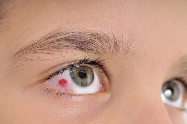
A pterygium (pronounced ter-ij-ee-um) is a wing shaped growth of tissue across the cornea, which is the clear window at the front of the eye. It is sometimes known as a “pearl“ because it looks white.

 It nearly always forms on the part of the cornea, which is visible when the eye is open. It is most often occurs in people who have lived in a hot dusty country or have worked outdoors for many years. It may be due to drying of the eye. It is not a cancer, but it can get slowly larger with time.

How does a pterygium affect the eye? 

In the early stages the eye may feel uncomfortable and look slightly red but vision is
unaffected. However, if the pterygium grows a lot, it may blur the vision, although this is
unusual.

What treatment is there for pterygium? 

If the pterygium is small, no treatment is required. If the eye is uncomfortable, lubricating
drops and / or ointment may help. Hypromellose or Liquifilm drops and Simple or Lacrilube
ointment are suitable. These can be obtained from your GP or bought at your local
pharmacy and can be used long term if needed.

If the pterygium advances until it is at the edge of the pupil or if it is enlarging and very
uncomfortable, it is best to have it surgically removed. 

What is the surgery for pterygium? 

This is usually performed under local anaesthesia as a day case in the operating theatre at
the Eye Unit. You can eat and drink normally before the operation .The eye is numbed with
drops and an injection and the eyelids are held open for you. The pterygium is scraped off
the cornea and the sclera (white of the eye). A piece of conjunctiva from under your upper
lid is removed and grafted onto the bare sclera, but the cornea is left to heal by itself. Only
absorbable stitches are used. These do not have to be removed but will dissolve and fall
out over the next few weeks. The eye will be covered with a pad.

What happens after the operation? 

You will be offered a drink before you leave the hospital. You will be given drops to use in
the eye once the dressing is removed. These drops need to be continued for several
weeks and you should get a repeat prescription from your GP if you think you will run out.
Do not stop the drops until the clinic doctor tells you to.
The eye may feel quite sore for a few days. You can to take painkilling tablets regularly.
The hospital will give you a small supply – ask you GP for more if needed.

You may need several follow up visits to clinic. The first one will be arranged before you
leave hospital. You may need at least a week off work so please ask the hospital for a
certificate if you need one.

Surgical removal is advisable in cases where: 

  • Redness due to numerous blood vessels in the pterygium makes the eye look bloodshot. (Topical medications can reduce this temporarily); 

  • The leading edge of the pterygium pulls the cornea, distorting its shape causing astigmatism; 

  • Normal vision is threatened by a large pterygium growing over the pupil;
    Persistent irritation, tearing and discomfort are experienced; 

  • Pterygium interferes with the wearing of contact lenses; 

  • Appearance of pterygium is unsightly.    

What problems are there after surgery? 

  • Pain - this should settle within a few days with painkillers, but please contact the
    hospital if it is getting worse despite regular medication. 

  • Redness. The eye may look redder for a few days after surgery but will gradually
    improve with time. 

  • Side effects from drops. Occasionally an allergy develops due to the drops or a
    pressure problem in the eye. The clinic doctor will check for these problems. 

  • The eye may still not have a perfectly smooth surface after surgery and lubricating
    drops may still be required. 

  • Scarring of the eye surface and eye muscles can occasionally cause restricted
    movement of the eye and double vision. Further treatment would probably help. 

  • The pterygium could come back again. This is much less common with modern
    surgery, but is occasionally very troublesome. Re-operation may be possible

The exact cause of pterygium isn’t known. One explanation is that too much exposure to ultraviolet (UV) light can lead to these growths. It occurs more often in people who live in warm climates and spend a lot of time outdoors in sunny or windy environments. People whose eyes are exposed to certain elements on a regular basis have a higher risk of developing this condition. These elements include: 

  • pollen 
  • sand
  • smoke
  • wind            eResearch by Navid Ajamin -- spring 2013 

Other risk factors include having light skin and light eyes.

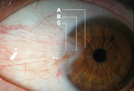
Diagnosing a pterygium is straightforward. Your eye doctor might be able to diagnose this condition based on a physical examination using a slit lamp. This lamp allows your doctor to see your eye with the help of magnification and bright lighting. If your doctor needs to do additional tests, they might include:

  • a visual acuity test, which involves reading letters on an eye chart
  • corneal topography, which is used to measure curvature changes in your cornea
  • photo documentation, which involves taking pictures to track the growth rate of the pterygium

If possible, avoid exposure to environmental factors that can cause a pterygium. You can help prevent the development of a pterygium by wearing sunglasses or a hat to shield your eyes from sunlight, wind, and dust. Your sunglasses should also provide protection from the sun’s UV rays. If you already have a pterygium, limiting your exposure to the following can slow its growth:

  • wind
  • dust
  • pollen
  • smoke
  • sunlight

It can also help prevent them from coming back if you’ve had any removed.

Reference:  

  • Pterygium - patient information University Hospital Southampton - NHS Foundation Trust  
  • healthline.com/health/pterygium#treatments6
  • rodgerdavies.com.au/pterygium

See Also:

  • Pterygium   emedicine.medscape.com

  • Pterygium   Patient Education Video (18+)

  • Pterygium   nlm.nih.gov

ناخنک یا پتریژیوم pterygium ، یک ضایعه گوشتی مثلثی شکل است که از سفیدی چشم (ملتحمه) روی سیاهی چشم (قرنیه) کشیده می‌شود. معمولا در طرف بینی چشم است. این ضایعه ناشی از رشد خوش‌خیم بافت پیوندی و رگهای ملتحمه است. ناخنک معمولاً ابتدا به صورت یک سفیدی روی ملتحمه ایجاد می‌شود که به تدریج به سمت قرنیه رشد می‌کند. به علت وجود رگهای زیاد معمولاً رنگ ناخنک صورتی یا قرمز است. ناخنک گاهی ملتهب می‌شود و باعث سوزش و خارش و اشکریزش می‌شود. گاهی اوقات ناخنک رشد زیادی می‌کند و تا وسط قرنیه می‌رسد و جلوی دید را می‌گیرد. به علاوه حتی ناخنک‌های کوچکتر هم می‌توانند با تغییر شکل قرنیه باعث ایجاد آستیگماتیسم شوند و دید افراد را تار کنند.

در ناخنک‌های خیلی بزرگ که وسط قرنیه را هم درگیر کرده‌اند ممکن است لازم باشد علاوه بر اقدامات فوق از پیوند قرنیه لایه‌ای برای اصلاح شکل قرنیه استفاده شود.

پينگوکولا (Pinguecula)

پينگوکولا که در بالغين فوق‌العاده شايع است. ضايعه به‌صورت ندول‌هاى زردرنگى در هر طرف قرنيه (به‌طور شايع‌تر در طرف داخل) در سطح بين پلک‌ها ديده مى‌شود. از بافت الاستيک و هيالن تشکيل مى‌شود و به‌ندرت اندازه آنها افزايش مى‌يابد. التهاب آنها شايع است. درمانى لازم نيست؛ ولى در مورد التهاب آن (پينگوکوليت)، ممکن است استروئيد ضعيف موضعى يا داروهاى ضدالتهاب غيراستروئيدى موضعى تجويز شود.

لازم به ذکر است که برخی از تومورهای چشمی ممکن است در ابتدا با ناخنک اشتباه شوند. پس در مواردی که ناخنک رشد خیلی سریعی دارد و یا مکرراً عود می‌کند.حتماً باید با چشم پزشک مشورت کنیم.

علت ناخنک و پیشگیری

اشعه ماوراء بنفش خورشید (UV) در ایجاد ناخنک مؤثر است. همچنین باد شدید و گرد و خاک با تحریک چشم در ایجاد ناخنک مؤثر می‌باشند.

بنابراین توصیه می‌شود افرادی که در مناطق آفتابی زندگی می‌کنند و مدت زیادی در معرض تابش آفتاب هستند از کلاه لبه‌دار و عینک آفتابی استفاده کنند.در افرادی که ناخنک دارند معمولاً ورود آب به چشمها (در موقع شستن دست و صورت یا حمام کردن) باعث تحریک چشم و ایجاد سوزش و قرمز شدن چشمها می‌شود. بنابراین این افراد باید دقت کنند که حتی الامکان آب وارد چشمشان نشود.

درمان

اگر ناخنک کوچک باشد و ظاهر ناخوشایندی نداشته باشد و باعث قرمزی و سوزش چشمها نشود نیاز به درمان خاصی ندارد.
اگر ناخنک گهگاه ملتهب شود و سوزش و قرمزی چشم ایجاد کند مصرف قطره‌های اشک مصنوعی به صورت مکرر ممکن است باعث تسکین علائم شود. در مواردی که ناخنک التهاب شدیدی دارد می‌توان با نظر چشم پزشک یک دوره درمان با قطره‌های استروئیدی یا سایر قطره‌های ضد التهاب انجام داد.

در مورد ناخنک‌های بزرگتر که از لحاظ ظاهری و زیبایی اهمیت دارند و یا با ایجاد آستیگماتیسم باعث تاری دید شده‌اند می‌توان با جراحی ناخنک را برداشت. البته اگر ناخنک به صورت ساده برداشته شود احتمال عود آن زیاد است و به خصوص در افراد جوان و افرادی که ناخنک ملتهب دارند در نیمی از موارد، عود دیده می‌شود. در این افراد بهتر است علاوه بر برداشتن ناخنک از روشهای تکمیلی مثل پیوند ملتحمه یا استفاده از داروهای خاص (مثلا میتومایسین) در حین جراحی استفاده شود.


Pterygium
A pterygium is a growth of pink fleshy tissue. It will usually start close to your nose and grow slowly towards the centre. Often there are no other symptoms, but a pterygium can irritate your eye and cause redness.If a pterygium grows onto your cornea (which is the layer in front of your iris and pupil), it can cause astigmatism and blurry vision

Symptoms may include: eResearch by Navid Ajamin -- spring 2010

  • Blurry vision
  • Itchy eyes
  • Feeling of something being in the eye
  • Gritty sensation
  • Burning, redness, and irritation

You can treat the irritation caused by a pterygium by using lubricating eye drops, and sometimes anti-inflammatory eye drops, which you can get from your optometrist or GP. If it's affecting your vision, or if the appearance worries you, you can have surgery to remove it.

Slit-lamp photos of different grades of pterygium and results of lymphatic vessel endothelial hyaluronan receptor 1 immunohistochemistry for the three grades of pterygia: A and D: Grade 1; B and E: Grade 2; C and E: Grade 3. Microvessels were dramatically increased in Grades 2 and 3 pterygia. Lymphatic vessel endothelial hyaluronan receptor 1 (LYVE-1) was positive for lymphatic vessels but negative for blood vessels. Red arrows point to lymphatic vessels and blue arrows, blood vessels (original magnification=100X).

Usually you'll have to get this done privately, but if it's severe it may be publicly funded. Talk to your GP or optometrist about your options.

Pinguecula
A pinguecula is a white or creamy colour, and doesn't grow across your cornea. It can cause irritation, redness, discomfort, and dryness.

Related image
You can also treat a pinguecula with lubricating eye drops or anti-inflammatory drops, which you can get from your optometrist or GP.

It's very rare to have a pinguecula surgically removed, but this can happen if it's very uncomfortable or you're worried about how it looks.

Usually you'll have to get this done privately, but if it's severe it may be publicly funded. Talk to your GP or optometrist about your options.[2]

A Pterygium is a usually harmless condition affecting the conjunctiva of the eye – often on the side closest to the nose. If it grows across the cornea, it can cause scarring and sometimes loss of vision. The growth may also distort the shape of the cornea, causing vision problems.

Pterygium images

One or both eyes can be affected and it’s more common in people between 20 and 40 years.

The exact cause of pterygium isn’t known. One explanation is that too much exposure to ultraviolet (UV) light can lead to these growths. It occurs more often in people who live in warm climates and spend a lot of time outdoors in sunny or windy environments. People whose eyes are exposed to certain elements on a regular basis have a higher risk of developing this condition. These elements include: pollen, sand, smoke and wind.

Diagnosing a pterygium is straightforward. Your eye doctor may diagnose this condition based on a physical examination using a slit lamp. This lamp allows your doctor to see your eye with the help of magnification and bright lighting. If your doctor needs to do additional tests, they may include:

  • Visual acuity test. This test involves reading letters on an eye chart.
  • Corneal topography. This medical mapping technique is used to measure curvature changes in your cornea.
  • Photo documentation. This procedure involves taking pictures to track the growth rate of the pterygium.

What treatments can help with pterygia?

Artificial teardrops, which are available without prescription from pharmacies, lubricate the surface of the eye to improve comfort and redness. They can be used safely on a regular basis. If a pterygium becomes particularly inflamed non-steriodal or steriod anti-inflammatory drops may be prescribed for a short period, under supervision of an eye specialist. Decongestant drops, such as ‘Clear Eyes’ are not recommended as they can cause rebound redness, making the eyes worse. Protecting the eyes from ongoing ultraviolet light exposure may reduce the chance of a pterygium enlarging, so sunglasses should be worn when outside. When symptoms persist despite medical treatment, surgical removal of the pterygium may be required.

Image result for pterygium yu[road

What does pterygium surgery involve?

Surgery may be recommended if a pterygium is causing persistent discomfort, redness or cosmetic concerns, and if it is growing progressively larger or interfering with vision or contact lens wear. Surgery can be performed on one eye or both eyes at the same time.

Pterygium surgery is generally done under local anaesthetic as a day-stay procedure. It takes about half an hour for one eye and is not particularly uncomfortable. The pterygium is excised from the surface of the eye, and then a small piece of healthy conjunctival tissue, from under the upper lid, is transplanted onto the exposed white of the eye. Although traditionally held in place with stitches, Eye Doctors use fibrin glue in the majority of cases, to make the surgery quicker and reduce the irritation on the eye afterwards. The transplant helps the eye to heal with an improved cosmetic appearance and reduces the risk of pterygium recurrence, to about one in fifty.

What happens after surgery?

Following surgery a patch is placed on the eye overnight and drops are prescribed, usually about four times daily for four-six weeks. Painkillers are also recommended in the first one-two days, because the eye may be quite uncomfortable. The eye may look red for up to a month after surgery and there may be mild swelling or discomfort during this time, as the eye heals.

Eye drops may be used to treat symptoms, such as inflammation, mild pain, itching or a feeling of having grit in the eye. Treatment for a minor irritation includes eye drops or ointments that help to lubricate and soothe the cornea. In more severe cases, you may be prescribed a short course of steroid eye drops.

When outdoors, wear good-quality, wrap-around sunglasses as recommended by an optometrist. Choose sunglasses that meet the Australian Standard and have an EPF UV rating of either 9 or 10.

A wide-brimmed hat will also protect your eyes from sun and wind exposure.

A pterygium reduces the vision in several ways:

  1. Distortion of the corneal optics. This begins usually when the pterygium is greater than 2mm from the corneal edge (limbus).
  2. Disruption of the tear film. The tear film is the first lens in the eye. Pterygia are associated with eyelid inflammation, called Blepharitis.
  3. Growth over the corneal centre, which leads to dramatic reduction of vision.
  4. Induced anterior corneal scarring, which often remains after surgical removal.

Key points about pterygium

  • Men are usually more likely to develop a pterygium than women.
  • A pterygium is a type of noncancer growth on your eye. It is often only a minor problem.
  • Many people don’t have any symptoms. But you might have symptoms such as eye irritation, itching, burning or redness.
  • You might have blurred vision if your growth covers a large part of your cornea.
  • You might only need treatments such as eye drops to reduce discomfort.
  • Some people need surgery to remove their growth, if it is causing major symptoms. But it often grows back after surgery.

Reference:

  1. daneshju.ir
  2. healthinfo.org.nz
  3. youngeyes.com.au/blog/pterygium_126s79
  4. en.wikipedia.org/wiki/Pterygium
  5. researchgate.net
  6. healthline.com/health/pterygium#diagnosis
  7. oceyeinstitute.com/articles/92630-eye-doctor
  8. cedars-sinai.org/health-library/diseases-and-conditions/p/pterygium.html
  9. eyedoctors.co.nz/services/pterygium
nicetoview.blogfa.com
عینک eyewear وبلاگ تخصصی عینک شامل مجموعه مطالب پزشکی است که اطلاعات مفیدی در رابطه با عینک , چشم، لنز، سلامتی چشم و راه های پیشگیری از بیماریهای چشمی، کنترل و درمان آن را در اختیار شما کاربر محترم می گزارد.

Always Be Healthy
همیشه سلامت باشید

Navid Ajamin نوید عجمین
eMail: navid.aj@outlook.com
موضوعات وب
پیوندها

unicef for every child

شمارنده