ابزار وبمستر

قرمزی چشم یا التهاب ملتحمه، التهاب و عفونت پرده شفافی است که زیر پلک ها و قسمتهایی از کره چشم را پوشانیده است.  علل قرمزی چشم معمولاً عفونتهای ویروسی یا باکتریایی است و یا می تواند حساسیتی بوده و در نوزادان می تواند ثانویه به عدم ایجاد سوراخ مجاری اشکی بطور کامل باشد.


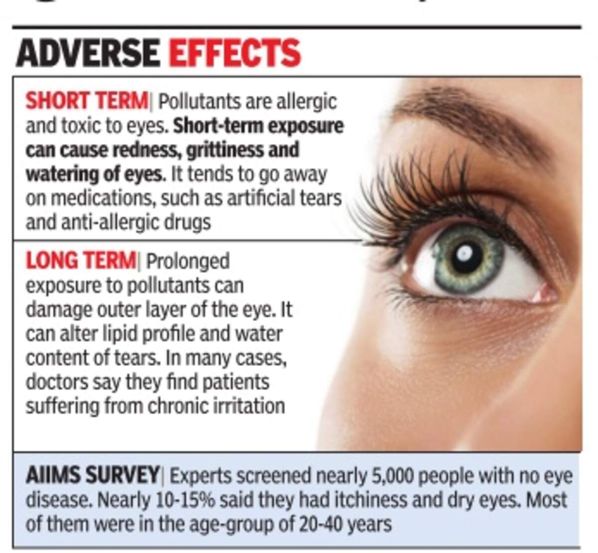
این بیماری ممکن است منجر به بروز احساس وجود خارجی در یک یا هر دو چشم شده ، به گونه ای که خود فرد احساس می کند قادر به بیرون آوردن جسم خارجی نیست، زمانی که فرد از خواب بیدار می شود، ممکن است چشم وی پر از ترشحات باشد. سفیدی چشم نیز به نظر قرمز آمده و فرد منظره ای را می بیند که قبلاً در چشم وجود نداشته است.

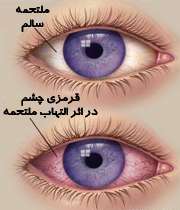
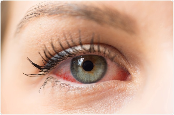
وجود التهاب سبب می شود که عروق کره چشم در مقایسه با قبل بیشتر مشخص شوند و همین عامل سبب می شود که سفیدی چشم به نظر قرمز رسد.قرمزی چشم اصطلاحی است که معمولاً برای بیان وجود التهاب ملتحمه به کار می رود.

بنظر می رسد که بروز التهاب علاوه بر ایجاد قرمزی برروی بینایی نیز گاهی تأثیر می گذارد.اگر فردی دچار قرمزی چشم شد با انجام اقدامات خاصی می تواند شکایات خود را برطرف نماید.ولی از آنجایی که قرمزی چشم می تواند مسری باشد، لذا باید هرچه سریعتر تحت درمان قرار گیرد.این درمان سریع بخصوص در سنین قبل از مدرسه که کودک می تواند به التهاب ملتحمه باکتریایی یا ویروسی مبتلا شود بسیار مهم است.

علائم و نشانه ها

          شایعترین علائم و نشانه های قرمزی چشم شامل خارش و یا قرمزی در یک یا هر دو چشم ، تاری دید و حساسیت به نور، وجود انواع ترشحات در یک یا هر دو چشم است که در طی شب منجر به تشکیل دلمه در چشم ها می شود و نهایتاً فرسودگی چشم خواهد بود.

علل

 باکتریها،ویروسها و حساسیت می توانند منجر به بروز قرمزی چشم شوند اما شایعترین علت ویروسها می باشند. گاهی اوقات نوزادان ثانویه به عدم باز شدن کامل مجرای اشکی نیز
می توانند مبتلا به قرمزی چشم شوند.
در ورم ملتحمه باکتریایی یا ویروسی، معمولاً یک یا هر دو چشم  مبتلا می شوند این دو فرم باکتریایی  و ویروسی می توانند با سرماخوردگی ارتباط داشته باشند، و هر دو فرم بسیار مسری هستند. این فرمها معمولاً در اطفال شایعتر بوده، اما بالغین نیز به همان نسبت می توانند مبتلا شوند.

در ورم ملتحمه حساسیتی معمولاً هر دو چشم مبتلا می شوند و معمولاً ثانویه به مواد حساسیت زا نظیر گرده گلها ایجاد می شود. در پاسخ به مواد حساسیت زا بدن شروع به ساختن موادی میکند که ایمنوگلوبولین E نام دارند . این مواد سبب تحریک سلولهای انتهایی شده و منجر به ترشح مواد دیگری گشته که آنجا موجب بروز به التهاب میشوند . از جمله این مواد هیستامین است. که آزاد شدن هیستامین در بدن میتواند منجر به بروز علائم حساسیت، نظیر قرمزی چشم ها شود. فرد مبتلا به قرمزی چشم حساسیتی ممکن دچار خارش شدید چشم ها شـود و علائم دیگری نظیر خارش بدن ، عطسه و ترشحات آبکی از بینی پیدا کند، بعلاوه ممکن است پلکها تا حدی دچار تورم شوند .

فاکتورهای خطر :

عوامل خطر که شاخص ابتلا را افزایش میدهند شامل تماس با مواد حساسیت زا و به همان میزان تماس با فردی که مبتلا به ورم ملتحمه باکتریائی یا ویروسی شده است میباشد . فرمهای ویروسی و باکتریایی معمولاً بسیار مسری بوده و فرد مبتلا میتواند تا 7 الی 14 روز بعد از شروع علائم سبب سرایت بیماری به دیگران شود .

چه زمانی باید به پزشک مراجعه نمود ؟

          قرمزی چشم یک حساسیت تحریک کننده بوده اما در اکثر مواقع برای بینایی بی ضرر است و به ندرت نیاز به درمان فوری و شدید پیدا می کند. ولی از آنجایی که این بیماری می تواند تا 2 هفته بعد از شروع علائم مسری باشد لذا تشخیص و درمان به موقع بسیار مهم می باشد. کودکان مبتلا به ورم ملتحمه باکتریایی باید تا شروع درمان از سایر کودکان دور نگه داشته شوند. اما فرم ویروسی تنها تا چند روز مسری هستند. اگر مشکلی در ارتباط با زمان بازگشت کودک خود به مدرسه دارید حتماً با پزشک در میان گذارید. گاهی اوقات ورم ملتحمه سبب بروز عوارضی در قرنیه می شود که همین امر نیاز به درمان به موقع را مسجل می سازد.

غربالگری و تشخیص

          برای تشخیص عامل بیماری ممکن است پزشک از ترشحات نمونه ای تهیه کرده و به آزمایشگاه بفرست تا بدین ترتیب عامل بیماری مشخص شده و درمان بهتری صورت گیرد. اگر بیمار کودکی باشد که مرتباً دچار قرمزی چشم می شود. باید از نظر وجود انسداد در مجاری اشکی توسط پزشک مورد معاینه و ارزیابی قرار گیرد.

درمان

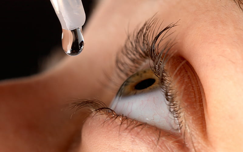
  اگر عفونت باکتریایی وجود داشته باشد نیاز به استفاده از قطره های آنتی بیوتیکی بوده تا حد ممکن عفونت هر چه سریعتر درمان یابد. به منظور پیشگیری از عود عفونت باید دوره درمان بطور کامل طی شود. معمولاً عفونتهای ویروسی نیز به خودی خود در طی یک هفته درمان می شوند. اگر فرم حساسیتی وجود داشته باشد، ممکن است از انواع قطره های چشمی جهت درمان استفاده شود که شامل آنتی هیستامین ها، ضد اختقانها، کورتونها و قطره های ضدالتهابی می باشند.

پیشگیری

          داشتن یک وضعیت بهداشتی خوب بهترین راه کنترل قرمزی چشم است .

 زمانی که عفونت تشخیص داده شد باید اقدامات ذیل انجام شود.

  • هرگز با دست خود چشمهایتان را نمالید.
  •  هر زمان که ممکن است و بصورت مکرر دستهای خود را بشویید.
  • هر روز حوله ها و لباسهای خود را بشویید و اجازه ندهید کسی دیگری از آنها استفاده کند.
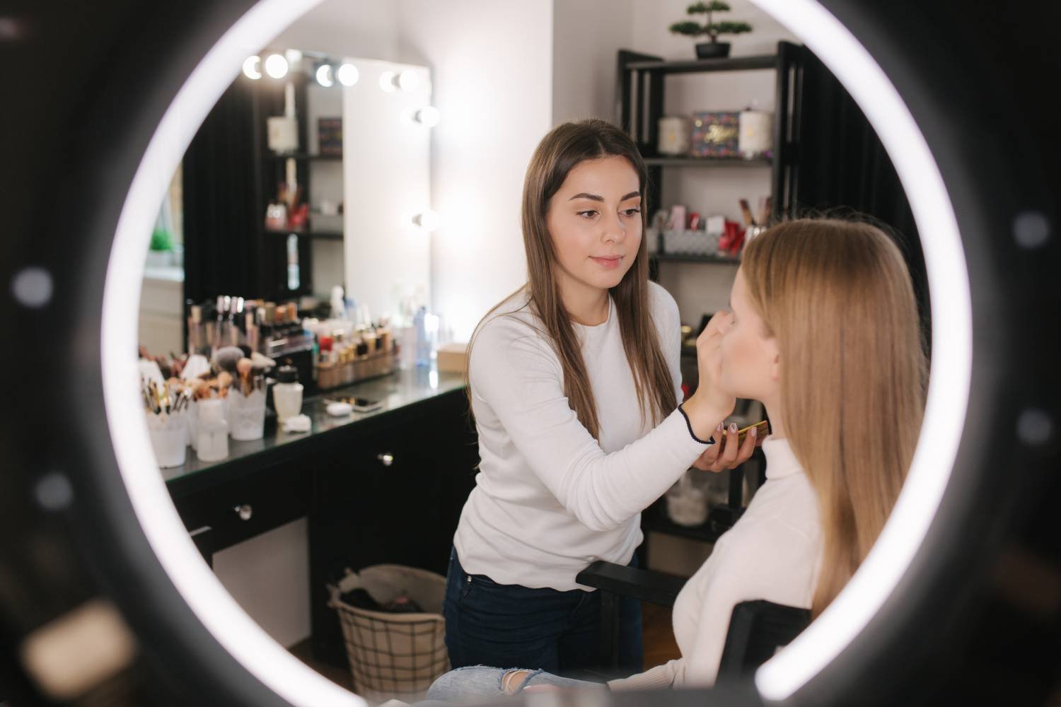
  • وسایل آرایشی چشم های خود را دور اندازید و به کسی دیگر اجازه ندهید که از آنها استفاده کند و هرگز به دیگران اجازه ندهید که از وسایلی که با چشم های شما تماس داشته اند، استفاده کنند.
  • به توصیه های پزشک خود در ارتباط با نحوه محافظت از لنزهای تماسی چشم خود عمل نمایید.

          اگر کودکتان مبتلا به قرمزی چشم  بوده و یا قرمزی چشم در مدرسه آنها شیوع پیدا کرده است از تماس آنها با سایرین ممانعت کنید. بسیاری از مدارس کودکان مبتلا را به خانه می فرستند.

          نوزادان بسیار مستعد ابتلا به عفونتهای چشمی باکتریایی موجود در کانال زایمان هستند. این باکتریها در مادر هیچگونه علامتی ایجاد نمی کنند.گاهی اوقات این باکتریها در نوزادان منجر به بروز عفونتهای چشمی بسیار شدیدی می شوند که نیاز به درمان فوری دارند. وجود همین اختلال سبب شده است که بلافاصله بعد از تولد از قطره های آنتی بیوتیکی چشمی نظیر اریترومایسین استفاده شود.

مراقبت از خود

          فرد می تواند با استفاده از کمپرسهای گرم برروی چشم مبتلا از شدت علائم آن بکاهد. بدین منظور از یک پارچه تمیز استفاده نموده و آنرا بعد از قرار دادن در آب ولرم و فشردن آن ، آنرا به آرامی بر روی چشم های خود قرار دهید.

          در موارد حساسیتی از مالیدن چشم هایتان خودداری ممانعت کنید، چرا که سبب رها شدن بیشتر هیستامین خواهد شد،در عوض می توان از کمپرس سرد برای تسکین علائم استفاده نمود. در این موارد از قطره های چشمی آنتی هیستامین و منقبض کننده های عروقی استفاده نمائید.

منبع: مركز پژوهش هاي سلامت خليج فارس  www.pghrc.ir

معمولا افرادی که با کامپیوتر زیاد سر وکار دارند در هنگام کار با کامپیوتر بعد از مدتی دچار سوزش ، خستگی ، چشم درد و سردرد میشوند .بنابراین لازم است جهت جلوگیری از ایجاد این مشکلات نکات زیر را رعایت نمایید.

١- رعایت فاصله استاندارد که بهترین فاصله حدود ۴٠ تا ۵٠ سانتیمتری است .

٢- نور محیط بایستی نور کافی و استاندارد باشد استفاده از لامپ های با نور سفید و زرد توام با هم بهترین نور میباشد البته مقدار نور به اندازه ای زیاد نباشد که چشم دچار خیرگی شود .

٣- صفحه مونیتور به شکلی قرار داده شود که نور منبع روشنایی و یا نور محیط بیرون به طور مستقیم به آن نتابد .

۴- مونیتور باید درست در مقابل چشم و در راستای محور بینایی هر دوچشم باشد .

۵- جهت جلوگیری از خشکی چشم تعداد پلک زدن بایستی نسبت به مواقع عادی بیشتر باشد .

۶- از مونیتور های استاندارد که تشعشعات کمتری دارند استفاده شود .

٧- سعی کنید چشمان خود را به مدت طولانی روی صفحه مونیتور خیره نکنید و در هنگامی که نیاز به نگاه کردن به مونیتور ندارید چشم خود را از مونیتور برداشته وبه فاصله های دور تر نگاه کنید .

٨- معمولا بعد از حدود دو ساعت کار با کامپیوتر حدود ١۵ تا ٣٠ دقیقه استراحت نمایید و در این فاصله یک نوشیدنی شیرین و یا یک شیرینی سالم مانند خرما _ بیسکویت یا عسل استفاده کنید .

٩- کنتراست و روشنایی مونیتور را به شکل استاندارد تنظیم نمایید .

١٠- در صورتی که تمامی نکات را به درستی رعایت کردید و باز هم دچار مشکل شدید ممکن است مشکلی در سیستم بینایی شما وجود داشته باشد مثلا چشم شما ضعیف باشد و یا چشمان شما دچار عدم تعادل عضلانی باشد بنابراین بهتر است جهت کنترل سیستم بینایی به متخصص بینایی سنجی و یا چشم پزشک مراجعه نمایید .

١١- افرادی که نمره چشم آنها دوربینی و یا استیگماتیسم است حتما بایستی از عینک استفاده نمایند . افراد نزدیک بین ممکن است در نمرات کم نیازی به استفاده از عینک نداشته باشند و یا در نمرات بالا هم ممکن است به نمره کمتری نیاز داشته باشند بنابراین در این مورد با متخصص خود مشورت نمایید .


What are the symptoms?

Computer vision syndrome symptoms include:

  • Eye discomfort (most common). This can feel like dryness, watering, itching, burning or the sensation of something in your eye.
  • Blurred vision that comes and goes. You may notice it when refocusing your eyes from near to distance or distance to near.
  • Sensitivity to bright lights.
  • Trouble keeping your eyes open.
  • Headache behind your eyes.

Many people with computer vision syndrome also develop aches and pains elsewhere in their bodies. This can happen when your posture isn’t ideal or you stretch and strain your neck to see your screen. Symptoms can include pain in your:

  • Neck.
  • Shoulders.
  • Back.

What causes computer vision syndrome?

Extensive use of digital devices causes computer vision syndrome. Some research shows that continuous screen use for at least two hours can be enough to trigger symptoms. The longer you spend staring at screens, the more likely you are to develop symptoms.

The screens themselves aren’t fully to blame. Spending a long time focusing on close-up objects can strain your eyes, whether you’re looking at a screen or a printed page. But the screens add another layer of strain to your eyes. Here’s why:

  • Constant refocusing. When you use screens, you have to constantly focus and refocus to see the print (which is made of pixels, or tiny dots). Constantly moving your eyes in this way can strain them.
  • Screen contrast levels. Often, there’s a low contrast level between the letters you read on a screen and their background. This can make your eyes work harder.
  • Inadequate blinking. Normally, you naturally blink about 18 to 22 times per minute. You need to blink enough to keep your eyes lubricated. But when using a computer, most people only blink three to seven times per minute. Screen use may also cause incomplete blinking. This means you only partly close your eye when you blink. Not blinking fully or often enough can cause the surface of your eyes to dry out.

What are the risk factors for this condition?

You face an increased risk of developing computer vision syndrome if you use computers or other digital devices for at least four hours per day.

Your symptoms may be more severe than usual if you:

  • Have dry eye disease.
  • Have uncorrected or under-corrected refractive errors, like nearsightedness.
  • Have eye conditions that affect your eyes’ ability to focus, like convergence insufficiency.

Reference: Computer Vision Syndrome: Symptoms, Treatment & Prevention (clevelandclinic.org)

Before we elaborate on the pink eye remedy, we have to find out what is pink eye (conjunctivitis)?

Pink eye is an inflammation of conjunctiva - the membrane that covers the eye and lines the inner surface of the eyelid.

Related image

The three most common types of pink eye are:

  • viral,

  • allergic, and

  • bacterial

Each requires different pink eye remedy.

Other than the allergic type, pink eye is typically contagious.

What are the signs and symptoms of pink eye?

Various combinations of the following symptoms may be present in cases of pink eye:

  • Itching eye

  • Redness in eye

  • Photophobia (sensitivity to light)

  • Swelling of the lids and/or discharge from the eyes The consistency of possible discharge may range from watery to purulent (pus-like), depending on the specific cause of the pink eye.

What causes pink eye (conjunctivitis)?

Pink eye (conjunctivitis) is often caused by viruses. It can also be caused by bacterial infections, allergies, chemicals, irritation from contact lenses or pollutants, as well as eye injury. Viral and bacterial pink eye are very contagious.

Bacterial or viral infections
These may occur when micro organisms are introduced or transmitted into the eye through contaminated hands, the use of washcloths or towels, cosmetics, use of false eyelashes or through the extended usage of contact lenses.

It is also quite common for a minor pink eye to accompany a viral cold or flu. Although bacterial infections and some of viral infections, particularly herpes, are not very common, they are potentially serious. Both types of infection are contagious.

Irritants
Another major cause of pink eye is the presence of irritants, which may take the form of pollutant or particles in the air, smoke, soap, hairspray, makeup, chlorine, cleaning fluids, among other. Some people may also get pink eye due to a seasonal allergic response to grass and pollen.

Pink Eye Remedy

It usually takes about a few days or up to two weeks for pink eye remedy to clear up the eye problem. Pink eye that occurs due to an allergy may continue for as long as the offending pollen or irritant is present. With such conditions, symptoms are likely to recur each year or on a regular basis, even with proper pink eye remedy.

Pink eye remedy may vary depending on the cause. Eye health medication in the form of ointments, eye drops or pills may be used to eliminate the infection in the eye, or to alleviate and relieve the allergic symptoms and discomfort. In the case of pink eye that occurs due to a viral cold or flu, your doctor may recommend that you be patient and let the viral infection run its course.

These are some of the steps you should take in the pink eye remedy:

  • Apply a cool compress to the infected eye or eyes about three to four times a day for 10-15 minutes. Make sure you use a clean washcloth or towel each time. This should help to reduce itching and swelling and alleviate some of the discomfort.

  • Wash your hands frequently and be sure to keep them away from your eyes in order to reduce or prevent recontamination.

  • Avoid rubbing your eyes. This may contribute to the spread of the infection and increase the irritation.

  • Wear sunglasses if your eyes are sensitive to the light.

  • Avoid exposure to the irritants that may be causing the pink eye.

  • Dispose of old eye makeup if the culture for bacteria, taken by the doctor, turns out to be positive.

  • Change your pillowcase each night. Alternatively, you can change your pillowcase every other day and turn it over nightly.

  • Avoid wearing contact lenses while you are using medication on your eyes, or if you feel discomfort in the eyes. Clean your contact lenses thoroughly before you begin using them again.

Can pink eye (conjunctivitis) be prevented?

Although many kinds of pink eye (conjunctivitis) are hard to prevent, you can take some measures to decrease the risk of re-contamination or spreading it to someone else .

Here are some eye health measures you can take to prevent re-contamination:

  • Do not share eye makeup or cosmetics of any kind with someone else. As far as possible, avoid sharing washcloths or towels.

  • Wash your hands frequently and keep them away from the eyes.

  • Wear protective goggles when you go swimming or when you have to work in a place that is exposed to chemicals that may irritate the eyes.

  • Do not use eye health medication, such eye drops, ointment, etc., which have not been prescribed for you. Someone else's prescription is not be suitable for you.

  • Avoid swimming in stagnant lakes or ponds.

  • Do not save remaining eye health medication for future use. Dispose of expired medication.

Red Eyes: Prevention & Cool Tips on How to Take Care!

Image result for red eye

A red eye is a very common condition, an eye that appears red due to illness or injury. Fortunately, many a time, the red eye is of short duration, not very troublesome, and resolves by itself.


causes of red and bloodshot eyes
Simple red eyes usually clear by remedies such as washing with plain cold water, applying cold packs to closed eyes, taking a short period of rest, etc.

Commonest cause is conjunctivitis, either due to allergy, or due to infection. Allergies can cause itchy, red eyes that appear swollen. Infection, on the other hand, can cause stickiness of the eye lids or eye lashes with some discharge in addition. While the very mild may clear by themselves, others will need specialist treatment.

Other causes of red eye, which need proper treatment are, keratitis (injury/infection of cornea), scleritis & episcleritis (involvement of white layers of eye).

Glaucoma or iritis can also cause redness of the eye, and these conditions would need immediate treatment. Acute glaucoma, if treatment is delayed, can lead to permanent loss of vision.

To prevent delay in treatment, any one of the following feature indicates that an urgent consultation with an ophthalmologist is important:

Along with redness of eyes, if there is:

(1) pain in or around the eyes.
(2) watering from the eyes.
(3) photophobia, or inability/hypersensitivity to tolerate light.
(4) diminution of ability to see clearly, which was good until the onset of red eye.

Lubricating eye drops and general soothing eye drops usually give some relief in almost all the conditions. That could be a reason sometimes when we may delay seeking specialist consultation.
One big danger is in using steroid eye drops, while very essential in treating some of the conditions mentioned above, has exactly the opposite and very harmful result in some other conditions. Hence, only a well trained ophthalmologist can identify these conditions quickly and advise accordingly.

Eye haemorrhage: Causes and treatments

Eye redness occurs when the vessels in your eye become swollen or irritated. Redness of the eye, also called bloodshot eyes, can indicate the presence of several different health problems. While some of these problems are quite innocent, others are serious and require proper medical attention.

Red eyes may be caused by anything from a poor night’s sleep to a more serious medical condition. But Mark Mifflin, M.D., an ophthalmologist with University of Utah Health Care’s Moran Eye Center, warns that attempts to whiten eyes often do more harm than good.

A subconjunctival hemorrhage often occurs without any obvious harm to your eye. Even a strong sneeze or cough can cause a blood vessel to break in the eye. You don't need to treat it. A subconjunctival hemorrhage may look alarming, but it's usually a harmless condition that disappears within two weeks or so.

To safely reduce redness of the eye, the following courses of action should be followed:

A professional eye exam. Many factors, including dry eye, eyelid inflammation, allergy, skin type, smoking, and exposure to dust or chemicals can cause or contribute to eye redness. Preservative free artificial tears or prescription eye drops prescribed by your eye care provider can help. Also, getting enough sleep is important, as is eating a variety of fruits and vegetables, and protecting the eyes from UV rays and dry air.

Avoid any eye drops or other medications that promise to reduce redness. “Generally these medicines temporarily narrow blood vessels. The effect may last for several hours, but they are always followed by rebound dilation,” says Mifflin. He also says that nearly all of these eye drops contain medications or preservatives which can be toxic to the surface of the eye when used repeatedly.

Be aware: discoloration of the white of the eyes can sometimes be a sign of a serious medical condition. For example, a yellow tinge could signify liver disease, or a dramatic increase in red might be due to an abnormality of the blood vessels inside the head or neck. Once again, consulting your optometrist is a good place to start.

Reference:

  • eye-exercises-for-good-vision.com
  • dejonghoptometry.co.za/red-eyes-night-driving-tips
  • practo.com/healthfeed/red-eyes-prevention-cool-tips-on-how-to-take-care
چشم یکی از اندام هایی است که شاید در نگاه اول به نظر بیاید سلامت آن چندان با تغذیه مرتبط نیست، ولی مشکلات چشمی ‌مانند آب مروارید (کاتاراکت)، آب ‌سیاه (گلوکوم)، خون ‌ریزی‌های درون چشمی ‌و نیز کم‌بینایی و نابینایی در برخی موارد، عوارض بیماری‌هایی مانند فشار خون بالا، دیابت و مشکلات عروقی هستند که در اثر تغذیه ناصحیح، چاقی و شیوه زندگی ناسالم به وجود می آیند.


چشم‌ها، شبکه‌ای از سلول‌های حساس هستند که برای عملکرد خود به مقادیر کافی مواد مغذی‌ نیاز دارند.

کمبود‌های تغذیه‌ای در درازمدت روی بینایی آثار سوء خواهد گذاشت و به همین علت در سراسر جهان در عرصه‌ علم چشم پزشکی، علاقه زیادی به بررسی نقش تغذیه و مکمل‌های غذایی در سلامتی چشم‌ها و بهبود بینایی وجود دارد.

 ویتامین‌های دوستدار چشم

ویتامین‌های A، C، E سه آنتی‌اکسیدان معروف هستند که نقش آنتی‌اکسیدانی و نیز نقش کلیدی در واکنش‌های متابولیک و عملکردی چشم دارند.

 ویتامین A

نقش بسیار مهمی ‌در سلامت چشم دارد و برای تطابق فرد به تغییر نور ضروری است و هنگام استفاده از نور چراغ، کامپیوتر، تماشای تلویزیون و انجام کارهای ظریف چشمی ‌مصرف بیشتری در بدن دارد. کمبود شدید این ویتامین منجر به اختلالاتی مانند کاهش بینایی در شب و شب‌کوری می گردد.

گزروفتالمی ‌یا خشکی قرنیه، شب‌کوری، کاهش بینایی در شب و نابینایی در کودکی همگی از عوارض کمبود ویتامین A هستند و متاسفانه افراد زیادی را در کشورهای در حال توسعه دچار کرده است.


آمارها نشان می‌دهد که هرساله نیم میلیون کودک در سراسر جهان کور می‌شوند که ۷۰ درصد علت آن، کمبود شدید ویتامین A در رژیم غذایی است.

بررسی‌های انجام شده در کشور ما، حاکی از وجود کمبود ویتامین A به خصوص در مناطق محروم می‌باشد. بر اساس گزارشات بین‌المللی در زمینه شیوع کمبود ویتامین A در کشورهای شرق مدیترانه، ایران از نظر کمبود در حد متوسط قرار دارد.

ویتامین A و پیش‌سازهای آن در مواد غذایی مانند میوه و سبزی‌هایی به رنگ زرد و نارنجی مثل هویج، زرد آلو ، انبه، شیر و لبنیات، جگر، ماهی، زرده تخم مرغ و همچنین انواع سبزی‌‌های برگ سبز یافت می‌شود.

ویتامین‌های گروه B

‌اعمال مختلفی به عهده دارند؛ از جمله در شکل‌گیری گلبول‌های قرمز و رساندن اکسیژن به سراسر بدن نقش دارند.

ویتامین B1 (تیامین):

در عملکرد سریع ماهیچه چشمی ‌و تعادل در حرکات چشم نقش دارد. افراد الکلی علایم کمبود شدید این ویتامین را نشان می‌دهند.
در سالمندان نیز کمبود ویتامین B1 دیده می‌شود.

منابع سرشار این ویتامین:

سبزی‌ها، مخمر و نوشیدنی‌های تخمیری(ماءالشعیر) ، گوشت و مرغ و ماهی، جگر، تخم مرغ، غلات سبوس دار و موز هستند.

ویتامین B2 (ریبوفلاوین):

در تنظیم سیستم بینایی چشم و ایجاد تعادل و حساسیت بینایی در چشم نقش دارد. در افراد دیابتی دفع ادراری این ویتامین افزایش یافته و در نتیجه میزان نیاز به‌ این ویتامین بیشتر خواهد بود.
برخی داروها مانند کلروپرومازین و باربیتورات‌ها میزان نیاز به ویتامین B2 را افزایش می دهند.

کمبود آن در موارد اولیه عوارضی چون سوزش چشم، ترس از نور، ریزش اشک و همچنین احتمال سریع ‌تر ابتلا به آب مروارید را موجب می‌شود.

این ویتامین نیز در غذاهایی مانند:شیر و لبنیات، گوشت‌های بدون چربی، جگر، سبزی‌ها، میوه‌ها، غلات سبوس دار یافت می‌شود.

ویتامین C :

این ویتامین که عملکرد ضد پیری دارد، در سلامت استخوان‌ها و بافت‌های بدن نقش دارد. همچنین موجب سلامت دهان و لثه‌ها، خونسازی و جریان خون مناسب در بدن به ویژه در چشم‌ها می‌شود.
تحقیقات نشان داده است، افرادی که در سنین بالا دارای عدسی چشمی ‌سالم و شفاف بوده‌اند، در طول زندگی از مقادیر کافی ویتامین C استفاده ‌کرده‌اند.

ویتامین C در سبزیجات و میوه های تازه و بیشتر در مرکبات (پرتقال، گریپ فروت ، انواع لیمو و نارنگی)، کیوی ، انواع توت ، گوجه فرنگی ، فلفل دلمه ای، هندوانه ، طالبی ، توت فرنگی ، سیب زمینی و سبزیجات برگ سبز مثل کلم بروکلی موجود است.

ویتامین E :

در دفاع بدن و شکل‌گیری گلبول‌های قرمز نقش دارد و کمبود آن به اختلالات ته‌چشم از جمله تخریب نقطه زرد چشم و بیماری تخریب ماکولا (از بین رفتن سلول های ناحیه مرکز بینایی که در سنین بالا اتفاق می افتد و سبب کاهش متوسط تا شدید بینایی می گردد) می‌انجامد.

روغن‌های گیاهی مثل روغن بادام زمینی ، سویا، ذرت، گلرنگ (کانولا)، آفتابگردان و جوانه‌گندم مهم‌ترین منابع غذایی ویتامین E به ‌شمار می‌روند. سایر منابع شامل:
دانه‌های روغنی، بذرها، غلات کامل(سبوس‌دار) و سبزی‌های برگ سبز است.

۴۰ درصد تصادفات به علت دید ناكافی، ۱۲ درصد به علت ندیدن علائم رانندگی، ۱۰ درصد به دلیل برف و باران، ۳ درصد مه و دود، ۳ درصد نور شدید و ۱۱ در صد نیز بر اثر عوامل دیگر رخ داده است. كه با توجه به این آمار اهمیت سلامت چشم و دید خوب در رانندگی مشخص می شود.

Image result for cell phones while driving

داشتن چشمان سالم از ضروریات یك رانندگی ایمن است . افراد ممكن است در حالت سكون، دید بسیارخوبی داشته باشند، اما در رانندگی با سرعت بالا شرایط به كلی تفاوت دارد. دید راننده زمان حركت اتومبیل، ممكن است بر اثر شرایط وعوامل خارجی به میزان زیادی مختل شود .

دمای هوا، میزان گرد و غبار، نور شدید، مه، برف و باران و رعد و برق، تمیز نبودن شیشه جلو یا شیشه چراغ ها، دود سیگار یا حتی كثیف بودن شیشه عینك راننده نمونه هایی از این عوامل و شرایط هستند.

  • Texting and driving makes an accident 23 times more likely.
  • Dialing your phone increase your chances of an accident by 2.8 times.
  • 1 in 5 drivers confess to surfing the web while driving.
  • Smartphones are the most common form of distraction for drivers.
  • Making even the most basic text takes a minimum of 5 seconds of your attention off of the road when you text and drive.
  • In addition to causing 330,000 injuries each year, it’s estimated that about 11 teens die every day as a result of texting and driving. And this is just teens – this is about 4000 total deaths per year in the United States

What kind of visual function is necessary for driving? Visual acuity and field of vision (visual field) are the most important factors for safe driving. Vision regulations for driving vary from state to state, so check local laws to find out what your requirements are.

While driving, the visual system is on alert and constantly under pressure. A good vision is essential in order to perfectly visualize the route and to be able to anticipate the different events. Good acuity and a wide visual field are therefore necessary when driving.

As research shows 1 in 5 drivers in the world has uncorrected poor vision and 90% of the information needed to drive comes from vision. The interaction between eyeglasses and the interface systems in the car, both day and night, is obviously crucial.

طبق گزارش‌های شركت های بیمه ای بین سال های ۱۹۹۸ تا ۲۰۰۵ علت تصادفات خودروها در سرعت بالا به شرح زیر بوده است :

۴۰ درصد تصادفات به علت دید ناكافی، ۱۲ درصد به علت ندیدن علائم رانندگی، ۱۰ درصد به دلیل برف و باران، ۳ درصد مه و دود، ۳ درصد نور شدید و ۱۱ در صد نیز بر اثر عوامل دیگر رخ داده است. كه با توجه به این آمار اهمیت سلامت چشم و دید خوب در رانندگی مشخص می شود. به همین منظور، توصیه می شود رانندگان هر سال دید چشم خود را كنترل كنند و با معاینه سالانه چشم، از سلامت دید خود هنگام رانندگی مطمئن شوند . بهتر است در این معاینات تست های كوررنگی و شب كوری هم انجام شود.

جالب است بدانید رانندگان با ۱۰ سال سابقه رانندگی حدود ۱۰ درصد و رانندگان با ۲۰ سال سابقه رانندگی حدود ۲۰ درصد دیدشان را از دست می‌دهند و این نشان می‌دهد كه رانندگان حرفه‌ای باید بیشتر مراقب چشمانشان باشند.

Image result for driving in snow

The visual requirements for driving include acuity of at least 20/60 or better in at least 1 eye with a single lens system; binocular horizontal field of vision of at least 35 degrees to the left and right side of fixation; and binocular vertical field of vision of at least 25 degrees above and below fixation.

Related image

Good eyesight is a basic requirement for safe driving. Poor vision increases the risk of collisions due to the driver’s inability to recognise and react in time to a hazard or the behaviour of other road users.

Image result for driver visual field

However, poor vision is believed to be massively underreported in government crash causation data due to the difficulty in determining if eyesight was to blame. Some casualties are likely to occur because drivers are unaware they have a vision problem and have neither corrected it nor reported it to the Driver and Vehicle Licensing Agency (DVLA). Untreated eye conditions can occur gradually over time. In extreme cases, someone can lose up to 40% of their vision without being aware they have a problem.

Estimates from the Royal College of Optometrists suggests 2-3% of drivers have vision below the minimum standard.

Drivers with visual field defects have double the incidence of road crashes and traffic violations compared to drivers with a full visual field, and almost half people with visual field loss are unaware of the problem.

But despite this, many drivers do not get their eyes tested regularly or even at all.

some terms and Definitions:

  • Visual Acuity: The ability to see clearly.
  • Distance Vision: The ability to see in the distance.
  • Depth Perception: Vision that gives objects their three-dimensional appearance and that enables a person to judge the relative distance between two objects.
  • Central Vision: The 3-percent cone at the center of your focus. Focusing on objects is done with the help of central vision.
  • Peripheral (Fringe) Vision: The area of vision to the left and right of the area of central vision.
  • Color Vision: A crucial aspect in traffic safety is color vision. Color vision is the ability to see colors.
  • Night Vision: The ability to see in low and variable light conditions.
  • Glare Vision: The ability to see when there is a rapid increase in light.
  • Glare Recovery: The ability of your eyes to quickly adjust from headlights back to the dark.
  • Vehicle-Reaction Time: The time it takes a vehicle to respond.
  • Visual Lead: The distance you can see ahead of your vehicle.
  • Visual Control Zone: The zone where you identify objects/conditions that may require a response or continuous attention.
  • Response Zone: The zone where you begin to respond to what you perceive.
  • Margin of Safety: Areas of roadway large enough to allow you the space, time, and visibility needed for safe movement at anytime.

Central cronic vision. You might find this hard to believe but our sharpest, clearest vision, called central conic, is only about 3 to 5 degrees .

Central vision is the most important part of a person's vision. It is used to read, drive, and see pictures or faces. Good central vision allows a person to see shapes, colours, and details clearly and sharply. An area of the retina (the lining of the back of the eye) called the macula provides central vision.

Central vision and fringe vision are both about of your field of vision. Central vision is what is directly ahead of you and 10-degrees out from that, while fringe vision is part of your peripheral vision that helps to monitor your zone condition that is defined by your central vision.

Related image

For safe driving, fleet drivers must have their eyes on the road, hands on the wheel, and full attention and focus on the task at hand. These three types of distracted driving – visual, manual and cognitive – are what can be the most risky.

Yes, you may still be able to drive a car or motorcycle if you only have one eye or sight loss in one eye (sometimes called "monocular vision"). This is provided that: the DVLA is satisfied that you have sufficient sight in your other eye (including a normal field of vision).

میدان دید راننده هنگام رانندگی بسیار با اهمیت است . هرچه میدان دید بیشتر باشد، كنترل خودرو هنگام خطر راحت تر صورت می گیرد. میدان دید ممكن است تحت تاثیر عوامل متعددی كاهش پیدا كند. خستگی، هیجان، ترس، نگرانی، بی‌احتیاطی، مصرف داروهای آرام بخش، سیگار كشیدن و مصرف مشروبات الكلی از این عوامل هستند. توصیه می‌شود رانندگان قبل از مصرف هرگونه دارو با پزشك مشورت كنند و در صورت داشتن اثرات خواب آور و كاهش هوشیاری از رانندگی اجتناب كنند.

بیشتر از ۹۰ درصد از مجموع عواملی كه شرایط رانندگی را در كنترل دارند در اختیار چشم است. هرچه چشم سالم‌تر باشد و راننده هنگام رانندگی دقت كافی داشته باشد، احتمال بروز حادثه به میزان زیادی كاهش پیدا می‌كند.

Image result for driving in dark

رانندگی در شب به خاطر وجود نور شدید و ناگهانی كه به چشم راننده می‌رسد قدرت تطابق چشم را تحت تاثیر قرار می‌دهد. عمل تطابق همان آماده شدن چشم برای پذیرش شدت‌های مختلف نور در مقاطع زمانی مختلف است و درست در همین لحظات است كه احتمال بروز حادثه به بیشترین حد خود می‌رسد.

در گزارش علت غرق شدن كشتی مسافربری تایتانیك، به كم شدن تطابق چشم ناخدای كشتی در تاریكی دریا اشاره شده كه سبب ناتوانی او در دید كوه یخی كه كشتی با آن برخورد كرده، شده است.

حتی هنگام روز كه راننده از محیط روشن به محیط تاریك یا از محیط تاریك به محیط روشن وارد می شود (مثلاً تونل‌ها) این حالت اتفاق می‌افتد و كاهش دید به علت عدم تطابق احتمال بروز حادثه را بیشتر می‌كند.‌[1]

A 1985 study by K. Rumar, using British and American crash reports as data, found that 57% of crashes were due solely to driver factors, 27% to combined roadway and driver factors, 6% to combined vehicle and driver factors, 3% solely to roadway factors, 3% to combined roadway, driver, and vehicle factors, 2% solely to vehicle factors and 1% to combined roadway and vehicle factors. eResearch by Navid Ajamin -- winter 2010

Visual acuity is usually recorded as:

  • "Uncorrected," which is without glasses or contact lenses
  • "Best corrected," which is with the best possible glasses or contact lens prescription

What is the minimum eyesight standard for driving?

The legal eyesight standard means that you must be able to read a number plate from 20 metres. You must not have been told by a doctor or optician that your eyesight is currently worse than 6/12 (decimal 0.5) on the Snellen scale. If you are in any doubt, you should discuss with your optician or doctor.

How many meters is the eye test?

For uncorrected visual acuity, you will be asked to remove your glasses or contact lenses and stand or sit 20 feet (6 meters) from the eye chart. You will keep both eyes open.

Human factors in vehicle collisions include all factors related to drivers and other road users that may contribute to a collision. Examples include driver behavior, visual and auditory acuity, decision-making ability, and reaction speed.[2]

Reference:

  • salamat.ir روزنامه سلامت
  • media.group.renault.com
  • en.wikipedia.org/wiki/Traffic_collision
  • brake.org.uk/facts-resources/15-facts/491-eyesight
  • Visual acuity test: MedlinePlus Medical Encyclopedia medlineplus.gov/ency/article/003396.htm
  • A good vision for a safer driving - Essilor Pro-Eyecare (essilor-proeyecare.eu)
  • State Vision Screening and Standards for License to Drive lowvision.preventblindness.org
  • The Importance of Good Vision for Driving & Minimum Vision Requirements epermittest.com
  • Driving safety: an accident is likely to occur 23 more times with cell phone use – The Andersons (andersonscanada.com)

این روزها بحث آلودگی هوا و وجود ذرات ریز معلق در آن، از جمله مهم‌ترین موضوعاتی است که بر سلامتی ما سایه ‌افکنده است. شاید تاثیر آلودگی‌ هوا بر ریه و قلب مشخص‌تر از دیگر اندام های بدن باشد و کمتر کسی از تاثیر این آلودگی‌های زیست‌محیطی بر روی چشم اطلاع داشته باشند. این مشکل در کلان‌شهرها به‌ویژه در تهران بارزتر است.

تقریبا همه تاثیر آلودگی هوا را در بدن خود حس می‌کنند. در هوای آلوده، دود ناشی از سوخت خودروها، ریزگردها، مونوکسیدکربن، دی‌اکسیدنیتروژن، ازن، آلاینده‌های آلی، ذرات غیرعادی دیگر مثل آزبست، همه و همه تاثیرات مضر جبران‌ناپذیری روی بدن انسان می‌گذارند. البته آثار آلودگی هوا بر افراد، با مشکل قلبی و ریوی قابل مشاهده است. تنگی نفس و کبودی لب‌ها از علایم آن می‌باشد.

ریزگردها ذراتی بسیار ریزتر از شن و به قطر چند میکرون هستند که به راحتی در ریه رفته و حتی از آن عبور می کنند و در طولانی مدت در عروق ریز رسوب کرده و باعث التهاب عروق می‌گردند (شبیه به آترواسکلروز) و باعث کم‌شدن کیفیت خون‌رسانی به چشم ها می‌شوند که درست مثل افزایش میزان مونوکسیدکربن خون، باعث خاکستری دیدن رنگ‌ها می‌گردند.

Image result for pollution affects eyes

وقتی خون‌رسانی و اکسیژن‌رسانی به چشم مختل شود، در طولانی مدت باعث ایجاد آب مروارید می‌گردد (درست مثل عوارض سیگار).

قدرت چسبندگی مولکول های مونوکسیدکربن به گلبول‌های قرمز خون، 200 برابر مولکول های اکسیژن است. در هوای سالم 6 ساعت و با کپسول اکسیژن خالص سی‌دقیقه طول می‌کشد تا مونوکسیدکربن از خون برود و خون پاک شود.

غیر از مونوکسیدکربن و دی‌اکسید نیتروژن، سرب نیز در هوا وجود دارد. سرب با غلظت 60 میکروگرم در دسی‌لیتر خون، سبب افزایش ورم مغزی می‌شود که علایم آن سردرد و کاهش دید است و حاصل مسمومیت مزمن آن کم‌خونی، افسردگی و کند ذهنی می‌باشد.

اشک ریزش

ذرات شیمیایی موجود در هوا مثل نیترات‌ها و ازون، سبب ایجاد سوزش چشم ناشی از اسیدی شدن سطح اشک می‌گردند و قرمزی و اشک‌ریزش ایجاد می‌کنند (مثل بوی پیاز پوست کنده که با ایجاد اسید سولفوریک در چشم، اشک ریزش ایجاد می‌کند).

Image result for Air pollution can also damage the eyes
The Adverse Effects of Air Pollution on the Eye

Studies have also linked high air pollution levels to increased occurrences of dry eye syndrome. Dry eye syndrome is a chronic condition in which your eyes can’t produce the right amount of tears to stay wet. This lack of moisture makes your eyes feel gritty, scratchy, or itchy, or produces a burning sensation.

Another study has shown some correlation between the levels of oxidants in the air and eye irritation at certain times of the year. The presence of aerosol is also suggested to be a critical factor. Oxidants can dissolve in the tear film and acidify it, further irritating the mucous membrane of the eye.

Commonly found air pollutants in metropolitan cities include:

  • Carbon monoxide
  • Nitrogen dioxide
  • Sulfur dioxide
  • Arsenic, asbestos, benzene, lead, chlorofluorocarbons, particulate matter and dioxin

Symptoms in the Eye

Image result for eye in air pollution

The frequently observed symptoms of air pollution-induced eye problem include:

  • Burning and redness
  • Irritation
  • Watering
  • Discharge
  • Allergy with severe itching, redness, discharge, swelling of the eyes and difficulty in opening them
  • Increased risk of infections
  • Itching
  • Gritty sensation
  • Visual difficulties including refractive errors and impaired color vision
  • Cataracts
  • Cancers
  • ...

The treatment of eye symptoms without infection or allergy is simply to cool the eyes by washing them gently with clean water, followed by a cool compress. Lubricating eye drops and using sunglasses are also helpful options. Contact lenses and eye makeup should be avoided until the eyes are healed completely.

Image result for kids in air pollution

While you can’t always prevent dry eye syndrome as acquiring of this condition increases with age, there are many steps you can take to minimise your risk of infections.

These include: eResearch by Navid Ajamin -- winter 2009

  • The golden rule, of course, would be to avoid exposure to harmful pollutants. On days when the pollution levels are such that there is a public health warning, please stay indoors, especially in the early hours of the morning when the pollution levels are at their peak. In case you cannot avoid exposure to the environment and have to step out, make sure you wear protective eyeglasses which will minimize your exposure to the pollution causing agents.
  • Wearing sunglasses or goggles while outdoors.
  • Avoiding vigorous eye rubbing in case of irritation.
  • Taking plenty of fluids to stay hydrated - if you don’t have enough water in your body, you won't be able to produce the amount of tears you need for pain-free eyes.
  • Using lubricating eye drops given by eye specialists.
  • Not wearing contact lens and eye makeup if eyes are feeling sore.
  • Not smoking since cigarette smoke quickly dries out your eyes and cause constant eye irritation.
  • Installing a high-quality air filter at home to keep outdoor air pollutants out of your nest.
  • Have a healthy diet rich in Omega 3 fatty acid including lots of green leafy vegetables, carrots, spinach, almonds, walnuts, berries and fish which are extremely good for the eyes.
  • Avoid the excessive use of screen devices, including mobile phones and laptops. If essential, ensure adequate periods of rest to avoid eye fatigue, dry eyes, and computer vision syndrome.

نفس کشیدن زن باردار در هوای آلوده باعث کند ذهنی جنین او به علت افزایش میزان سرب، مونوکسیدکربن و آلاینده‌های آلی در خون مادر می‌گردد. استفاده از منابع غذایی چربی امگا 3 ، ویتامین C و E ، آب نارنج، آب انار و چای سبز در پاک سازی خون از این آلاینده ها بسیار موثر است.

اگر موتور سواری، بدون عینک محافظ انجام شود، به علت خیس بودن چشم ها، ذرات آلاینده‌ هوا ، در تمام مسیر فیلتر شده و روی چشم می‌نشینند و آلودگی هوا چند برابر در چشم رسوب می‌کند و عوارض زیادی بر جای می‌گذارد که قرمزی، سوزش، تغییر رنگ سفیدی چشم و کدر شدن آن، ضخیم شدن و قرمز شدن لبه پلک از عوارض آن است.

Beware! 5 Major Eye Problems Caused by Air Pollution

  1. Air Pollution Causes Dry Eye Syndrome
  2. Air Pollution Increases the Risk of Strabismus
  3. Air Pollution Causes Allergic Conjunctivitis in Children
  4. Air Pollution Causes Acute Uveitis
  5. Air Pollution Causes Open-Angle Glaucoma

To summarize, it can be said that air pollution contributes to a wide range of eye disorders. Dry eyes are the most common nuisance in big cities.

Children are particularly susceptible to pollutants as they can develop strabismus as well as allergic conjunctivitis. Fine particles can also acute uveitis while exposure to industrial pollutants can lead to glaucoma.

Hence, it can be seen that the eyes are the most vulnerable to damage caused by air pollution.

In fact, children and the elderly are more prone to developing diseases as their eyes are quite sensitive to any kind of particles.

Also, if you have a history of allergies, it is always advisable to protect your eyes, particularly during the daytime.

Reference:

  • parsiteb.com
  • eye7.in/eye-care-in-air-pollution
  • news-medical.net/health/Air-Pollution-and-Eye-Health.aspx
  • airhealth.in/health-problems/beware-5-major-eye-problems-caused-by-air-pollution
  • zeenews.india.com/health/air-pollution-how-it-affects-your-eyes-helpful-tips-to-protect-your-vision-1948647

در صورتي كه از فرآورده هاي آرايش چشم استفاده مي كنيد بهتر است نكات زير را به منظور حفظ ايمني خود رعايت كنيد:

  1. اگر اين فرآورده باعث بروز هر گونه التهاب و غيره گردد فوراً استفاده از آن را قطع كنيد و در صورتيكه اين واكنش ها ادامه پيدا كرد به پزشك مراجعه كنيد.
  2. هنگامي كه در چشمها و اطراف آن عفونت و يا التهاب وجود دارد از مصرف اين فرآورده تا بهبودي كامل خودداري كنيد.
  3. باكتري هاي موجود بر روي دست شما هنگامي كه به چشم انتقال پيدا مي كنند مي توانند باعث بروز عفونت در چشم گردد بنابراين قبل از بكار بردن اين فرآورده ها حتماً دستهاي خود را بشوييد.
  4. اطمينان حاصل كنيد كه هر وسيله يا موادي كه بكار مي بريد كاملاً تميز باشد.
  5. اين فرآورده را بطور مشترك با هيچ كس مصرف نكنيد.
  6. فرآورده هاي آرايشي چشم را هميشه تميز نگه داريد و اجازه ندهيد توسط گرد يا غبار آلوده شود.
  7. از مصرف فرآورده هاي كهنه جداً خودداري كنيدكارخانه هاي توليد كننده معممولاً توصيه مي كنند كه فرآورده هاي ريمل چشم 2 تا 4 ماه بعد از خريداري دور ريخته شود.
  8. ريمل چشم و ساير فرآورده هاي حاوي حلال در صورت خشك شدن بايد دور ريخته شود. به هيچ وجه از آب يا ساير حلالها براي مرطوب كردن اين فرآورده ها استفاده نكنيد چرا كه با اين كار باكتري ها به علت از بين رفتن سيستم مواد نگهدارنده قادر به رشد و تكثير مي شوند.
  9. معمولاً شرايط مناسب نگهداري اين فرآورده ها در جاي خشك و خنك مي باشد.
  10. هنگام بكار بردن يا خارج كردن اين فرآورده بسيار مواظب باشيد كه در كره چشم و ساير نواحي حساس خراشيدگي ايجاد نشود. هيچ وقت در حالت سيار ( در اتومبيل و غيره) از اين فرآورده هاي استفاده نكنيد.
  11. هيچ يك از انواع فرآورده هاي آرايشي را در نواحي نزديك چشم استفاده نكنيد مگر اينكه اين فرآورده ها مخصوص استفاده در نواحي اطراف چشم باشد.بطور مثال از فرآورده خط لب به جاي خط چشم استفاده نكنيد چرا كه با اين كار ممكن است چشم خود را در معرض آلودگيهاي انتقال يافته از اطراف دهان و يا رنگهاي غير مجاز قرار دهيد.
  12. يكي از فرآورده هاي رنگي كه در بعضي قسمتهاي جهان قرار دارد سرمه مي باشد اين فرآورده معمولاً حاوي نمكهاي فلزات سنگين ( سرب و ....)، رنگهاي غير مجاز مي باشند. لازم به به ذكر است كه بر روي بسته بندي بعضي فرآورده هاي آرايشي چشم اصطلاحاً از عنوان سرمه استفاده مي شود در حاليكه اين فرآورده ها سرمه واقعي نمي باشند بناربراين در صورتيكه داراي مجوزهاي لازم بهداشتي باشند قابل مصرف مي باشند.
  13. هنگام خريد از سلامت كالا و بهداشتي بودن آن مطمئن شويد. بطور مثال اگر در هنگام خريد با فرآورده اي مواجه شديد كه اجزاي تشكيل دهنده آن بر روي بسته قيد نشده باشند اين احتمال وجود دارد كه فرآورده مجوزهاي لازم بهداشتي را دارا نباشد. يكي از راههايي كه در آن احتمال انتقال آلودگي وجود دارد استفاده از تسترهاي مشترك در مراكز فروش، آرايشگاهها و .... مي باشد. بنابراين درصورتي كه احتياج به تست كردن يك فرآورده در هنگام خريد دارد حتماً از فرآورده هاي يكبار مصرف استفاده كنيد.

Image result for eye makeup hygience


Reference: fdo.mui.ac.ir

Healthy habits can lower the risk of age-related macular degeneration. If there is a family history of AMD, you should take necessary precautions like consulting an ophthalmologist. As the disease mostly affects adults above 50, they should make periodical visit to the ophthalmologist. Taking nutritional supplements after consulting a doctor can be very helpful for middle-aged people and those with family history of AMD.

The first symptoms of AMD include:

  • Reduced or blurry vision at the center in one or both eyes
  • Visual distortion – Lines appear wavy
  • Dark spots in the center
  • Colors can fade in a few cases
  • Letters might disappear suddenly when reading
  • Difficulty in adapting to dimly lit places


Quitting smoking, or never starting, is an important way to prevent AMD. Having a healthy lifestyle and lowering cholesterol can help lower your risk for AMD and also help prevent the dry form of the disease from progressing to the wet form, which can cause permanent vision loss.

Researches have already established a direct relation between smoking and AMD. Lutein, an antioxidant, plays a major role in protecting the retina from ultraviolet rays. Tobacco hampers the absorption of lutein. By stopping to smoke, you are not only saving your eyes but also avoid the risk of several other diseases.


People with high blood pressure are more prone to AMD. Regular check up of blood pressure and treating it is very essential for avoiding AMD.

Pollution has damaged the natural protection against sun’s rays. So it is wise to wear sun glasses for protection from ultraviolet rays.

A high fat diet is an invitation for AMD. A diet high in fruits and vegetables and antioxidants can prevent the disease to an extent. Including green vegetables and fish also reduces the risk.

Regular exercise and maintaining an optimum weight are also crucial for preventing AMD.

Check Your Vision Daily To Detect AMD

Amsler Grid can be used to check your vision at home. It is quite similar to a graph paper and has a dark dot in the center. By using the Amsler Grid, you can detect early signs of AMD. Missing area, blurring, wavy or broken links while watching Amsler Grid can alert you against AMD. Early detection of AMD is very vital for controlling it.


An Amsler Grid can be easily printed or you can watch it in your computer screen.

  1. While looking at Amsler Grid, make sure that you wear your glasses if you have one.

  2. Sit or stand about 15 inches away from the grid.

  3. Look at the dark dot in the middle.

  4. While focusing on the dark dot, you should be able to clearly see the lines.

  5. If you see any missing lines or if they are blurred or wavy, immediately contact an ophthalmologist.

  6. If there is no problem when you watched the grid with both eyes, repeat the exercise by closing one eye at a time. This is because AMD can affect one eye but the other eye makes up for it.

Types of Age-Related Macular Degeneration

What Causes Macular Degeneration?

Age-related macular degeneration (AMD) can rob people or their central vision, impacting their ability to read and drive. Fortunately, over the past 10 years research has uncovered some of the clues to what may be causing the disease, and this has helped shape efforts to prevent and treat AMD. eResearch by Navid Ajamin -- winter 2010

Early-Stage AMD

All AMD patients start out with early-stage (early) AMD, which often shows no noticeable symptoms. Eye doctors detect little white spots in the retina called drusen that can occur with advancing age. The retinal cells become less efficient at performing “housekeeping” tasks and small “garbage” deposits develop. The causes of early AMD are thought to involve oxidative stress and inflammation.

Oxidative stress is a disturbance in the balance between the production of very reactive oxygen-containing molecules that can adversely interact with other molecules inside our cells, and our body’s ability to neutralize these molecules. It can be caused by bright light, a poor diet with not enough antioxidants, and too much iron in the retina. The resulting inflammation can contribute to a number of age-related diseases, including age-related macular degeneration. Antioxidants are molecules present in cells that can prevent these harmful reactions.

People who inherit certain genes in the “complement cascade,” which is part of the immune system, have a higher risk for AMD, probably due to inflammatory damage to the retina.

Patients with early AMD may maintain good vision for their entire lives, or they may progress to late AMD. Changes in vision can be monitored at home with an Amsler grid or the ForeseeHome Monitor®.

Related image

Late-Stage AMD

Late-stage (late) AMD comes in two forms: wet AMD, or the dry AMD condition known as geographic atrophy. Wet AMD is always considered a late stage of AMD. In this condition, abnormal blood vessels sometimes grow behind the macula, the central part of the retina. Fluid leaks and vision is distorted.

In advanced dry AMD, there are regions of the retina where cells waste away and die (atrophy). Sometimes these regions of atrophy look like a map to the doctor who is examining the retina, hence the term geographic atrophy.

Geographic atrophy is caused by the death of light-sensitive cells known as photoreceptors, and their support cells known as retinal pigment epithelium cells or RPE. The area of atrophy usually expands slowly over time until it involves the entire central retina (macula). This causes a blind spot in the center of the visual field.

Also, if another part of the retina, called Bruch’s membrane, is damaged, new, abnormal blood vessels can invade the retina in a type of healing response gone wrong. These blood vessels leak blood and fluid into the retina, causing it to become wet. This fluid in the retina immediately disrupts vision and, over time, can lead to damaged retinal scar tissue. The Age-Related Disease Studies found that getting certain vitaminsexternal icon and minerals every day may slow the progression of the disease from the early or middle stages to the later stages. Specifically, combinations of the following vitamins can reduce risk of late AMD by 25%:

  • Vitamin C
  • Vitamin E
  • Beta-carotene
  • Zinc
  • Copper

Green, leafy vegetables have large amounts of many of these vitamins.

If AMD progresses to later stages, your eye doctor may use other therapies, such as injections and laser treatment.

If vision loss does occur, there are vision rehabilitation services and devices to help you use your remaining vision as well as you can. Speak to your eye doctor about new technologiesexternal icon such as magnifiers and telescopic glasses, and ask about resources for low vision in your area.

Ways to prevent age-related macular degeneration:

1. Quit smoking

"Rules 1, 2 and 3 are stop smoking," says Rosenthal. Smoking may double a person's risk of developing AMD. And the habit exposes you to dangerous free radicals, unstable molecules that can cause cellular damage and keep nutrients from reaching the retina. The sooner you can quit, the better.

2. Know your family history

People with a first-degree relative affected by AMD have a much greater risk of developing it, too. Those with family ties to the disease should be vigilant for potential symptoms that include difficulty recognizing faces, struggling to adapt to low light and seeing straight lines that appear wavy.

3. Eat leafy greens

Load up your plate with spinach, kale and Swiss chard, among other green veggies. "They have a lot of antioxidant vitamins," Rosenthal says. Those nutrients help protect against cellular damage from free radicals, which can contribute to eye disease.

4. Take supplements

Patients with deficient diets might consider multivitamins. And people at risk of advanced AMD should ask their doctor about a specialized blend of supplements known as AREDS. The macular degeneration vitamins are "not a treatment or cure but can decrease your risk of getting the more severe forms of AMD," Rosenthal says.

5. Wear sunglasses

Such eyewear offers protection from UV and blue light that can cause retinal damage from repeat exposure (the American Macular Degeneration Foundation recommends wearing a pair with a "UV 400" label). "Lifetime use is probably helpful," Rosenthal says.

6. Maintain a healthy blood pressure and weight

Poor blood circulation due to hypertension can also restrict blood flow to the eyes, thus contributing to AMD. Losing weight is a proven way to lower blood pressure; even small gains help — especially if you already have hypertension.

7. Test yourself with an Amsler grid

The tool that helps doctors detect vision problems related to macular damage can be used at home. If, after staring at the paper grid, you notice that the central part of your vision in one eye has become darker or the grid lines are wavy, call your doctor. Keep your Amsler grid in a place that reminds you to check it daily.

Reference:

  • cl66.org
  • brightfocus.org/macular/causes
  • cdc.gov/visionhealth/resources/features/macular-degeneration.html
  • michiganmedicine.org/health-lab/7-healthy-habits-help-prevent-macular-degeneration
  • myassuredhomenursing.com/age-related-macular-degeneration-symptoms-prevention

Cataracts [1]

Cataracts are a common age-related vision problem. About 20.5 million Americans age 40 and older have cataracts, and the older a person gets the greater the risk for developing cataracts. Women are more likely to develop cataracts than men, and African Americans and Hispanic Americans are at particularly high risk.

Pediatric cataracts
Congenital cataracts: Causes, types and treatment

In addition to age, other factors may increase the risk of cataract development.

These include: Diabetes، Smoking، Over-exposure to sunlight

Symptoms

During the early stages, cataracts may have little effect on vision. Symptoms vary due to the location of the cataract in the eye (nuclear, cortical, or posterior subcapsular). Depending on the type and extent of the cataract, patients may experience the following symptoms:

  • - Cloudy vision

  • - Double or blurry vision

  • - Glare and sensitivity to bright lights

  • - Colors appear faded

  • - Difficulty reading due to reduced black-white contrast [2]

  • - Sudden nearsightedness

  • - Changes in the way you see color, especially yellow

  • - Problems driving at night because oncoming headlights are distracting

  • - Problems with glare

  • - Sudden temporary improvement in close-up vision

Types of cataracts include: eResearch by Navid Ajamin -- winter 2010

  • Age-related cataracts. As the name suggests, this type of cataract develops as a result of aging.

  • Congenital cataracts. Babies are sometimes born with cataracts as a result of an infection they had before they were born, or they may develop during childhood.

  • Secondary cataracts. These may develop as a result of other diseases, like diabetes, or long-term exposure to toxic substances, certain medications (such as corticosteroids or diuretics), ultraviolet light, and radiation.

  • Traumatic cataracts. These can form after injury to the eye.

A cataract can be congenital (a person is born with it) or can be acquired or developmental (it appears later in childhood or in adulthood).

Common causes include:

  • heredity (inheriting a cataract from a parent)
  • trauma (injury) to the eye
  • radiation (treatment for cancer)
  • medication (for example steroids)
  • other illnesses or conditions (for example Down syndrome)
  • infection (for example German measles).

In many cases, however, the cause of a cataract is not known.

Cataracts do not go away by themselves, except in a few rare situations.

Mild cataract

If the cataract is small and does not affect your child's vision, the doctor may decide not to treat it at all or may prescribe special eye drops to dilate (widen) the pupil to allow more light to enter the eye. If your child needs eye drops, make sure you carefully follow the instructions for putting them in.

Serious cataract

If the cataract is more serious, your child will need surgery to remove it. Cataracts cannot be removed with medication or lasers.

? What Causes Cataracts

Key points

  • A cataract is a cloudy area over the lens of the eye. In children under nine, it stops the eye's vision from developing normally and can cause amblyopia.
  • If the cataract is mild, the doctor may decide to prescribe eye drops to make the pupil bigger or not to treat it at all if the vision is not affected. If the cataract is serious, the only way to treat it is to remove all or some of the lens during surgery.
  • After surgery, your child will need a corrective device such as a contact lens, intra-ocular lens or, in rare situations, eye glasses. Your child will also need to wear a patch to help both eyes develop equally strong vision.
  • Your eye doctor will need to check your child's vision regularly after surgery. It is extremely important that you keep all follow-up appointments.
  • Please call the eye doctor if your child has increased pain, swelling or sensitivity to light after surgery or if their corrective device causes any redness, swelling, discharge, pain or other unusual symptoms.[3]

https://images.ctfassets.net/u4vv676b8z52/52KX6iIvA4DAYtVAwe7wGg/f1406e4fd3848132980ea5fdbb9982bf/cataract-types-678x446.gif?fm=jpg&q=80

Other cataract types include:

  • Pediatric cataracts. Pediatric cataracts affect babies and children. Babies may be born with cataracts (congenital), or the cataracts may form sometime after birth. Pediatric cataracts typically run in families, but they can also happen due to eye injuries or other eye conditions. Babies and children with pediatric cataracts need prompt treatment to prevent problems like amblyopia (lazy eye).
  • Traumatic cataracts. These cataracts form when something injures your eye. Treatment for this type is more complicated because structures around the lens may also need repair.
  • Secondary cataracts. These are cloudy patches that form on your lens capsule, or the membrane that covers your lens. Another term for this condition is posterior capsular opacification. It’s a common but easily treatable complication of cataract surgery.
Understanding early-onset cataracts

Types of age-related cataracts

There are several types of age-related cataracts. Their names refer to their location in your lens. To understand these types, it helps to learn a bit about the anatomy of your lens.

Your lens is made up of a few layers. You can think of them like the layers of an apple:

  • The nucleus is in the center of your lens. It’s like the core of the apple.
  • The cortex is the layer that surrounds the nucleus. It’s like the fruit you eat that surrounds the core.
  • The lens capsule is the thin membrane covering the cortex. It’s technically not part of the lens itself, but is instead like a close-fitting skin that covers and protects the lens. The lens capsule is a bit like the apple skin.

Eye care providers classify cataracts based on where they form in your lens. Often, people have more than one type at the same time. That’s because it’s common for cloudy patches to form in multiple areas of your lens. The three most common types of age-related cataracts are:

  • Nuclear sclerotic cataract, which forms in the nucleus.
  • Cortical cataract, which forms in the cortex.
  • Posterior subcapsular cataract, which forms in the posterior cortex. “Posterior” in this context means the “back” of your lens. This is the part that’s closest to your retina (which is behind your lens). “Subcapsular” means the cataract forms at the outer edge of the cortex, just beneath the lens capsule.
myths about childhood cataracts

What can be mistaken for cataracts?

Glaucoma and cataracts are both common degenerative eye conditions and potential causes of blindness. Both conditions can be age related and eventually lead to reduction in sight, which is why they often get confused.[13]

Is cataract genetic?

Cataracts can be hereditary, as parents may pass down genetic mutations that cause the disease. However, most cases of cataracts are due to the cumulative effects of lifelong environmental exposures and aging. Cataract surgery is an effective and safe treatment for the condition.[12]

What common disease affects the lens?

Cataracts. Refractive errors like astigmatism, farsightedness (hyperopia), nearsightedness (myopia), and age-related loss of up-close focusing (presbyopia). Glaucoma. Age-related macular degeneration.[11]

What causes cataracts in pregnancy?
Disease factors suffered by the mother such as low blood sugar, lack of oxygen, and hypothermia. Infectious factors in the body attack pregnant women such as chickenpox, influenza, and polio as well as metabolic disorders.[8]

Causes of acquired cataracts [7]

Cataracts that develop in children after they're born are known as acquired, infantile or juvenile cataracts.

Causes of this type of cataracts can include:

Why cataracts develop in young people
  • galactosaemia – where the sugar galactose (which mainly comes from lactose, the sugar in milk) cannot be broken down by the body
  • diabetes – a lifelong condition that causes a person's blood sugar level to become too high
  • eye trauma – as a result of an injury to the eye or eye surgery
  • toxocariasis – a rare parasitic infection that can sometimes infect the eyes, spread from animals to humans via their infected faeces

But most of these problems are either rare or do not usually cause cataracts to develop in children.

At what age do cataracts usually start?

The proteins in your eye’s lens start to break down around age 40. But you typically won’t notice symptoms until age 60 or later. Certain medical conditions, like diabetes, may cause you to have symptoms sooner.

How common are cataracts?

Cataracts are very common in the U.S. and globally. According to the World Health Organization, about 17% of people around the world have cataracts that cause problems with their vision. However, the prevalence varies widely by country and region. There’s a higher prevalence in middle-income and low-income nations where people often have more risk factors and limited access to cataract treatment.

In the U.S., nearly 1 in 5 people age 65 to 74 have cataracts that affect their vision. More than 50% of people over age 80 either have cataracts or had surgery to remove them.[4]

What deficiency causes cataracts?

?Does screen time lead to pediatric cataracts

Vitamin B2 and vitamin B3 are needed to protect glutathione, an important antioxidant in the eye. Vitamin B2 deficiency has been linked to cataracts. Older people taking 3 mg of vitamin B2 and 40 mg of vitamin B3 per day were partly protected against cataracts in one trial.[5]

What increases the risk of cataracts?

Family history of cataracts at a young age. Years of excessive exposure to the sun and UV rays. Smoking. Obesity.[6]

What is the most common cause of congenital cataracts?

Genes and genetic conditions

Recent research suggests genetic causes are responsible for the majority of bilateral congenital cataracts in the UK. Cataracts can also be associated with conditions caused by chromosome abnormalities, such as Down's syndrome.[7]

What systemic diseases cause cataracts?

Alcohol intake and nutritional status may play a role in cataract formation. Cataract has been associated with many systemic diseases mainly diabetes mellitus, hypertension, obesity, chronic kidney disease and autoimmune disease. Cataract is also a hallmark of many metabolic disorders and syndromes.[9]

What are the biochemical causes of cataract?

Conclusions: Hyperglycemia, hypernatremia, hypokalemia and hypocalcemia can independently increase the patients' risk to cataracts. Corrections in these biochemical parameters may reduce cataract incidence.[10]

Reference:

  1. clarian.org/ADAM/doc/In-DepthReports/10/000026.htm
  2. webmd.com/eye-health/cataracts/health-cataracts-eyes
  3. aboutkidshealth.ca/Article?contentid=837&language=English
  4. my.clevelandclinic.org/health/diseases/8589-cataracts-age-related
  5. peacehealth.org/medical-topics/id/hn-1019009
  6. stanfordhealthcare.org/medical-conditions/eyes-and-vision/cataract/risk-factors.html
  7. nhs.uk/conditions/childhood-cataracts/causes
  8. jec.co.id/en/article/5-causes-of-cataracts-in-children-congenital-cataracts
  9. pubmed.ncbi.nlm.nih.gov/33426783
  10. pmc.ncbi.nlm.nih.gov/articles/PMC9851602
  11. my.clevelandclinic.org/health/diseases/eye-diseases
  12. medicalnewstoday.com/articles/are-cataracts-hereditary
  13. guysandstthomasspecialistcare.co.uk/news/glaucoma-vs-cataracts-whats-the-difference

به لحاظ ویژگی خاصی که چشم دارد ، باید مراقبت های ویژه ای در مورد آن به عمل آورد . برای مثال ورود مواد خارجی به داخل چشم ، چه از طریق هوا و یا استفاده از فراورده های آرایشی چشمی ، می تواند موجب بروز حساسیت و قرمزی چشم شود .

Things to Know About Children's Eyes and Vision

What is eye care hygiene?

Eye hygiene refers to a set of measures for maintaining the normal functioning of your eyes and preserving their health. Ophthalmologists recommend some simple rules, not just in a situation where certain vision problems appear, but for you to follow daily and make sure your eyes stay in good shape.

Properly organize the work process and workplace

  1. every hour take short breaks (about 5-7 minutes) from working at the computer
  2. school-age children shouldn’t be continuously in front of the screen for longer than 30-40 minutes. Children under 2 years of age are not recommended to use a computer, tablet or smartphone.
  3. set up the right lighting in the workplace
  4. it is optimal that the desktop is in front of the window. The light from additional artificial light sources (table lamps) for right-handers should fall on the left side, for left-handers - on the right. Lamps with a yellow tint of light, which is close to natural, are preferred. The lamp’s power should be greater than 60 watts.
  5. adjust the brightness of the computer screen. The monitor should not be brighter than the general lighting in the room.
  6. position the monitor at arm's length, just below eye level
  7. choose a comfortable computer chair that will provide support for your back

Eye care tips for children

Stick to special rules when reading books

  1. read in good light
  2. do not read in the dark with a flashlight
  3. keep the book at least 30 centimeters away from your eyes
  4. place the book on a flat surface
  5. do not read in moving vehicles - due to shaking, an additional strain is created on the eyes.
  6. protect your eyes from UV exposure with polarized sunglasses
  7. do not touch your eyes with dirty hands
  8. visit an ophthalmologist regularly and check your eye health
  9. use artificial tear preparations if your daily activity is associated with intense visual strain. They can be used for prevention, as well as when symptoms of dry eyes appear (redness, burning, a feeling of "sand" in the eyes).
Why do my eyes burn when I wear mascara

همچنین باید اشاره داشت که محصولات آرایشی چشم مانند ریمل را باید با دقت تمام به کار برد و باید مراقبت باشیم تا از این طریق میکروب به محیط چشم وارد نشود از طریق پاک کردن آرایش صورت به طور کلی و به ویژه چشم ، یکی از نکاتی است که حتما باید رعایت شود . شستشو با محلول های چشمی هم می تواند از خروج مواد زائد ، موثر واقع شود .

از طرفی باید اشاره داشت ، کاربرد عینک های آفتابی در جاهایی که نور آفتاب شدید است و انعکاس آن چشم را آزار می دهد و یا عینک های محلولی در زمانی که گرد و غبار و ذرات معلق بسیار کم است ، می تواند چشم را محفوظ بدارد . نکته دیگر در زمینه بهداشت چشم است این است که از خیره شدن به منابع نوری ، به ویژه خورشید در ساعاتی از روز که شدت تابش آن زیاد است و همچنین لامپ هایی با نور زرد باید خوداری کرد .

از طرفی همان طور که نور شدید ، چشمان را آزار می دهد نور کم هم می تواند اثرات نامساعدی بر جای گذارد به ویژه که ما مدام از چشمان خود کار می کشیم ، گفتنی اینکه بهتر است نور سفید به جای نور زرد استفاده شود چون چشمان را کمتر خسته می کند . نکته دیگری که باید در مورد بهداشت عمومی چشم به آن اشاره داشت استراحت چشم است . در چنین مواقعی بهتر است چشمان را حتی برای چند ثانیه کوتاه بسته و به چشم استراحت داد . بدین ترتیب می توان مانع پلک زدن های متوالی

چشم شد و از خستگی عمومی آن کاست .

از دیگر موارد که در خسته کردن چشم بسیار موثر است نگاه کردن به صفحه تلویزیون یا مونیتور کامپیوتر است ، نگاه کردن به این صفحات باعث خشک شدن چشم می شود و همین امر باعث قرمز شدن و ایجاد سوزش می شود ، بهتر است از خیره شدن به مدت طولانی به این صفحات خوداری کنید . و هر چند مدت یکبار چشمان خود را بسته و یا به یک نقطه دور دست نگاه کنید .

شستشوی چشم با آب سرد برای بر طرف کردن خشکی سطح چشم و از بین بردن سوزش بسیار موثر است .

Here are 10 eye hygiene tips for better eye health:

How to Get Paint Off Skin
  1. Wash your hands regularly
  2. Avoid touching or rubbing your eyes
  3. Protect your eyes from the sun
  4. Wear protective eyewear
  5. Take frequent breaks from screens
  6. Keep your contact lenses clean
  7. Use proper lighting when reading or working on screens
  8. Eat a nutritious diet for eye health
  9. Avoid smoking
  10. Get regular eye exams

Reference:

  • bcpars.net
  • annettasmitoptometrists.co.za
  • vizols.com/blog/eye-hygiene-how-to-care-for-your-eyes
  • totalvisionsolanabeach.com/10-eye-hygiene-tips-for-better-eye-health

Photophobia is a symptom of abnormal intolerance to visual perception of light.

As a medical symptom, photophobia is not a morbid fear or phobia, but an experience of discomfort or pain to the eyes due to light exposure or by presence of actual physical sensitivity of the eyes, though the term is sometimes additionally applied to abnormal or irrational fear of light such as heliophobia.The term photophobia comes from the Greek φῶς (phōs), meaning "light", and φόβος (phóbos), meaning "fear". Photophobia is a common symptom of visual snow.

photon a wave or particle

Light sensitivity, or photophobia, is an intolerance of light. Some only feel discomfort from bright lights, while others in extreme cases can not stand any type of light. Sources can range from sunlight, fluorescent light, incandescent light or flames of candles or fires. Some people tend to squint or close their eyes if their sensitivity is too strong. There are many different reasons why someone could have a sensitivity to lights, but the biggest issue is the underlying cause, as photophobia is a symptom, not a condition or disease. Photophobia is known to happen to all ages, young and old.

Photophobia is classified as an extreme sensitivity to light. Photophobia is not a disease on its own. It is usually a symptom caused by another condition. It can be extremely painful, frustrating and debilitating at times.

When exposed to bright light, look for the following:

  1. the inability to be in the sun without squinting
  2. searching out dark or shady areas for relief
  3. nausea or dizziness
  4. headaches or migraines
  5. eye pain

Some of these symptoms can be normal if they are mild and are not associated with pain. If they become significant enough that you avoid the sun or alter your habits, it is time to see a doctor.

Symptoms of Photophobia

There are a few obvious symptoms to recognize your sensitivity to light has increased, such as:

  1. Discomfort
  2. Need to close eyes
  3. Need to squint
  4. Burning
  5. Excessive tearing

In some cases, there might not be any sort of symptoms except the sensitivity to light itself. People have reported nothing one day, then sensitivity the next day. Each individual is unique and experiences different symptoms. Again, it depends on the underlying cause. In other cases, people will suffer many other types of symptoms, depending on the condition or disease that is causing the light sensitivity.

Glare-control sunglasses, like these by Corning, are helpful if you are sensitive to sunlight or even strong indoor lighting.

Causes of Photophobia

There are several different reasons why someone might be suffering from photophobia or sensitivity to light. It’s not a disease, disorder, problem or condition. In fact, it’s a symptom of many different diseases, disorders, problems and conditions. For example, an infection or inflammation that irritates the eyes can cause photophobia. Also, it can be a symptom of an underlying disease such as a viral illness or a severe headache or migraine.

People can be sensitive to light for many different reasons. It doesn’t always occur because of an eye condition, and sometimes there isn’t a cause at all – some people are just more sensitive to light than others.

Image result for melanopsin

IPRGC cells or melanopsin cells transduce light into pain.

Some eye diseases cause this symptom, including:

  • Dry eye
  • Uveitis (swelling of the inside of your eye)
  • Keratitis (swelling of your cornea, the clear layer that covers the colored part of your eye)
  • Iritis (swelling of the colored ring around your pupil)
  • Cataracts (cloudy coverings over the lenses of your eyes) Corneal abrasion (a scratch on your cornea)
  • Conjunctivitis (inflammation of the conjunctiva, the clear tissue that sits over the white part of your eye)
  • Damage to your retina, the light-sensitive layer in the back of your eye Blepharospasm (a condition that makes your eyelids close uncontrollably)

Image Credit: BestPhotoPlus / Shutterstock

Photophobia may also affect some people who have these mental health conditions:

  1. Agoraphobia (a fear of being in public places)
  2. Anxiety
  3. Bipolar disorder
  4. Depression
  5. Panic disorder

Some things which can make you light sensitive include:

Medications taken for other conditions – for example tetracycline (an antibiotic), and digitalis (a drug used for heart problems).

People who suffer or have suffered with migraines tend to be more sensitive to light. Some find that special coloured tinted lenses can help. These coloured lenses are individually prescribed by some optometrists and can also be used to help people who experience glare from pages of text, such as people with dyslexia.

Meningitis can cause light to become painful quite quickly. If your light sensitivity comes on very suddenly or a child that you’re caring for becomes very light sensitive very quickly, this should be checked straight away by a medical professional in case it is the first sign of this more serious condition. eResearch by Navid Ajamin -- winter 2010

If you begin to experience light sensitivity, have your eyes checked by an optometrist (optician) – they can check that there is no underlying eye condition which may be causing this and may be able to suggest ways to help you cope.

Photophobia prevention at home

Once the underlying cause has been determined, a few practical tips can help minimize photophobia.- Use polarized sunglasses when outdoors

Wear a hat or cap when outdoors

Avoid bright fluorescent lights

Utilize natural light where possible for indoor settings

Control indoor lighting with dimmers and consider replacing any fluorescent or cool white LED light bulbs with a warm white LED light bulb or an incandescent light bulb.

Control the brightness on your screen by adjusting the settings on your TV, computer, phone and other devices

Wear light-filtering lenses or tinted lenses indoors At home is where we have the greatest ability to control our light environment.

Photophobia Treatment

The best treatment for light sensitivity is to address the underlying cause. Once the triggering factor is treated, photophobia disappears in many cases.

If you are taking a medication that causes light sensitivity, talk to your prescribing physician about discontinuing or replacing the drug.

If you're naturally sensitive to light, avoid bright sunlight and other harsh lighting sources. Wear wide-brimmed hats and sunglasses with ultraviolet (UV) protection when outdoors in daylight. Also, consider wearing eyeglasses with photochromic lenses. These lenses darken automatically outdoors and block 100 percent of the sun's UV rays.

For bright sunlight, consider polarized sunglasses. These sun lenses provide extra protection against glare-causing reflections of light from water, sand, snow, concrete roadways and other reflective surfaces.

In an extreme case, you may consider wearing prosthetic contact lenses that are specially colored to look like your own eyes. Prosthetic contact lenses can reduce the amount of light that enters the eye and make your eyes more comfortable.

Reference:

  • rnib.org.uk
  • webmd.com
  • axonoptics.com
  • en.wikipedia.org
  • sciencedirect.com/science/article
  • allaboutvision.com
  • michiganheadandneck.com

See also:

  • GUIDE TO PHOTOPHOBIA / LIGHT SENSITIVITY -- axonoptics.com

از هر 200 عمل ليزيك چشم در ايران يك عمل منجر به كوري وكاهش ديد در فرد ميشود دكتر هرمز شمش با بيان اين مطلب افزود متاسفانه به دليل ورود بدون كـنترل و محدوديت دستگاههاي گران قيمت ليزيك به داخل كشور و راه اندازي موج بزرگي از تبليغات مثبت بدون آموزش عوارض منفي اين عمل موجب شده تا در ايران بيشترين استـقبا ل از اين عمل نسبت به ساير كشورهاي دنيا وجود داشته باشد وي ابتلا به كوري و كاهش ديد را بهايي سنگين براي خلاصي ازعينك دانست و ادامه داد ا حتمال ابتلا به عوارض جزيي نظير مبدل شدن فرد نزديك بين به دوربيـن -ايجاد لك قرنيه-عفونت چشم- تـنگ شدن شبكيه-انسداد عروق چشمي- بسيار بيش از يك در 200 عمل است كه معمولا بسياري از افراد از اين موارد بي اطلاع بوده و انجام اين عملها را صد در صد موفق و بي عيب مي پندارند.

procedures

side effects statistics

side effects

surgery

risks & Candidates

complications

دكتر شمس افزود تنها كاري كه وزارت بهداشت ميتواند انجام دهد تدوين دستورالعمل مشخصي براي پزشكان است تا انجام اين عمل گران قيمت را براي هر فردي توصيه نكنند و فقط در مواردي كه مطمئن باشندمثبت وكارامد است از اين عمل استفاده شود .وي مدت انجام عمل ليزيك چشم را حدود 3 دقيقه و هزينه آن را بيش از دويسـت هزار تومان عنوان كرد و گفت گران بودن هزينه خريد و نگهداري دستگاههاي ليزيك چشم عمده دليل بالا بودن قيمت اين عملها است و همچنين بعضي از پزشكان براي درامد بيشتر تمايلي به آگاهي بخشي و آموزش عمومي نسبت به عوارض منفي و خطرناك اين عملها در جامعه ندارند.

eResearch by Navid Ajamin -- winter 2009

عوارض عمل لیزیک چشم

  • وجود مشکل دید به هنگام رانندگی در شب و سختی مطالعه
  • احساس خستگی در چشم
  • تاری دید و مه آلودگی دید
  • خشکی چشم
  • حساسیت به نور
  • بازگشت مقداری از عیب انکساری چشم
  • ایجاد انواع آستیگماتیسم نا پایدار و نا منظم
  • عدم بهبود کامل عیوب چشم ها
  • عفونت قرنیه
  • هاله بینی مخصوصا در شب
  • دوبینی در روزهای ابتدایی بعد از عمل
  • حساسیت به نور در هفته های اول که با عینک آفتابی کنترل می‌شود
  • نیاز مبرم به عینک آفتابی استاندارد
  • نیاز مبرم به عینک های طبی جهت استفاده از گوشی هوشمند و کامپیوتر
  • مشکلات دید در شب
  • امکان ایجاد آستیگماتیسم ناخواسته
  • امکان عفونت قرنیه در صورت عدم رعایت مراقبت‌ها
  • ایجاد بیماری های ناشناخته و متقاوت که بیماران گزارش می کنند مثل تغییر در رنگ ها و ...
  • ...

عوامل خطرساز جراحی:

تمامی افراد، واجد شرایط انجام لیزیک نمی باشند. شرایط خاص و عوامل آناتومیک می تواند خطر ابتلا به یک نتیجه نامطلوب را افزایش داده

این موارد بدین ترتیب می‌باشند:

  • قرنیه های بیش از حد نازک یا نامنظم
  • مردمک بزرگ
  • عیوب انکساری شدید
  • نوسان در بینایی
  • خشکی چشم
  • سن شما (بیش از 50) و کمتر از 18
  • باردار بودن ،پرستاری
  • داشتن اختلالات خودایمنی
  • ...

Complications that result in a loss of vision are very rare. But certain side effects of LASIK eye surgery are common. These include dry eyes and temporary visual problems such as glare. These symptoms usually clear up after a few weeks or months. Few people consider them to be a long-term problem.

Risks of LASIK surgery include:

  • Dry eyes. LASIK surgery causes a temporary decrease in tear production. For the first six months or so after your surgery, your eyes may feel unusually dry as they heal. Dry eyes can reduce the quality of your vision.

    Your eye doctor might recommend eye drops for dry eyes. If you experience severe dry eyes, your eye doctor may recommend additional management, including tear drain plugs or medicated eye drops.

  • Glare, halos and double vision. You may have a hard time seeing at night after surgery. This usually lasts a few days to a few weeks. You might notice increased light sensitivity, glare, halos around bright lights or double vision.

    Even when a good visual result is measured under standard testing conditions, your vision in dim light (such as at dusk or in fog) may be reduced to a greater degree after the surgery than before the surgery.

  • Undercorrections. If the laser removes too little tissue from your eye, you won't get the clearer vision results you were hoping for. Undercorrections are more common for people who are nearsighted. You may need another LASIK procedure within a year to remove more tissue.
  • Overcorrections. It's also possible that the laser will remove too much tissue from your eye. Overcorrections may be more difficult to fix than undercorrections.
  • Astigmatism. Astigmatism can be caused by uneven tissue removal. It may require another surgery, glasses or contact lenses.
  • Flap problems. Folding back or removing the flap from the front of your eye during surgery can cause complications, including infection and excess tears. The outermost corneal tissue layer may grow abnormally underneath the flap during the healing process.
  • Corneal ectasia. Corneal ectasia, a condition in which the cornea is too thin and weak, is one of the more-serious complications. The abnormal cornea tissue is unable to maintain its shape, which can lead to cornea bulging and worsening vision.
  • Regression. Regression is when your vision slowly changes back toward your original prescription. This is a less common complication.
  • Vision loss or changes. Rarely, surgical complications can result in loss of vision. Some people also may not see as sharply or clearly as previously.

Conditions that increase risks

Certain health conditions can increase the risks associated with LASIK surgery or make the outcome less predictable.

Doctors may not recommend laser refractive surgery for you if you have certain conditions, including:

  • Autoimmune disorders, such as rheumatoid arthritis.
  • A weakened immune system caused by immunosuppressive medications or HIV.
  • Constantly dry eyes.
  • Recent changes in vision due to medicines, hormonal changes, pregnancy, breastfeeding or age.
  • Inflammation of the cornea, lid disorders, eye injuries or eye diseases, such as uveitis, herpes simplex affecting the eye area, glaucoma or cataracts.
  • Disorders of the cornea, including keratoconus or corneal ectasia.

LASIK surgery is usually not recommended if you:

  • Have an eye disease that causes the cornea to thin and bulge, such as keratoconus.
  • Have a family history of keratoconus or other corneal ectasia.
  • Have good overall vision.
  • Have severe nearsightedness.
  • Have very large pupils or thin corneas.
  • Have age-related eye changes that cause vision to be less clear.
  • Participate in contact sports that may be associated with blows to the face.

If you're considering LASIK surgery, talk to your doctor about your questions and concerns. Your doctor will discuss whether you're a candidate for the procedure or other similar procedures.

During the procedure

LASIK surgery is usually completed in 30 minutes or less. During the procedure, you lie on your back in a reclining chair. You may be given medicine to help you relax. After numbing drops are placed in your eye, your doctor uses an instrument to hold your eyelids open.

A suction ring is placed on your eye just before cutting the corneal flap. This may cause a feeling of pressure, and your vision may dim a little.

Your eye surgeon uses a small blade or cutting laser to cut a small hinged flap away from the front of your eye. Folding back the flap allows your doctor to reach the part of your cornea to be reshaped.

Using a programmed laser, your eye surgeon reshapes parts of your cornea. With each pulse of the laser beam, a tiny amount of corneal tissue is removed. After reshaping the cornea, the surgeon lays the flap back into place. The flap usually heals without stitches.

During the surgery, you'll be asked to focus on a point of light. Staring at this light helps you keep your eye fixed while the laser reshapes your cornea. You may notice a distinct odor as the laser removes your corneal tissue. Some people describe smelling an odor similar to that of burning hair.

If you need LASIK surgery in both eyes, doctors will generally do the procedure on the same day.

Special situations

Pregnancy: Hormonal changes associated with pregnancy can lead to changes in the corneal curvature. This change in turn can cause the eye power to fluctuate. So if you are planning to get pregnant in the next 1 year, you should consider postponing the Lasik surgery. The right time is after the pregnancy and period of lactation.

Diabetes: People with diabetes can develop fluctuations in blood sugar levels. This in turn can cause fluctuations in eye power. So for diabetics the rule of thumb is no LASIK unless there is strict control of blood sugar levels for the past several years.

Stability of numbers

Most people tend to achieve a stable eye power by the age of 20-23 years. It is important that eye power is stable prior to considering Lasik. Stable eye power signifies 2 things.

Firstly it signifies that the growth phase of eye has completed and hence the possibility of future increases in eye power is lesser.

Secondly it suggests that the eye is healthy and there are no eye diseases or other external factors like diabetes, hormonal changes etc influencing the eye power.

The truth is, having LASIK doesn't guarantee you'll never have to wear glasses or contacts again. But chances are, you may not need them for a very long time, depending on how old you are when you have your procedure done.

Almost everyone over 40 struggles with at some point, called presbyopia. From the Greek words “presbys” and “opia,” meaning “old man” and “eye” respectively, presbyopia is when the lens in one’s eyes loses flexibility as they age, which affects their ability to focus on things up close. There comes a day when your arm just isn’t long enough — as hard as you might try — to get your phone far enough away from your face so you can actually read your texts. At that point, people typically need reading glasses.

So while LASIK may help correct your vision, it can’t stop the aging process. People who have had LASIK will eventually experience presbyopia too, and may need glasses or contacts.

It is important to realize that presbyopia will eventually affect everyone regardless of LASIK. The age of onset to become presbyopic will not be different if you wear glasses/contacts, have LASIK or are naturally 20/20 at distance.

Reference:

  • mayoclinic.org/tests-procedures/lasik-eye-surgery/about/pac-20384774
  • rochestereyecenter.com/blog/cataract-surgery-rochester-ny-0
  • dragarwal.com/blog/all-about-lasik/is-lasik-permanent

See also:

به دلیل وجود بسیاری از آلاینده ها و گرد و غبار در جو و هوای کنونی کشور به این نکات مهم توجه فرمائید.

* کودکان و سالخوردگان و بیماران ریوی و خاص حتما از رفت و آمد در خیابان های شلوغ و آلوده اجتناب کنند.

* منزل و جاهایی که فضای سبز سالمی داشته باشد بهترین مکان برای عموم مردم است.

* در حد امکان از تردد در سطح شهر اجتناب کنید.

* بیماران ریوی و همه کسانی که حساسیت به اینگونه آلاینده های زیست محیطی دارند حتما از ماسک (فروش در تمامی داروخانه های معتبر در سطح کشور) استفاده کنند .

«چنانچه شما در این وضعیت بیماری تان حادتر شده است حتما به پزشک معالج خود مراجعه نمائید»

* ترجیحا استفاده از کولرهای گازی به جای کولرهای آبی و بدانید گرم بودن محیط مشکلات این بیماران را بیشتر می کند.

The Air Quality Index (AQI) associates a numerical value of particulates with a color and an advisory warning. (AirNow)

On days when pollution levels are low, you don’t need to be worried about going outside. Children shouldn’t be stopped from going to school or taking part in games. But even on these days, it’s a good idea to avoid spending long periods of time in places where pollution levels build up, such as busy roads – particularly if you have a lung condition.

As air pollution levels rise, people with lung conditions are at an increased risk of becoming ill and needing treatment. When levels are high, the government will issue an air pollution alert. If you or your child has a long-term lung condition, it’s sensible to take extra precautions on these high pollution days:

  • Reduce or avoid strenuous, outdoor exercise. If you have a lung condition, exercise has many benefits, so if possible, keep doing your exercise indoors in a well-ventilated room or gym.
  • Stay away from pollution hotspots such as main roads and busy road junctions.
  • Try to get to work a little earlier before rush hour has begun and levels of pollution have built up.
  • If you cycle, run or walk as part of your commute, use back streets away from the bulk of vehicle congestion.
  • Make sure you carry your reliever inhaler with you if you use one.
  • If you have asthma, use your preventer inhaler regularly.

If you find your condition is getting worse, or if you’re getting wheezy or coughing from walking outside, get in touch with your doctor. If you’re out and about, you could also call into any chemist, where a pharmacist can also give you advice.

Anyone who experiences discomfort such as sore eyes, a cough or a sore throat should consider reducing their levels of physical activity outdoors.

Reference:

  • How to Breathe Easy in Polluted Cities citylab.com
  • How can I protect myself from air pollution? blf.org.uk

See also:

  • How to Help Stop Pollution wikihow.com

As summer approaches, new research from optometrists in Australia shows that motorists with a colour vision defect (colour blindness) need to choose the tints of their sunglasses with care. The wrong choice could result in either misreading traffic lights or in significant delay in reading them, with obvious potentially dangerous consequences.

The study shows that some sunglass tints, currently permitted for wear by drivers and riders, cause a measurable decrease in the ability of colour defective observers to detect and recognize traffic signals. This is prima facie evidence of an increased risk in the use of these lenses.

Colour defective drivers are already known to have problems detecting and recognizing road traffic signals relative to people with normal colour vision, in particular longer reaction times and incorrect recognition of signals. Colour defective drivers were found to have significantly worse performance when wearing sunglasses than colour normals. Combinations of signals and sunglasses of similar colours are of particular concern. Drivers wearing lenses with green or green/yellow tints are liable to have problems identifying amber and green traffic lights; those wearing red/brown tints may have trouble with amber and red lights.

Sunglasses come in a wide range of colourations; for this study, clear, grey, green, yellow-green, yellow-brown, red-brown were examined. The yellow/green, the red/brown and the yellow/brown tints pass the current European standards for sunglasses, despite being implicated in decreased performance for people with a colour vision defect in this study.

Prevalence of colour vision deficiency varies according to ethnic origin, with the majority caused by a genetic defect, on the X chromosome, which results in males being far more likely to suffer than females. The most common form of colour deficiency is red/green colour blindness, although blue/yellow is also well-known. Some studies put the prevalence of red/green colour blindness as high as 10% (US white males). Given that young males are already identified as relatively high risk drivers, this research identifies a serious area of concern for road users and the authorities responsible for driver safety. eResearch by Navid Ajamin -- summer 2009

Statistics from Brake, the road safety charity in the UK, show that one in eight driving licence holders is under the age of 25 - yet more than a quarter of drivers killed are from this age group. It also shows that young male drivers have much higher accident rates than young females and that males aged 17-20 are seven times more at risk than all male drivers.

The European Council of Optometry and Optics recommends that motorists have regular eye examinations and ask their optometrist for professional advice about sunglasses.

Reference: American Academy of Optometry

Presbyopia

Presbyopia is a vision condition in which the lens loses its flexibility, making it difficult to focus on close objects. During the early and middle years of life, the crystalline lens of the eye has the ability to focus both near and distant images by getting thicker for near objects and thinner for distant objects. When this ability is lost, presbyopia results.

Presbyopia Symptoms

This eye condition undergoes gradual development and the initial signs and symptoms might begin to appear when you are in your 40s. Some of those include:

  • Inclination to place reading material farther and farther away to be able to see the letters clearly

  • Blurriness in vision at normal reading distance

  • Eyestrain, often leading to headaches, after getting engaged in reading or other close-up work

  • Tiredness or exposure to dim lighting further aggravates these symptoms

  • Blurry close vision that starts after age 40

  • Difficulty adjusting focus when switching from near to distance vision

  • Eye fatigue along with headaches when doing close work

Causes of presbyopia:

  • Age: As we mature, the lenses in the eyes lose some of their elasticity, and without elasticity they lose some of their ability to change focus for different distances. The lens is controlled by the ciliary muscle. As this muscle encounters increased resistance it works harder to control the lens. Eventually the ciliary cannot control the lens and reading becomes difficult. Presbyopia may seem to occur suddenly, but the actual loss of flexibility takes place over a number of years. Long before an individual is aware that seeing close up is becoming more difficult, the lenses in the eyes have begun losing their ability to flatten and thicken. Only when the loss of elasticity impairs vision to a noticeable degree is the change recognized. Presbyopia usually becomes noticeable in the early to mid-forties.

  • Genetics

  • Stress

  • Personality type

  • Diet

Treatment of presbyopia

Reading glasses, contact lenses and visual improvement therapies are used by many for the treatment of presbyopia.

Diet and supplementation can also be of help.

Can young children have presbyopia?

Young children naturally have soft lenses, but there are medical conditions that can make it difficult for them to pull those lenses into focus. Like presbyopia, this often shows up as trouble seeing up close. In children and young people, this is called accommodative insufficiency.

At what age does presbyopia start?

Overview. Presbyopia is the gradual loss of your eyes' ability to focus on nearby objects. It's a natural, often annoying part of aging. Presbyopia usually becomes noticeable in your early to mid-40s and continues to worsen until around age 65.

Can presbyopia be temporary?

There is no way to stop or reverse the normal aging process that causes presbyopia. However, presbyopia can be corrected with eyeglasses, contact lenses or surgery. People who have trouble seeing both near and far may benefit from progressive lenses. In the future, presbyopia may be treated with eye drops.

How can I prevent presbyopia?

Presbyopia cannot be prevented any more than aging can be prevented. However, general eye health practices may delay the progression of presbyopia and reduce your risk for developing more serious eye conditions.

Some things you can do to ensure overall eye health include:

  • Regular eye exams
  • Eating a healthy, balanced diet rich in leafy vegetables, fish, protein, and citrus fruits or juices
  • Taking eye vitamins
  • Exercising
  • Limiting alcohol
  • Not smoking
  • Controlling chronic health conditions that could negatively impact your vision such as diabetes or high blood pressure
  • Wearing sunglasses
  • Wearing safety glasses when participating in activities that pose the risk of eye injury
  • Wearing the right strength of prescription eyewear

Reference:

  • advancedfamilyeyecare.com/presbyopia
  • irisvision.com/presbyopia-common-eye-condition-related-to-aging
  • aao.org › eye-health › diseases American Academy of Ophthalmology
  • goodrx.com › ... › Presbyopia
  • mayoclinic.org › presbyopia
  • agape1.com

Night blindness is a type of vision impairment also known as nyctalopia. People with night blindness experience poor vision at night or in dimly lit environments.Historically, nyctalopia, also known as moonblink, was a temporary night blindness believed to be caused by sleeping in moonlight in the tropics.Although the term “night blindness” implies that you can’t see at night, this isn’t the case. You may just have more difficulty seeing or driving in darkness.

Eyes vision: bad eye vision at night

Night blindness is a heterogeneous group of ophthalmological disorders that results in a diminished ability to see under scotopic (dim light) conditions.

There are both acquired and congenital forms of night blindness.

  • Acquired forms of night blindness include an insufficiency of vitamin A and paraneoplastic syndromes (melanoma-associated retinopathy and cancer-associated retinopathy).
  • Congenital forms include both stationary (in which the severity remains relatively constant throughout life) and progressive (in which severity increases over time) forms of night blindness.
  • Stationary forms can result from genetic mutations in rod photoreceptors or rod bipolar cells.
  • Progressive forms include retinitis pigmentosa, choroideremia, and gyrate atrophy.

Some types of night blindness are treatable while other types aren’t. See your doctor to determine the underlying cause of your vision impairment. Once you know the cause of the problem, you can take steps to correct your vision.

nyctalopia is a type of vision impairment. People with night blindness experience poor vision at night or in dimly lit environments. It is not a disease in itself, but rather a symptom of an underlying problem, usually a retina problem.

Your vision relies on light coming through your eye, hitting the retina at the back of the eye, being transmitted through the optical nerve, and processed by the brain.

? Will street lights damage night vision

At night, your pupils will get larger to allow more light in. The rod cells on your retina will collect more light, as they are more photosensitive than the cone cells. Your optic nerve will take this mostly black-and-white image to your brain, where it is interpreted into your surroundings.

What Causes Night Blindness?
A number of eye conditions can cause night blindness, including:

  1. Nearsightedness or blurred vision when looking at faraway objects.
  2. Cataracts or clouding of the eye's lens.
  3. Retinitis pigmentosa, which occurs when dark pigment collects in your retina and creates tunnel vision.
  4. Vitamin A Deficiency
  5. Zinc Deficiency
  6. Dry Eyes
  7. Diabetes

Are the Symptoms of Night Blindness?

To identify night blindness, the American Academy of Ophthalmology suggests that people consider the following questions:

  • Is moving around the house in dim light a challenge?
  • Is driving at night increasingly difficult?
  • Is it tricky to recognize faces in dim light?
  • Does it take an abnormally long time to adjust to a light room after being in the dark?
  • Does it take a long time to see in a darkened room after being in the light?

Other symptoms may also occur with night blindness. The nature of these symptoms will depend on the underlying cause but may include:

  • headaches
  • eye pain
  • nausea
  • vomiting
  • blurry, or cloudy vision
  • sensitivity to light
  • difficulty seeing into the distance

The sole symptom of night blindness is difficulty seeing in the dark. You're more likely to experience night blindness when your eyes transition from a bright environment to an area of low light, such as when you leave a sunny sidewalk to enter a dimly lit restaurant. You're likely to experience poor vision when driving due to the intermittent brightness of headlights and streetlights on the road.

How can I Prevent Night Blindness? eResearch by Navid Ajamin -- spring 2009

You can't prevent night blindness that's the result of birth defects or genetic conditions, such as Usher syndrome. You can, however, properly monitor your blood sugar level and eat a balanced diet to make night blindness less likely.

For many people around the world, night vision can be a problem. There are many root causes of night blindness, but the common symptom is the inability to see well or clearly at night.

Different causes of this condition will require different approaches to treatment. Getting regular eye exams to diagnose some issues before night blindness occurs can help to reduce your risk.

People with poor night vision typically are not able to see well in the dark. They are, however, able to see perfectly well during the day, even though transitions from bright environments to dim ones, such as when entering a darkened hall from the sunny outdoors, may be challenging.

One key to seeing at night is a healthy amount of rhodopsin, which is an eye pigment in the retina responsible for night vision. It is used specifically by the photoreceptor cone cells to perceive light, while the rods, on the other hand, are highly sensitive to darkness. Rhodopsin enables us to quickly adapt our vision from a dark room to a light room.

Vitamin A is an essential component of rhodopsin, so a deficiency in vitamin A can result in poor night vision. Though vitamin A deficiency is rare in industrial nations, there are other reasons vitamin A intake may be compromised, including:

  • Iron deficiency can affect vitamin A uptake.
  • Small-bowel bypass surgery may reduce vitamin A absorption.
  • Excess alcohol consumption impairs absorption.
  • Medications can affect fat absorption (Xenical) or cholesterol (statins).
  • Low fat diets may be low in vitamin A.
  • Zinc deficiency is associated with decreased release of vitamin A from the liver.
  • Other conditions such as fibrosis, pancreatic insufficiency, and inflammatory bowel disease affect how vitamin A is utilized in the body.

For people whose night vision begins worsening due to other related eye conditions, the vision loss can occur very slowly over time. It can also occur quickly, depending on how acute the eye condition is, such as with untreated retinal bleeding. Cataracts, for example, tend to develop slowly, but for a smaller percentage of the population, they can evolve rapidly.

Symptoms vary (based on the individual) and can include any or all of the following:

  • Weak vision in dim light
  • Difficulty seeing during night driving
  • Slow vision adaption between bright and dim light conditions (such as taking a longer time than other people to adjust to indoor lighting when coming from the bright outdoors)
  • Fifty to seventy percent of people with night blindness also have nystagmus and strabismus, as well as low vision and myopia.

Only your doctor can provide an adequate diagnosis of any signs or symptoms, and whether they are night blindness symptoms, or symptoms of another disorder. The determination of the cause of night blindness should be fairly easy with a full, dilated exam and targeted tests, such as an optical coherence tomography scan (commonly known an OCT scan). This is particularly important so that the eye doctor has a history of test results with the patient.

If the night blindness is genetic, your doctor may administer a test called an electroretinogram, which determines the function of the retina and therefore its proper classification.

Reference:

  • healthline.com/health/vision-night-blindness
  • en.wikipedia.org/wiki/Nyctalopia
  • nvisioncenters.com/education/night-blindness
  • researchgate.net/publication/284826657_Night_Blindness
  • lybrate.com/topic/night-blindness-causes-and-symptoms
  • naturaleyecare.com/eye-conditions/night-blindness
  • medicalnewstoday.com/articles/324004.php#what-are-the-symptoms

See also: Identifying a gene for canine night blindness

CVS چیست؟ مجموعه ای از علائم چشمی و بینایی است كه بر اثر كار با كامپیوتر ایجاد می شود.تقریباً 3/4 كسانیكه زیاد با كامپیوتر كار می كنند گرفتار این علائم هستند. به نظر می رسد با فراگیرتر شدن بكارگیری كامپیوتر در محل های كار و حتی در خانه ها تعداد كسانیكه ازCVS رنج می برند رو به افزایش باشد.

Computer Vision Syndrome is the name given to eye problems caused by prolonged computer use including: Eye irritation (Dry eyes, itchy eyes, red eyes) Blurred vision. Headaches. Backaches.American Optometric Association (AOA) defines CVS or digital eye strain as a group of eye and vision-related problems that results from prolonged usage of computers, tablets, e-readers, and cell phones which causes increased stress to near vision in particlular. CVS:Computer Vision Syndrome

CVS also has symptoms that cause discomfort to the eyes of children such as dryness or a burning sensation. Excessive use of screens may result in myopia, or near-sightedness, at a younger age than would otherwise be expected. Poor posture while using screens can lead to neck, shoulder, and back strain.

علائم CVS:

مهمترین علائم عبارتند از: خستگی چشم، خشكی چشم، سوزش، اشك ریزش و تاری دید، همچنین ممكن است سبب درد گردن و شانه ها نیز بشود.

چشم انسان حروف چاپی را بهتر از حروف نمایش داده شده بر روی مانیتور می بینند. علت این امر این است كه حروف چاپی كنتراست بیشتری با صفحه سفید زمینه داشته و لبه های آنها واضح تر است حال آنكه در مورد صفحه مانیتور چنین نیست و لبه ها به وضوح حروف چاپی نیستند بلكه حروف از یك مركز با كنتراست بالا شروع شده و به تدریج كم رنگ تر می شوند و پس از تبدیل به خاكستری كمرنگ ناپدید می گردند. بنابراین لبه های حروف بر روی صفحه مانیتور وضوح حروف چاپی را ندارد.

یكی از مهمترین دلایل خشكی و سوزش چشم هنگام كار با كامپیوتر كاهش میزان پلك زدن است بطوریكه افراد هنگام كار با كامپیوتر تقریباً 5/1 حالت عادی پلك می زنند. این مساله به همراه خیره شدن به صفحه مانیتور و تمركز بر روی موضوع كار سبب می شود تا پلك ها مدت بیشتری باز بمانند و در نتیجه اشك روی سطح چشم سریعتر تبخیر می شود.

11 توصیه برای كاهش علائم CVS

1- سعی كنید بطور ارادی پلك بزنید. این كار سبب می شود سطح چشم شما با اشك آغشته شده و خشك نشود.

در صورتیكه مشكل شما شدید باشد می توانید از قطره های اشك مصنوعی استفاده كنید.

2- مركز مانیتور باید حدود 10 تا 20 سانتیمتر پایین تر چشمان شما باشد. این وضعیت علاوه بر اینكه باعث می شود پلك ها پایین تر قرار گیرند و سطح كمتری از چشم در معرض هوا باشد، از خستگی گردن و شانه ها نیز می كاهد. در این موارد هم باید مانیتور را در ارتفاع مناسب قرار داد و هم ارتفاع صندلی را نسبت به میزكار تنظیم كرد بطوریكه ساعد شما هنگام كار با keyboard موازی با سطح زمین باشد.

3- مانیتور خود را طوری قرار دهید كه نور پنجره یا روشنایی اتاق به آن نتابد، هنگام كار با كامپیوتر سعی كنید پرده ها را بكشید و روشنایی اتاق را نیز به نصف وضعیت معمولی كاهش دهید. اگر از چراغ مطالعه بر روی میز خود استفادهمی‌كنید آن را طوری قرار دهید كه به صفحه مانیتور یا چشم شما نتابد. همچنین می توانید از صفحه های فیلتر نیزبر روی صفحه مانیتور استفاده كنید. تابش نور به صفحه مانیتور سبب كاهش كنتراست و خستگی چشم می شود. این مساله بخصوص زمانیكه زمینه صفحه تیره باشد شدیدتر خواهد بود.

4- به چشمان خود استراحت دهید. سعی كنید هر 5 تا 10 دقیقه چشم خود را ازمانیتور برداشته و به مدت 5 تا 10 ثانیه به نقطه ای دور نگاه كنید، این كار سبب استراحت عضلات چشم می شود. همچنین به شما وقت می دهد پلك بزنید و سطح چشم شما مرطوب شود.

5- اگر مجبورید كه متناوباً به یك صفحه نوشته و مانیتور نگاه كنید (خصوصاً در مورد تایپیست ها) ممكن است چشم شما خسته شود زیرا باید تطابق خود را تغییر دهد. برای جلوگیری از این مساله سعی كنید صفحه نوشته شده را در حداقل فاصله و هم سطح با مانیتور قرار دهید. برای اینكار می توانید ازCopyholder استفاده كنید.

6- فاصله مانیتور با چشمان شما باید 50 تا 60 سانتی متر باشد.

7- روشنایی و كنتراست مانیتور خود را تنظیم كنید. میزان روشنایی مانیتور باید با روشنایی اتاق هماهنگی داشته باشد. یك روش برای تنظیم روشنایی مانیتور ایناست كه به یك صفحه وب با زمینه سفید (مثل این صفحه) نگاه كنید. اگر سفیدی صفحه برای شما مثل یك منبع نور است روشنایی مانیتور زیاد است و باید آن را كم كنید. در مقابل، اگر صفحه كمی خاكستری به نظر می رسد روشنایی را زیاد كنید. در مجموع روشنایی باید در حدی باشد كه چشمان شما احساس راحتی كنند. كنتراست مانیتور باید حداكثر باشد تا لبه حروف بیشترین كنتراست را با نوشته خود پیدا كند.

8- مشخصات دیگر مانیتور خود را تنظیم كنید، كیفیت نمایش تصاویر بر روی مانیتور به سه عامل بستگی دارد:Resolution،Refresh Rate وDot Pitch.

Refresh Rate نشاندهنده فركانس تجدید تصویر بر روی مانیتور است. فركانس پایین می تواند برای چشم خسته كننده باشد و فركانس های خیلی پایین سبب پرش تصویر می شوند. بهترین Refresh Rate حدود 70 هرتز یا بیشتر است. Resolution یا وضوح تصویر كه بهRefresh Rate نیز بستگی دارد به تراكم پیكسل های تصویر بر روی مانیتور گفته می شود. هر چه تعداد پیكسل ها بیشتر باشد جزئیات بیشتری از تصویر دیده می شود. بطور كلی هر چه Resolution بیشتر باشد بهتر است ولی باید به Refresh Rate نیز توجه داشت. گاهیResolution بالا Refresh Rate پایین دارند بنابراین باید وضعیتی را انتخاب كرد كه هر دو بیشترین تعداد را داشته باشند.

9 - Dot pitch برsharpness تصویر موثر است و هر چه عدد آن كمتر باشد تصویرSharpتر است. بیشتر مانیتورها Dot pitch بین 25/0 تا 28/0 میلی متر دارند. 28/0 میلی متر یا كمتر عدد مطلوب است.Refresh Rate وResolution را در ویندوز می توانید در Properties Display تنظیم كنید ولیDot pitch قابل تنظیم نیست. eResearch by Navid Ajamin -- spring 2009

10 - اگر علی رغم رعایت توصیه های گفته شده بازهم دچار علائمCVS هستید می توانید از عینك های مخصوص استفاده كنید زیرا گاهی مشكل در دید متوسط است. ما بطور معمول كمتر از دید متوسط استفاده می كنیم زیرا بیشتر اوقات یا اشیاء دور را نگاه می كنیم و یا اشیاء نزدیك. ولی مانیتور كامپیوتر دقیقاً در فاصله ای از چشم قرار می گیرد كه مربوط به دید متوسط است. اگر شما عینكی هستید عینك شما به احتمال زیاد برای كار با كامپیوتر مناسب نیست زیرا دید متوسط را اصلاح نمی كند. برای دریافت عینك مناسب كامپیوتر به چشم پزشك مراجعه كنید.

11- هنگام كار با كامپیوتر سعی كنید گردن خود را راست نگهداشته و شانه را عقب دهید. قوز كردن هنگام كار طولانی با كامپیوتر سبب دردهای گردن و شانه ها می شود. اگر پشتی صندلی شما قابل تنظیم است آن را طوری تنظیم كنید كه كاملاً به پشت شما بچسبد. همچنین ارتفاع صندلی خود را طوری تنظیم كنید كه كف پاها روی زمین قرار داشته و زانوی شما در زاویه 90 درجه قرار داشته باشد.keyboard وMouse باید پایین تر از آرنج و نزدیك دستان شما قرار داشته باشد.

و نهایتاً اینكه: اگر همچنان بر اثر كار با كامپیوتر دچار مشكلات چشمی هستید بهتر است به یك چشم پزشك مراجعه كنید.

See also:

Safety and the Sun
 
 
The sun is dangerous:
  • It makes ultraviolet light, that can cause sunburn and other skin damage to people who are not wearing sun protection. Many people learn this the hard way, but it might save a little grief and improve people's chances with skin cancer if children were warned.
  • It is too bright to look at. Looking at the sun focusses its light on one part of the retina, and tries to heat it to the temperature of the surface of the sun. Eclipses of the sun and other events that bring the sun to our attention tempt people to look directly at it.  This is dangerous.  We should warn people not to look at the sun.
Lenses and the sun make a dangerous combination. The lenses we provide can easily burn a hole in a piece of paper, or start a small fire.  The image of the sun on a white card is a very bright spot, so that just looking at it will give "spots before the eyes" for many minutes afterwards. For these reasons we recommend that you avoid doing optics investigations with lenses or mirrors in any place where direct sunlight is available. Wait for a cloudy day, or until the sun is not streaming into your classroom windows! Don't look at the sun!

تنبلي چشم نوعي اختلال بينايي است كه اگر به موقع تشخيص داده و درمان نشود ، فرد را از يك چشم بسيار كم بينا يا نابينا مي كند و با تشخيص به موقع ، به راحتي و كاملا قابل درمان است.

چشمان برخي كودكان ، با آنكه سالم به نظر مي رسند اما از سلامتي و ديد كافي برخوردار نيستند .
رشد و تكامل مركز بينايي در مغز از دوران جنيني تا حدود ده سالگي ادامه دارد ، اما حداكثر سرعت رشد آن تا سه سالگي است .

Classification and Types

  • Stimulus deprivation or amblyopia of disuse
  • Amblyopia secondary to nystagmus
  • Anisometropic amblyopia
  • Isoametropic amblyopia
  • Strabismic amblyopia
  • Meridional amblyopia
  • Idiopathic amblyopia
  • Organice amblyopia

نتيجه تكامل دستگاه بينايي ، در اين دوران ، ديد واضح و كامل است . در اين مدت ، بخصوص در سه سال اول زندگي ، هر عاملي كه باعث اشكال ديد ، در يك يا هر دو چشم كودك شود ، رشد و تكامل دستگاه بينايي را متوقف مي سازد و در نتيجه باعث كاهش ميزان بينايي طفل مي شود . معني اين سخن آن است كه امكان دارد چشمي را كه از نظر ظاهر كاملا طيعي است از بينايي كامل و طبيعي برخوردار نباشد.
در صورتي كه اشكال ديد كودك ، قبل از پنج سالگي درمان شود ، حتي اگر چشم در اين مدت تنبل شده باشد با درمان ، بينايي كامل بدست مي آيد .
پس از پنج سالگي ، هر چه درمان چشم تنبل بيشتر به عقب بيافتد ، احتمال به دست آوردن بينايي كامل كمتر مي شود . به طوري كه پس از هفت تا ده سالگي ، درمان هيچ تاثيري در بينايي نخواهد داشت.
آمبليوپي نوعي كاهش بينايي است كه در ان نمي توان در چشم و يا در راههاي عصبي بينايي هيچ گونه عيب ارگانيك (ساختماني)واضحي براي آن يافت و در واقع از مواردي است كه نه مريض چيزي مي بيند ونه دكتر در چشم مريض چيزي مي بيند از نقطه نظر باليني زماني تشخيص آمبليوپي مسجل مي گردد كه حتي با بهترين تصحيح انكساري نيز نتوان ديد بيمار را افزايش داد و در روي تابلوي اسنلن حداقل دو رديف اختلاف ديد وجود داشته باشد در اينجا مي توانيم بگوييم چشمي كه ديد كمتري دارد دچار آمبليوپي است با شرط اينكه در ته چشم نيز نتوان هيچ گونه ضايعه ارگانيكي كه مسئول كاهش بينايي باشد يافت نمود.

How Does Amblyopia Occur?
While it is essentially the inability of the eye to focus clearly, the causes are much more detailed and widespread. According to the National Eye Institute, it is caused by a number of conditions, including:

  • Strabismus:  The misalignment of the eyes. Strabismic amblyopia is the most frequent cause. In this situation, lazy eye occurs as a result of the brain bypassing the visual information being received from the eye that is misaligned.
  • Cataract: The clouding of the front part of the eye. This form of lazy eye is known as deprivation amblyopia and results from light being unable to enter and focus in the eye.
  • Blurry vision: Refractive error caused by the eye’s inability to focus light on the retina, often referred to as being nearsighted, farsighted, or astigmatism. This form of lazy eye is known as refractive amblyopia and occurs when the brain disregards the information being received from the eye that has the refractive error; over time, the condition develops as a result of underutilization of the weaker eye.[2]

عوامل ايجاد كننده تنبلي چشم
اشكالات مادرزادي در ساختمان چشم ، مانند آب مرواريد و پايين افتادگي پلك باعث ميشود كه چشم نتواند تصوير واضحي از اشياء دريافت كند و بتدريج دچار تنبلي شود . در مورد كاتاراكت بهترين درمان عمل جراحي است كه اگر پس از تولد تا سه ماهگي هرچه سريعتر صورت بگيرد ، نتيجه بهتري خواهد داشت . اما پس از سه ماهگي نتيجه چندان رضايت بخش نخواهد بود.
در نزديك بيني ، دور بيني و آستيگماتيسم ، اگر ديد هر دو چشم بسيار كم باشد ، هر دو چشم و آگر تفاوت دي آنها زياد باشد ، چشم ضعيف تر نمي تواند تصوير واضحي از اشياء دريافت كند و دچار تنبلي مي شود .
لوچي يا انحراف چشم نيز مي تواند باعث دو بيني شده در نتيجه تصوير واضحي روي شبكيه تشكيل نشود . بدين ترتيب مغز بتدريج براي حذف دو بيني ، تصوير چشم منحرف را حذف كرده و تنبلي چشم ايجاد خواهد شد.


پيشگيري و درمان
تنها راه پيشگيري از تنبل چشم تشخيص بموقع عوامل ايجاد كننده آن است و چون بسياري از اين عوامل براي خانواده ها ناشناخته است ، چشم همه كودكان بايد حداقل سه بار قبل از دبستان در زمانهاي مختلف معاينه شود :

 سه ماهگي -  دو تا سه سالگي - پنج تا شش سالگي

همچنين اگر كودكي انحراف چشم دارد ، والدين بايد به محض تشخيص ، او را نزد متخصص برده و تحت درمان قرار دهند.
درمان تنبلي چشم ، معمولا با بستن چشم سالم است ، تا چشم تنبل به كار بيافتد و تنبلي آن برطرف شود . مدت بستن چشم را متخصص تعيين مي كند و در فواصل مشخص نيز بايد بوسيله متخصص معاينه شود .
گاهي اوقات براي درمان عينك و در برخي موراد جراحي توصيه مي شود . اگر تنبلي چشم بر اثر بيماريهاي مادرزادي مانند آب مرواريد ، پايين افتادگي پلك يا ديگر عيوب ساختماني باشد بايد قبل از سه ماهگي درمان شود و اگر به علتهاي ديگر باشد ، درمان قطعي تا قبل از پنج سالگي امكانپذير است و بعد از آن درمان معمولا نتيجه خوبي ندارد .
گاهي اوقات مدت درمات چندسال است . در اين صورت والدين طفل بايد استقامت ، پشتكار و حوصله لازم را داشته باشند و از رفت و آمد نزد متخصص و ساير مشكلات جنبي آن خسته نشوند.
اگر متخصص توصيه كرد چشم كودك را ببنديد ، طبق دستور او عمل كنيد و چشم كودك را كمتر يا بيشتر از زمان تعيين شده نبنديد .
در صورت تشخيص تنبلي چشم ، حتما تا پايان ده سالگي پيگير وضعيت بينايي و تغييرات عيوب انكساري كودك باشيد.[1]

Reference: 1.parsiteb.com 2.eyeque.com/knowledge-center/facts-about-amblyopia-lazy-eye/

نتايج تحقيقات جديد نشان داده است كه مصرف مولتي ويتامين‌ها مي‌تواند در پيشگيري از كاهش قدرت بينايي و يا نابينايي موثر باشد آمارها حاكي از آن است كه در حال حاضر بيش از 30 ميليون فرد بزرگسال بالاي 40 سال به انواع مختلفي از بيماريهاي چشمي چون آب مرواريد، گلوكوم يا آب سياه و نارسايي چشمي ناشي از ديابت مبتلا هستند كه به همين خاطر در معرض خطر از دست دادن بينايي خود قرار دارند.

Image result for vitamin type

The 9 Most Important Vitamins for Eye Health -- healthline.com

1. Vitamin A
2. Vitamin E
3. Vitamin C
4. Vitamins B6, B9 and B12
5. Riboflavin
6. Niacin
7. Lutein and Zeaxanthin
8. Omega-3 Fatty Acids
9. Thiamine

اين وضعيت روز به روز وخيم‌تر مي‌شود و طبق پيش بيني‌ها ظرف 30 سال آينده شمار مبتلايان دو برابر خواهد شد.

بر اساس نتايج مطالعات اخير، هر چند جراحي براي برخي از بيماران يك راه حل به شمار مي‌آيد اما با تشخيص زودهنگام پيشگيري نيز امكان پذير است.

به گفته متخصصان؛ انواع مولتي ويتامين‌ها بويژه روي، ويتامين A ‌و C، بتاكاروتن و آنتي اكسيدانها مي‌توانند در اين زمينه نقش كمك كننده داشته باشند.

براساس مطالعات به روز شده با عنوان «مشكلات بينايي» در آمريكا كه از سوي انستيتو ملي چشم و مركز پيشگيري از نابينايي آمريكا، شمار مبتلايان به بيماري چشمي تحليل نقطه زرد ناشي از پيري از سال 2002 تاكنون 25 درصد افزايش يافته است.

هم چنين در اين دوره نرخ ابتلا به ساير بيماريهاي چشمي نيز به نسبت قبل افزايش قابل ملاحظه‌اي داشته است.

براساس تحقيقات مركز پيشگيري از نابينايي آمريكا، از دست دادن قدرت بينايي بر روي كيفيت زندگي شمار زيادي از مردم آمريكا تاثيرات نامطلوبي بر جاي گذاشته است.

پژوهشگران توصيه مي‌كنند كه يكي از راه‌هاي پيشگيري، افزايش سطح آگاهي مردم از راه آموزش است. هم چنين كارشناسان توصيه مي‌كنند كه براي پيشگيري از بروز بيماريهاي چشمي حتما با استفاده از عينك‌هاي آفتابي از چشمان خود در برابر پرتوهاي مضر فرابنفش محافظت كنيد، در نور مناسب مطالعه كنيد، آزمايشات منظم چشم را انجام دهيم، ‌رژيم غذايي سالم داشته باشيد، سيگار نكشيد و در نهايت هم از مولتي ويتامين‌ها مصرف كنيد.

image003

اگرچه چند نوع ويتامين، ماده معدني و ساير مواد مغذي هستند كه به طور خاص براي سلامت چشم‌ها مفيد مي‌باشند، مانند ويتامين‌هاي A ، C ، D ، بتاكاروتن، روي، ساير آنتي اكسيدانها و لوتئين، در مجموع داشتن چشم‌هاي سالم به بهره مندي از سلامت و بهداشت مناسب وابسته است. براي مثال ديابت يك عامل مهم در بروز بيماريهاي چشمي است در حالي كه داشتن يك رژيم غذايي سالم همراه با ويتامين‌هاي ب كمپلكس بويژه ويتامين B3 ‌مي‌تواند تاثير پيشگيرانه قابل توجهي داشته باشد.

ابتلا به بيماري تحليل نقطه زرد، با كمبود ويتامين A و گلوكوم با كمبود تيامين (ويتامين B1) و B12 در ارتباط است.

image004

در واقع تاثير ويتامين‌ها در حفظ سلامت چشم تا حدي است كه برخي از كارشناسان معتقدند كه حتي فقدان يك ماده مغذي نيز مي‌تواند باعث بروز نارسايي‌ها و مشكلات بينايي شود. به اين ترتيب مصرف مولتي ويتامين‌ها به جز آن دسته كه تنها براي مراقبت از چشم تجويز مي‌شوند، مي‌تواند براي حفظ بينايي موثر باشد.

از سوي ديگر، متخصصان انستيتو ملي رويال نابينايي در انگليس با اعلام نتايج تحقيقات اخير خود هشدار دادند كه چاقي نيز يك فاكتور اصلي و مهم در از دست دادن بينايي است.

اين متخصصان مي‌گويند: بيشتر مردم به اشتباه تصور مي‌كنند كه اگر هيچ مشكل آشكاري با بينايي‌شان نداشته باشند، همه چيز بايد به خوبي انجام گيرد.

پژوهشگران هم چنين تاكيد مي‌كنند كهمصرف ميوه‌ها نيز در حفظ سلامت چشم‌ها و بينايي تاثير بسزايي دارد؛ به طوري كه مصرف بيش از سه نوبت ميوه در روز مي‌تواند خطر بروز مشكلات چشمي را تا 36 درصد كاهش دهد.

Reference: isna.ir   hidoctor.ir   hamshahrionline.ir   healthline.com

See also: Vitamins slideshare_

Basically, smoking is extremely bad for your health and apart from having effect on your teeth, lungs and heart it also has a terrible effect on your Eyes. You may think that smoking is no way related to your eyes and it cannot effect them at all. But you are wrong.

Smoking enhances the release of Oxidants. Oxidants are chemical byproducts that can damage your cells. Cells around the eye are quite vulnerable, even they get damaged in due course of time.

Smoking has long been known to cause heart disease and lung cancer; however many people don't realize that smoking can lead to vision loss. Studies show smoking increases the risk of age-related macular degeneration, cataracts, glaucoma and diabetic retinopathy and Dry Eye Syndrome.

Macular Degeneration causes the deterioration of the retina of the eye. This may cause a gradual loss of vision. In non-smokers this is caused primarily due to age and also diabetes in some cases. But in smokers, it is more prevalent than non-smokers. Smokers are at twice the risk of Macular Degeneration than non-smokers. eResearch by Navid Ajamin -- autumn 2008

AMD is a multifactorial disease with age, systemic health, genetic and environmental risk factors influencing disease progression. The most important pathogenic factors leading up to AMD include oxidative stress, inflammation, and local production of angiogenic factors. A substantial body of literature suggests a role for oxidant injury to the RPE and local inflammation as putative mechanisms in the pathogenesis of AMD. However, to date, little is known about the molecular signal(s) linking oxidation to inflammation in this late-onset disease.

In AMD loss or dysfunction of retinal photoreceptors is the ultimate cause of vision loss. However, the initial pathogenic target of AMD is the retinal pigment epithelium (RPE), Brusch’s membrane (BrM), and choriochapillaris. Clinical manifestations of AMD may present in early or a late form. In early AMD (commonly known as dry degeneration) (Fig.B), variuos lipic-derived and protein-rich extracellular deposits, known as drusen, accumulate under the RPE. Ultimately, early AMD can progress to the late form of the disease; geographic atrophy (commonly known as advanced dry AMD) (Fig.C) or neovascular AMD (commonly known as wet or exudative AMD) (Fig.D). Geographic atrophy is characterized by death of RPE and photoreceptors (Fig.C).

Figure 1.

Cataract is another problem that comes as a part of smoking side effects. A study observed that in America, smokers are 60% more susceptible to age related cataracts. Posterior Subcapsular Cataract is 2.6 times more common in heavy smokers. In this type of cataract the cataract appears on the inner surface of the lens of the eye.

Glaucoma There is a strong link between smoking and high blood pressure, cataracts and diabetes all of which are risk factors for glaucoma.

Diabetic Retinopathy
Smoking can increase your chances of getting diabetes. It can also make managing diabetes more difficult for those who already have it. Complications of diabetes made worse by smoking include retinopathy, heart disease, stroke, vascular disease, kidney disease, nerve damage, foot problems and many others.

Dry Eye Syndrome
Dry Eye Syndrome is more than twice as likely to impact smokers as non-smokers.

Eye Irritation

Non-smokers often complain of eye irritation when exposed to tobacco smoking.

Tobacco fumes irritate and inflame the conjunctiva, a thin and clear membrane that covers of the white part of the eye (conjunctivitis).

Often causing bloodshot eyes. Does that mean smokers are spared from this irritation of eyes?

Of course not. In fact, they are putting themselves under the barrage of the smoke every time they lit up that cigarette.

Strabismus

Strabismus, more commonly known as ‘crossed eye’, is a condition in which the eyes are not aligned properly.

In 2010, a study on smoking and pregnancy appeared in The American Journal of Epidemiology.

It is discovered that children born to mothers who smoked during pregnancy have 38 percent higher risk of getting strabismus as compared to those who eschewed smoking while pregnant.

Smoking 10 or more cigarettes a day was associated with a 90 percent greater risk.

With every cigarette you smoke, you are deteriorating the condition of your eyes. Life is as black as your cigarette ash without eyes. Cigarettes are just not worth blindness. Make your mind and quit smoking today. Its now or never!

Reference:

  • smoking.ygoy.com/2007/10/17/smoking-is-bad-for-your-eyes
  • improveeyesighthq.com/smoking-and-eye.html
  • health.ny.gov/prevention/tobacco_control
  • intechopen.com/books

قطره هاي چشمي مي توانند در درمان بيماريها يا رفع ناراحتيهاي چشمي بسيار مفيد باشند. اما مهم است كه بياد داشته باشيد كه تمامي داروها مي توانند عوارض جانبي داشته باشند. داروها بيماريها را درمان مي كنند اما مي توانند گاهي اوقات مشكل ايجاد كنند.

قطره چشم، شکل مایع دارویی استریل به صورت حلال یا سوسپانسیون است که برای معالجه بیماری‌های چشم انسان یا حیوان استفاده می‌شود. ممکن است قطره چشم برای معالجه عفونت، آب سیاه، حساسیت و چشم خشک مصرف شود.

انواع قطره ها [3]
قطره های چشمی بنابر کاربردهایی که دارند به چند نوع تقسیم می شوند:

  1. قطره های ضد احتقان
  2. قطره های ضد التهاب
  3. قطره های ضد آلرژی
  4. قطره های آنتی بیوتیک
  5. قطره درمان گلوکوما (آب سیاه)
  6. قطره های کورتونی
  7. قطره های چند منظوره

بسياري از بيماريهاي چشمي را مي توان با قطره ها درمان كرد. اغلب اثر داروها در چشم باقي مي ماند و فقط مقدار كمي از آن به ساير نقاط بدن ميرسد بنابراين خطر كمي نسبت به داروهاي خوراكي در ايجاد عوارض جانبي دارند.

Eye drops or eyedrops are liquid drops applied directly to the surface of the eye usually in small amounts such as a single drop or a few drops. Eye drops usually contain saline to match the salinity of the eye. Drops containing only saline and sometimes a lubricant are often used as artificial tears to treat dry eyes or simple eye irritation such as itching or redness.

Non-prescription vs. prescription eye drops

Eye drops and ointments can be broken down into two categories: non-prescription eye drops and prescription eye drops.

You might see "prescription" abbreviated as "Rx." Non-prescription eye drops can also be called over-the-counter or "OTC" eye drops.

Non-prescription drops work well in many cases, and they usually cost less than their prescription counterparts. Doctors may choose to prescribe prescription eye drops for a more complex condition requiring a specific treatment that isn't available over the counter.

OTC eye drops may be available without a prescription, but it's always good practice to ask an eye doctor for help. Doctors are specially trained to help patients figure out the best form of treatment.

Eye drops have less of a risk of side effects than do oral medicines, and such risk can be minimized by occluding the lacrimal punctum (i.e. pressing on the inner corner of the eye) for a short while after instilling drops.

بعضي از عوارض قطره هاي چشمي موضعي هستند بدين معني كه روي چشم اثر مي كنند ولي در تمامي بدن اثري ندارند. اما مانند هرگونه داروي ديگري قطره هاي چشمي مي توانند سلامتي شما از جمله سلامتي چشمانتان را به خطر بياندازند.

داروها را صحيح استفاده كنيد:
وقتيكه پزشك شما دارويي را برايتان تجويز مي كنند بايد نکات مهم زير را درنظر داشته باشيد:

  • مطمئن شويد كه اسم دارو را مي دانيد. داروها همواره يك اسم ژنريك و يك اسم تجارتي دارند. اسم ژنريك را مي توان با حروف ريز زير نام دارو پيدا كرد.
  • مطمئن شويد كه ميزان دفعات مورد نياز استفاده از دارو را مي دانيد.
  • بروشور دارو كه حاوي جزئيات درباره عوارض جانبي و تذكرات لازم مي باشد را بخوانيد. چنانچه سئوالي داشتيد، با چشم پزشكتان تماس بگيريد.
  • چنانچه در هنگام استفاده از دارو ناراحتي برايتان ايجاد شد بلافاصله با چشم پزشكتان تماس بگيريد.

گاهي اوقات چشم پزشك مي تواند داروي ديگري را تجويز كند يا از روش ديگري براي درمان استفاده كند. اما گاهي اهميت قطره هاي چشمي بحدي است كه ممكن است شما مجبور باشيد بعضي از عوارض جانبي را در ضمن درمان تحمل كنيد.

حساسيت ها Can eye drops cause eye sensitivity

Conjunctivitis (pink eye) – symptoms and treatment - Southern Cross NZ

بياد داشته باشيد که هر گونه حساسيت دارويي را بايد به پزشک یا چشم پزشک معالجتان اطلاع دهيد. هر دارويي ميتواند باعث ايجاد حساسيت شود حتي قطره هاي ساده كه احتياج به نسخه ندارند چون بهر حال حاوي مواد شيميايي هستند. گاهي اوقات داروهائي هستند كه ممكن است در گذشته براي شما مشكل آفرين بوده اند. مهم است كه هر گونه واكنش حساسيتي را که در گذشته داشته ايد به پزشك بگوئيد. واكنشهاي حساسيتي نسبت به قطرههاي چشمي معمولا باعث ايجاد خارش ،تورم وايجاد دانه هاي قرمز در اطراف چشم مي شوند. در صورت مشاهده هر يک از اين عوارض متعاقب مصرف قطره هاي چشمي حتماً با چشم پزشک خود تماس بگيريد.

Eye drops are a key part of your ophthalmologist’s tool kit. Some of the common eye drops you might encounter are: [9]

  • Dilating drops during eye exams
  • Redness-relieving drops
  • Lubricating drops for dry eye
  • Itch-relieving (anti-allergy) drops
  • Numbing drops before surgery
  • Antibiotic drops for some infections
  • Pressure-lowering drops for long-term treatment of glaucoma

Because there are so many kinds of eye drops, it’s hard to generalize about them. But there are a few things that are true for all eye drops.

Different pharmacological classes of eye drops can be recognized by patients by their different colored tops. For instance the tops to dilating drops are a different color than anti-allergy drops.

Rinse eye drops

Eyes drops sometimes do not have medications in them and are only lubricating and tear-replacing solutions.

Drug administration

  • Dry eyes There is a wide variety of artificial tear eye drops that provide different surface healing strategies. One can find bicarbonate ions, hypotonicity, viscosity, and non-preserved types. They all act differently and therefore, one may have to try different artificial tears to find the one that works the best.
  • ]Steroid and antibiotic eye drops Steroid and antibiotic eye drops are used to treat eye infections. They also have prophylactic properties and are used to prevent infections after eye surgeries. They should be used for the entire time prescribed without interruptions. The infection may relapse if the use of the medication is stopped.
  • Glaucoma Eye drops used in managing glaucoma help the eye's fluid to drain better and decrease the amount of fluid made by the eye which decreases eye pressure. They are classified by their active ingredient and they include: prostaglandin analogs, beta blockers, alpha agonists, and carbonic anhydrase inhibitors. There are also combination drugs available for those patients who require more than one type of medication.
  • Allergies Some eye drops may contain histamine antagonists or nonsteroidal anti-inflammatory drug (NSAIDs), which suppress the optical mast cell responses to allergens including (but not limited to) aerosolized dust particles.
  • Pink eye Antibiotic eye drops are prescribed when conjunctivitis is caused by bacteria but not when it is caused by a virus. In the case of allergic conjunctivitis, artificial tears can help dilute irritating allergens present in the tear film.
  • Mydriatic eye drops These make the eye's pupil widen to maximum, to let an optometrist have the best view inside the eyeball behind the iris. Afterwards in sunny weather they can cause dazzling and photophobia until the effect of the mydriatic has worn off. In Russia, Tropicamide, a mydriatic eye drop, is used to some degree as an inexpensive recreational drug. Like other anticholinergics, when taken recreationally, tropicamide acts as a deliriant. According to one reporter, when injected intravenously, as is most often the case, the drug often "brings on suicidal feelings."

Eye drops or eyedrops are saline-containing drops used as an ocular route to administer.

Depending on the condition being treated, they may contain steroids, antihistamines, sympathomimetics, beta receptor blockers, parasympathomimetics, parasympatholytics, prostaglandins, nonsteroidal anti-inflammatory drugs (NSAIDs), antibiotics, antifungals, or topical anesthetics. Eye drops sometimes do not have medications in them and are only lubricating and tear-replacing solutions.

Eye drops have less of a risk of side effects than do oral medicines, and such risk can be minimized by occluding the lacrimal punctum (i.e. pressing on the inner corner of the eye) for a short while after instilling drops. Eye drops are also used for stopping itching and redness of the eyes. Prior to the development of single-use pre-loaded sterile plastic applicators, eye drops were administered using an eye dropper, a glass pipette with a rubber bulb.[6]

Step-By-Step Approach To Putting In Eye Drops

How to Put in Eye Drops - American Academy of Ophthalmology

  • Wash your hands with soap and water; then dry them with a clean towel.
  • If you are wearing contact lenses, remove them. The only exception is if you are using eye drops that are specifically formulated to remoisten your contacts or if your doctor advised you to use the drops in this manner.
  • Remove the dropper cap and look closely at the tip to make sure it's not cracked or otherwise damaged.
  • Do not touch the tip.
  • Either lie down or tilt your head back, and look up at the ceiling. Concentrate on a point on the ceiling, keeping your eye wide open.
  • Place one or two fingers on your face about an inch below your eye; gently pull down to create a pocket between your lower eyelid and your eyeball.
  • Use your other hand to hold the eye drop bottle, pointing the tip downward. Resting your hand on your forehead may help steady it.
  • Hold the bottle close to your eye (about an inch away). Be careful not to let the dropper touch your eye or eyelashes, since this can introduce bacteria and other organisms into the eye drops in the bottle.
  • Squeeze lightly to instill one drop inside your lower lid.
  • Remove your hands from your face, gently close your eyes and tilt your head down for a few seconds.
  • Try not to blink, as this can force some of the drop out of your eye before it has had a chance to be absorbed.Knowing how to apply eye drops properly will save you time, aggravation and, especially in the case of prescription drops, quite a bit of money.
  • To keep as much of the drop on your eye as possible, press lightly on the inner corner of your eyelid, next to your nose. A small duct that drains tears away from your eye and into your nose is located here. By pressing at this point, you close down the opening of this drainage duct, allowing the eye drop to remain on the surface of your eye longer. This technique also minimizes the funny taste you may get in your mouth after applying certain eye drops.
  • Use a clean tissue to absorb and wipe away any drops that spill out of your eye and onto your eyelids and face.
  • If you are using eye drops on both eyes, repeat this procedure for the secondeye.
  • Replace the cap of the bottle and screw it on securely. Never wipe the dropper tip with anything, as this may contaminate the drops.
  • Wash your hands to clean away any stray eye drops.[2]

As with any medication, it’s important that eyedrops be taken as directed. Missing doses or overusing drops can affect treatment.

You may want to move an eyedrop bottle from one location to another once it's been used. If you prefer, keep a log or draw up a chart and check off the dose whenever a drop is applied.

Mind the Expiration Date

Eye drops usually expire about one to two years after the manufacturing date. However, once your eye drops are opened, you should throw them away after three months of use, as there is a greater risk of contamination. You should never use eye drops after their expiration date, or after three months of use.[7]

When having a prescription filled, check the expiration date to make sure it won’t lapse during your treatment. If eyedrops do expire, speak to your doctor about whether they're safe to continue using or if you need a new prescription.

Don’t Self-Diagnose

Don’t treat yourself for red eyes. For minor concerns, if a condition does not improve in 24 to 48 hours, then you should absolutely follow up with the doctor to make sure and identify what you have.If you have more severe symptoms like vision loss, consult a doctor immediately.

Know What You’re Taking

Always double-check the bottle in your hand before putting drops in your eye. The worst mistake is actually confusing the eyedrop with the ear drop, and vice versa.Some people will put ear drops in their eyes, and sometimes that can be disastrous.[4]

Is it OK to put eye drops in the refrigerator?

  • Keep your eye drops in the refrigerator. (Note: Most eye drops are fine to store at temperatures between 40 and 60 degrees Fahrenheit once they are opened.) This way, you can feel the cool drop as it falls onto your skin. If you are using a gel-based eye drop, then keeping it cool may make it difficult to squeeze out the drop. These drops are best kept at room temperature.
  • Initially, you might want to practice these techniques with artificial tears instead of using your actual eye drop medication.
  • Wash your hands before putting in your eye drops.
  • Be careful not to let the tip of the dropper touch any part of your eye.
  • Make sure the dropper stays clean.
  • If you are putting in more than one drop, wait at least two to five minutes before putting in the next drop. This will prevent the first drop from being washed out by the second.
  • If the instructions say “Shake well before using,” this is the time to do it.
  • Start by lying down flat. Position the dropper directly over your eye and then close your eyes. Place a drop in the inner corner of your eyelid (the side closest to your nose). By opening your eyes slowly, the drop should fall right into your eye.
  • Close your eyes gently and wait a few moments.
  • Gently blot around your eyes to remove any excess [8]

Reference:

  1. doctor.ir drops
  2. allaboutvision.com/resources/applying-eye-drops.htm allaboutvision.com/buysmart/eye-drops.htm
  3. fa.wikipedia.org
  4. everydayhealth.com/sanjay-gupta/using-eyedrops-properly
  5. en.wikipedia.org/wiki/Eye_drop#Types_and_uses
  6. en.wikipedia.org/wiki/Eye_drop
  7. insider.com/do-eye-drops-expire
  8. visionaware.org/your-eye-condition/glaucoma/glaucoma-eye-drop-tips
  9. aao.org/eye-health/treatments/eye-drops-kinds-recommendations

Infant vision concerns the development of visual ability in human infants from birth through the first years of life. The aspects of human vision which develop following birth include visual acuity, tracking, colour perception, depth perception, and object recognition.[1]

At birth, babies can't see as well as older children or adults. Their eyes and visual system aren't fully developed. But significant improvement occurs during the first few months of life.

Steps in Infant Vision Development

The following are some milestones to watch for in vision and child development. It is important to remember that not every child is the same and some may reach certain milestones at different ages.

Birth to four months

  • At birth, babies' vision is abuzz with all kinds of visual stimulation. While they may look intently at a highly contrasted target, babies have not yet developed the ability to easily tell the difference between two targets or move their eyes between the two images. Their primary focus is on objects 8 to 10 inches from their face or the distance to parent's face.

  • During the first months of life, the eyes start working together and vision rapidly improves. Eye-hand coordination begins to develop as the infant starts tracking moving objects with his or her eyes and reaching for them. By eight weeks, babies begin to more easily focus their eyes on the faces of a parent or other person near them.

  • For the first two months of life, an infant's eyes are not well coordinated and may appear to wander or to be crossed. This is usually normal. However, if an eye appears to turn in or out constantly, an evaluation is warranted.

  • Babies should begin to follow moving objects with their eyes and reach for things at around three months of age.

Five to eight months

During these months, control of eye movements and eye-body coordination skills continue to improve.
Dep th perception, which is the ability to judge if objects are nearer or farther away than other objects, is not present at birth. It is not until around the fifth month that the eyes are capable of working together to form a three-dimensional view of the world and begin to see in depth.
Although an infant's color vision is not as sensitive as an adult's, it is generally believed that babies have good color vision by five months of age.
Most babies start crawling at about 8 months old, which helps further develop eye-hand-foot-body coordination. Early walkers who did minimal crawling may not learn to use their eyes together as well as babies who crawl a lot.

Nine to twelve months

  • At around 9 months of age, babies begin to pull themselves up to a standing position. By 10 months of age, a baby should be able to grasp objects with thumb and forefinger.
    By twelve months of age, most babies will be crawling and trying to walk. Parents should encourage crawling rather than early walking to help the child develop better eye-hand coordination.
    Babies can now judge distances fairly well and throw things with precision.

One to two years old

  • By two years of age, a child's eye-hand coordination and depth perception should be well developed.
    Children this age are highly interested in exploring their environment and in looking and listening. They recognize familiar objects and pictures in books and can scribble with crayon or pencil. [2]

What causes poor eyesight in children

Critical warning signs

  • Excessive tearing
  • Red or encrusted eyelids
  • White pupils
  • Extreme sensitivity to bright light
  • Constant eye turning

Vision problems

  • Strabismus
  • Nystagmus
  • Amblyopia
  • Photophobia
  • Tumor in the eye
  • Cataract [1]

Most parents or caretakers wonder whether your child needs an eye exam. Vision screenings performed by school nurses are not as complete as a comprehensive eye exam performed by an optometrist. Eye exams are extremely important for children, because vision problems are fairly common among preschoolers and school aged children. By identifying your child’s vision problems early, your child will have greater success with treatment. If left untreated, some eye conditions and diseases may stunt visual development and cause visual impairment.

According to the American Optometric Association, infants should have their first comprehensive eye exam by the age of six months. After that, children should receive additional eye exams at the age of three, and then at the age of 5 or 6. The American Optometric Association also recommends an eye exam every two years for school aged children who do not require vision correction. Children that do need glasses or contact lenses should have eye exams according to your optometrist’s recommendations.

What are common vision problems in children?

The 12 most common vision problems in children are:

  1. Myopia (short-sighted)
  2. Hyperopia (long-sighted)
  3. Amblyopia (lazy eye)
  4. Convergence Insufficiency
  5. Depth perception and 3D vision difficulties
  6. Double vision
  7. Eye fatigue
  8. Eye teaming problems
  9. Focusing difficulties
  10. Hand-eye coordination difficulties
  11. Strabismus (crossed-eyes)
  12. Visual processing dysfunction

Vision Development in Preschool and School-aged Children

Focus, tracking, depth perception, and other aspects of vision continue to develop throughout early and middle childhood. Convergence, the ability of both eyes to focus on an object simultaneously, becomes more fully developed by about age seven; this is one reason any problems a child has with focusing or eye alignment should be treated before that age.

Common Childhood Eye Diseases & Conditions

Amblyopia , strabismus (misaligned eyes) , Ptosis , "Cloudy" eyes , Conjunctivitis (pink eye) , Chalazion , Stye , Preseptal or Orbital Cellulitis , Blocked Tear Duct , Computer Vision Syndrome , Dry eyes , Headaches ... eResearch by Navid Ajamin -- summer 2008

Most children are naturally somewhat farsighted (hyperopic) but can see well at other distances. More pronounced myopia (nearsightedness) and astigmatism are thought to be inherited. There is some evidence from recent studies in the United States and Australia that the amount of time school-aged children spend outdoors, in natural light, may have some impact on whether they develop mild myopia.[3]

When scheduling an eye exam for your child, choose a time when he or she is usually alert and in a good mood. Be sure to tell us if your child has a history of delayed motor development, engages in frequent eye rubbing, fails to maintain eye contact, cannot keep visual fixation on an object, has poor eye tracking, or has failed a vision screening. We will also want to know about previous ocular diagnoses. Make sure to inform us of any family history of eye problems, such as nearsightedness, farsightedness, strabismus, amblyopia, or eye diseases.[4]

Reference:

  1. en.wikipedia.org
  2. aoa.org
  3. aao.org
  4. vernonoptometry.com
  5. optometrists.org

20/20 VISION

What does 20/20 vision mean?

20/20 vision is a term used to express normal visual acuity (the clarity or sharpness of vision) measured at a distance of 20 feet. If you have 20/20 vision, you can see clearly at 20 feet what should normally be seen at that distance. If you have 20/100 vision, it means that you must be as close as 20 feet to see what a person with normal vision can see at 100 feet.


Does 20/20 mean perfect vision?

No. 20/20 vision only indicates the sharpness or clarity of vision at a distance. There are other important vision skills, among them peripheral awareness or side vision, eye coordination, depth perception, focusing ability and color vision that contribute to your overall vision ability.

Is 15/15 vision better than 20/20?

No. 15/15 means normal sharpness of vision at 15 feet just as 20/20 indicates normal acuity at 20 feet. Most optometrists in Canada use 20 feet as the standard to express sharpness of vision.

Why do some people have less than 20/20 vision?

Visual acuity is affected by many factors. Less than optimum clarity may result from vision conditions like nearsightedness, farsightedness, or astigmatism, or from eye diseases.

Will clarity of vision vary with distance?

Some people can see well at a distance, but are unable to bring nearer objects into focus. This condition can be caused by farsightedness or presbyopia (a loss of focusing ability). Others can see items that are close, but cannot see those far away. This condition may be caused by nearsightedness.

If my vision is less than optimum, what can I do?

A comprehensive eye examination by a Doctor of Optometry should identify those causes, if any, that are affecting your ability to see well. In most cases, your optometrist can prescribe glasses, contact lenses or a vision therapy program that will help improve your vision. If the reduced vision is due to an eye disease, the use of ocular medication or other treatment may be needed. [1]

Eye Information : A-Z of Terms Explained [7]

Words your optometrist/optician might use...

The terms here are the most commonly used by your optician, most will only be of use in special cases or with people with 'complex' prescriptions. However, if you would like to know a little more about your eyes and the words used to describe their health please read on!

  • ABERRATION: Any defect or a distortion in any optical system.
  • ACCOMMODATION: The process by which the eye increases its power to focus on a specific object.
  • ACUITY (V.A. or visual acuity): Describes the clarity of vision.
  • AFOCAL: A lens or optical system with zero power.
  • AMBLYOPIA: (Also known as having a lazy eye) often occurs when the eye does not develop fully during early childhood and is not usually correctable via opticial means.
  • AMETROPIA: A defective refractive ability of the eye e.g short-sightedness or long-sightedness.
  • ANISOMETROPIA: Unequal refractice power in each eye, typically greater then 1.00D.
  • ANTIMETROPIA: A condition in which one eye is myopic and the other is hypermetropic.
  • APHAKIA: The ocular condition in which the lens of the eye is absent, or has been surgically removed.
  • AQUEOUS HUMOUR: Fluid that fills and separates the anterior (front) and posterior (back) of the human eye. It is a transparent gelatinous fluid.
  • ASPHERICAL: Not spherical, aspherical lenses have a curved surface.
  • ASTIGMATISM: When the cornea at the front of the eye is unevenly curved, often resulting in a rugby ball shaped eye, causing blurred vision.
  • BIFOCAL: A lens with two focal lengths, usually for distance/near vision.
  • BINOCULAR: Relating to both eyes.
  • BIOMICROSCOPE: An instrument designed for detailed examination of the eye, used particularly in contact lens practice. (Often referred to as a slit-lamp).
  • BLEPHARITIS: A chronic inflammation of the eyelid margins.
  • CANTHUS: Either corner of the eye where the eyelids meet.
  • CATARACT: Is when the clear lens inside your eye becomes cloudy or misty.
  • CONCAVE: A surface shaped like the inside of a sphere.
  • CONVERGENCE: Movement of the eyes turning inwards, i.e. towards each other.
  • CONVEX: Is when a lens is shaped outward.
  • CORNEA: The transparent anterior portion of the eye.
  • CRYSTALLINE LENS: The lens of the eye, which focusses light on to the retina.
  • DECENTRATION: The displacement, horizontal and/or vertical, of the centration point of a spectacle lens from the standard optical centre position.
  • DIOPTRE: The unit of measurement of refractive power of a lens.
  • DIPLOPIA: (Double vision) A condition where objects are seen 'double'.
  • DISC (OPTIC): The region of the retina, where the optic nerve joins the eyeball.
  • DISTORTION: Defect of an optical system resulting in an alteration of an objects original shape.
  • DIVERGENCE: Movement of the eyes, turning away from each other.
  • EMMETROPIA: An eye which gives perfect vision so that no glasses are required. Occurs when light from an object at infinity is sharply focussed on the retina.
  • FIXATION: Maintenance of visual gaze on a single location. The object is sharply formed on the retina.
  • FOCIMETER:An instrument that measures the power of a spectacle lens, or contact lens.
  • FUNDUS: The back of the eye. The fundus is viewed using an instrument called an ophthalmoscope.
  • GLAUCOMA: An eye disease characterised by increased intra-ocular fluid pressure which causes a restriction in field of vision.
  • GRAFT: Surgical procedure to replace damaged structures (e.g.cornea) with donor tissue.
  • HYDROGEL: A type of plastics material that contains water, commonly used in the manufacture of soft contact lenses.
  • HYPERMETROPIA (HYPEROPIA): Often referred to long-sightedness. When people are long sighted, they can see distance objects well but have difficulty focussing on objects that are close. This vision problem occurs when light rays entering the eye focuses behind the retina, rather than directly on it.
  • INJECTION (e.g. conjunctival ): Redness of the eye caused by the dilation (expanding) of superficial blood vessels in the sclera (white of the eye).
  • INTER-PUPILLARY DISTANCE: The horizontal distance measured between the pupil centres. Usually abbreviated to PD. Essential when making up spectacles.
  • IRIS: The coloured part of the eye surrounding the pupil.
  • IRITIS: A condition causing inflammation of the iris.
  • KERATITIS: An inflammation of the cornea.
  • KERATOMETER: An instrument used to measure the curvature of the front surface of the cornea. Often used in contact lens fitting.
  • LACRIMAL: Relating to the tears.
  • MEIBOMIAN GLANDS: Small glands in the eyelids known as meibomian glands are responsible for secreting oil that covers and protects the surface of the eye.
  • MIOSIS: Contraction of the pupil. (A drug that causes this is a miotic)
  • MONOCLE: A single lens, with or without a frame, worn by holding between the brow and the cheek.
  • MONOCULAR: Relating to one eye (or lens).
  • MULTIFOCAL: A lens with multiple powers eg a trifocal or varifocal lens.
  • MYDRIASIS: Dilation (enlarging) of the pupil. (A drug that causes this is a mydriatic)
  • MYOPIA: Often referred to short-sightedness. When people are short sighted, they can see near objects well but have difficulty focussing on objects that are far away. This vision problem occurs when light rays entering the eye focuses infront the retina, rather than directly on it.
  • NEAR VISION: The ability to read/carry out close work, usually at a distance of 33 - 40 cm.
  • NEUTRALIZATION: The process of determining the power of an unknown lens, using trial lenses of known power.
  • OCCLUDER: A device placed in front of an eye to effectively block vision.
  • OPACITY: Condition of a tissue or structure that is not transparent.
  • OPHTHALMOSCOPE: A hand held instrument for viewing the eye in particular the retina.
  • OPTICAL CENTRE: The point on a lens through which a ray of light will pass undeviated.
  • ORBIT: The bony socket of the skull where the eye is situated.
  • ORTHOPTICS: Is the diagnosis and non-medical management of abnormalities of binocular vision. This includes strabismus (squint), amblyopia (lazy eye).
  • PERMEABILITY: The ability of a material to allow the passage of a gas or fluid.
  • PHOTOCHROMIC: A lens made of a material that changes colour as a result of exposure to UV light and heat. For more information on photochromatic lenses please click here.
  • PRESBYOPIA: When the lens inside the eye loses some of its flexibility so that it becomes difficult to focus on close objects. Usually occurs with age.
  • PRISM: Is used in spectacles to correct a muscle imbalance.
  • PROSTHESIS: An artificial eye, or implant.
  • PUPIL: The opening within the centre of the iris. Thsi determines the amount of light which enters the eye.
  • PUPILLARY DISTANCE: This is the distance between the centre of the the pupils in each eye (measured in millimeters). Important in positioning the lenses of the glasses correctly in relation to the pupils so as to obtain the optimum vision.
  • REFLEX: Reaction of certain parts of the eye to a stimulus, e.g. the pupil reflex being the reaction of the pupil to a light stimulus.
  • RETINA: The light sensitive layer of cells lining the back of the eye. Often liked to the film of a camera.
  • RETINOSCOPE: Hand held instrument used to obtain an objective assessment of a patient's refractive condition (their prescription)
  • SALINE: A sterile solution of sodium chloride (salt) in water. Often used to rinse contact lenses
  • SCLERA: The white of the eye. A tough opaque fibrous tissue which serves as the eyes protective coat.
  • SCOTOMA: An area of partial or complete loss of vision surrounded by a normal field of vision.
  • SLIT - LAMP (SLIT-LAMP BIOMICROSCOPE): An instrument used to examine the eye under high magnification. Also used in contact lens practice.
  • STRABISMUS: A misalignment of the eyes so that the eyes are not directed towards the same point, when the patient is fixating. As images are formed at different points this often results in 'double vision'.
  • SURFACING: The process of generating, smoothing and polishing a spectacle lens surface to a given curvature.
  • TONOMETER: An instrument used for measuring the fluid pressure inside the eyeball (the intra ocular pressure (IOP)
  • TRIAL CASE: A case containing both spherical and cylindrical lenses which are either positive or negative in power. Used in testing eye sight.
  • TRIAL FRAME: An adjustable spectacle frame which can hold several lenses and is used in testing eye sight.
  • UNAIDED VISION: Vision without any form of correction i.e spectacles or contact lenses.
  • UNIOCULAR: Another expression for monocular, i.e. referring to one eye.
  • VERTEX DISTANCE:The distance (in mm) from the surface of the cornea, to the posterior surface of the spectacle lens or trial lens.
  • VISUAL ACUITY: The ability of the eye for seeing distinctly the details of an object at a specific distance.
  • VITREOUS HUMOUR: The clear gelly that fills the space between the lens and the retina of the eyeball.

Terms / Abbreviations

Meaning

Notes

dispensing optician

a person qualified to prescribe and dispense as well as to make glasses and contact lenses.

Dispensing opticians are trained to make up optical prescriptions written by Optometrists or Ophthalmologists. They also advise on, supply and fit glasses, and other optical aids to meet their patients' requirements.

A dispensing optician is a registered healthcare professional and plays various roles within an optical practice to help patients. The core role of a dispensing optician (DO) is to advise on, fit and supply the most appropriate spectacle frames and lenses for each person.[9]

optometrist

/ɒpˈtɒmɪtrɪst/

While opticians have received significant training, it is not to the same level as an optometrist who will have a degree in optometry. If an optometrist, having performed an eye test, recommends glasses or contact lenses, then an optician can help ensure that any visual correction provided works optimally.[8]

ophthalmologist

/ˌɒfθalˈmɒlədʒɪst/

DV: Distance VisionFar Vision
NV: Near VisionNear Vision
OD: Oculus DexterRight eye
OS: Oculus SinisteLeft eye
OU: Oculi UterqueBoth eyes
SPH: Spherical convergent or divergent refractive power+ / -
CYL: Cylindrical adding or subtracting power cylindrically+ / -
Axis: the angle in degrees 0..180
dioptre / diopter For example, a 3-dioptre lens brings parallel rays of light to focus at 1⁄3 metre.
Prism and BasePrism correction is measured in "prism diopters", and Base refers to the direction of displacement
Pupillary Distance (PD)is the distance between pupil centers, usually expressed in millimeters
Interpupillary Distance (IPD)It is written as two values if the prescription is for bifocals or progressive lenses

monocular pupillary distance ("monocular PD")

In countries such as the United Kingdom, PD measurement is not a legal requirement as part of the prescription and is often not included

Back vertex distance (BVD)

is the distance between the back of the spectacle lens and the front of the cornea

(the front surface of the eye)

This is essential in higher prescriptions (usually above ±4.00D) as slight changes in the distance between the spectacles and the eyes above this level can cause the patient to perceive a different power, leading to blur and/or other symptoms
SER: Spherical Equivalent RefractionEye care professionals use the term spherical equivalent refraction (SER) to refer to an eye's effective focusing power if only spherical aberration were present

SER = sph. + 1/2*cyl.

Distant vision

near vision.1

near vision.2

The DV portion of the prescription describes the corrections for distant vision. For most people under forty years of age, the NV or near-vision portion of the prescription is blank because a separate correction for near vision is not needed

for example :

Trifocals are mostly used by people with advanced presbyopia who have been prescribed 2 diopters or more of reading addition

The intermediate addition is normally half the reading addition. So, for someone with a distance prescription of -4 diopters and a reading addition of +3, the reading portion of their trifocals would have a net power of -1, and the intermediate segment would be -2.5 diopters. [3]

Corneal pachymetry

Corneal pachymetry is the process of measuring the thickness of the cornea. [5]A pachymeter is a medical device used to measure the thickness of the eye's cornea. It is used to perform corneal pachymetry prior to refractive surgery, for Keratoconus screening, LRI surgery and is useful in screening for patients suspected of developing glaucoma among other uses.[5]

eResearch by Navid Ajamin -- winter 2004

Description Term [6] Abbreviation

ACAnterior chamberfluid-filled space between the iris and the endothelium
AC 4/4Grade 4 anterior chamber angleopen angle between cornea and iris
AC 3/4Grade 3 anterior chamber angle
AC 2/4Grade 2 anterior chamber angle
AC 1/4Grade 1 anterior chamber angle
AC 0/4Grade 0 anterior chamber angleclosed angle between cornea and iris
AC/AAccommodative convergence / Accommodation ratiothe portion of the range of convergence that occurs in response to accommodation
AccAccommodationprocess of changing optical power to maintain focus as distance changes
AdAdvised
AddAddition
AITAfter-image transfer
ALTAlternating
ALT ETAlternating esotropia
ALT XTAlternating exotropia
ARCAnomalous retinal correspondence
A/VArteriole–venue ratio
BIOBinocular indirect ophthalmoscopy
BSVBinocular single vision
BVBinocular vision
BVDBack vertex distance
BVPBack vertex power
CDCentration distance
C/DCup–disc ratio
CFCount fingers vision – state distance
c/o or c.o.Complains of
CTCover test
c/uCheck up
CWClose work
ΔPrism dioptre
DDioptres
DCDioptres cylinder
DNADid not attend
DOBDate of birth
DSDioptres sphere
DVDistance vision
DVDDissociated vertical deviation
EFEccentric fixation
FBForeign body
FDFixation disparity
FFFoveal fixation
FHGFamily history of glaucoma
FMHFamily medical history
FOHFamily ocular history
F/UFollow up appointment
GHGeneral health
G(M)PGeneral (medical) practitioner
HAHeadaches
HARCHarmonious abnormal retinal correspondence
HMHand motion vision – state distance
HxHistory
IOLIntra-ocular lens
IOPIntra-ocular pressure
ISNTInferior, Superior, Nasal, Temporalrule used to assess optic disc appearance
KKeratometry

OS

Left eye
LHyperT or LHTLeft hypertropia
LHypoTLeft hypotropia
LOLenticular opacity
L/R FDL/R fixation disparity
L/RL hyperphoria
Left ETLeft esotropia
LVALow vision aid
MDUMallett distance unit
MNUMallett near unit
M.WingMaddox Wing
MRMaddox rod
NB: NADNo abnormality detected(is frequently used but is not recommended)
NCTNon-contact tonometer
NDNeutral density filter
NLPNo light perceptionNo light perception is considered total visual impairment, or total blindness; see Visual_impairment#Classification
NPCNear point of convergence or no previous correction
NRCNormal retinal correspondence
NVNear vision
NWTNormal wearing time
o symptomsZero symptoms
ϕHorizontal orthophoria
θVertical orthophoria
Horizontal and vertical orthophoria
OC'sOptical centres
Occ.Occupation
ODoculus dexter (right eye)
OHOcular history
OMBOculo motor balance
ONHOptic nerve head
OphOphthalmoscopy
OSoculus sinister (left eye)
OUoculus uterque (both eyes)
PDPupillary distance
PERRLAPupils equal, round, reactive to light and accommodation
PHPinholesee Pinhole occluder
PHNIPinhole No Improvementsee Pinhole occluder
PHVAPinhole Visual Acuity
PLPerception of light
POH / PrOHPrevious ocular history
PPAPeri-papillary atrophy
PtPatient
RAPDRelative afferent pupillary defect
ODRight eye
Ret.Retinoscopy
RHyperTRight hypertropia
RHypoT or RHTRight hypotropia
RNFLRetinal nerve fibre layer
RPERetinal pigment epithelium
RSOTRight esotropia
RxPrescription
SESpherical Equivalent
SLESlit lamp examination
SLMSlit lamp microscope
EPEsophoria
ETEsotropia
Supp.Suppression
VVision (unaided)
VAVisual acuity
VA OSLeft visual acuity
VA ODRight visual acuity
VDUVisual display unit
VFVisual field
VPSVariable prism stereoscope
WDWorking distance
X/12X months
X/52X weeks
X/7X days
XPExophoria
XTExotropia

Reference:

  1. foryoureyesonly.ca
  2. en.wikipedia.org/wiki/Eyeglass_prescription
  3. en.wikipedia.org/wiki/Trifocal_lenses
  4. careers.nhs.scot/careers/explore-our-careers/optometry/dispensing-optician
  5. wikipedia.org/wiki/Corneal_pachymetry
  6. en.wikipedia.org/wiki/List_of_optometric_abbreviations
  7. hdavisopticians.co.uk/eye-information-a-z-of-terms-explained
  8. specsavers.co.uk/eye-test/what-is-an-optometrist
  9. abdo.org.uk/for-the-public/what-is-a-dispensing-optician

اشعه هایی فرابنفش خورشید در ساعت 9 صبح و 2 تا 3 عصر برای چشم ها خطرناکترند . به نقل از ایتارتاس محققان ژاپنی اعلام کرده اند پرتوهای خورشید از بهار تا اواخر شهریور زیان آورترین اثرات را در ساعت 9 صبح و کمتر از آن در ساعت 2 تا 3 عصر بر چشم انسان می گذراند . در زمستان بیشترین تأثیر پرتوهای خورشید بر چشم در هنگام ظهر می باشد . قرار گرفتن طولانی مدت در معرض اشعه آفتاب بدون عینک های ضد آفتاب در این ساعت خطرکاهش بینایی ، آستیگماتیسم و بیماری های جدی چشمی را بیشتر می کند.

In brief: How much sun is too much?

What time of day are UV rays highest?

The higher the UV Index, the more protection you will need. UV radiation is particularly high in mountainous regions, at the equator and in regions where the ozone layer is thin, such as in Australia. In most places, UVB radiation is at its strongest between 11 a.m. and 3 p.m. because the sun is high in the sky then.

Light exposure regulates your circadian rhythm — your body's internal clock. Sunlight in the morning, preferably in the first 30 to 60 minutes after waking, has been shown to increase alertness, boost mood, lower stress, and improve sleep quality.

Natural light, especially in its early morning form, can help regulate our sleep cycles, elevate our mood, and even improve eyesight quality by decreasing the risk of nearsightedness (myopia) in children.

Reference:

  • insightvisionoc.com/eye-care/is-sunlight-good-or-bad-for-my-eyes
  • mazums.ac.ir دانشگاه علوم پزشکی مازندران
  • ncbi.nlm.nih.gov/books/NBK321117
  • uvindextoday.com/uv-index-scale

بیماری های چشمی رایج به ترتیب حروف لاتین شامل :

Acanthamoeba Keratitis * Adie's Pupil *
Age-Related Macular Degeneration * Amblyopia *
Angioid Streaks and PXE * Anophthalmia and Microphthalmia *
Astigmatism * Azoor *
Blepharitis * Cataracts *
Coloboma * Colour Vision Deficiency *
Conjunctivitis * Corneal Dystrophy *
Diabetes  * Diabetic Retinopathy *
Disorders of the Conjunctiva * Double Vision *
Drusen * Dry Eye *
DRY EYE – PUNCTUM PLUGS - David Crystal BSc MCOptom * Dyslexia  *
Entropion * Episcleritis *
Eyelid and Tear Gland Disorders * Farsightedness *
Fuchs Dystrophy * Glaucoma *
Herpes Simplex Infection * Herpes Zoster (Shingles) Eye Infections *
Iritis * Keratitis *
Keratoconus * Lattice Degeneration *
Macular Hole * Myopia - Nearsightedness * Nystagmus *
Ocular Myasthenia * Ocular Rosacea *
Photophobia * Posterior Vitreous Detachment *
Presbyopia * Ptosis *
Retinal Detachment * Retinitis Pigmentosa *
Sjogrens Syndrome * Solar Retinopathy *
Spots, Floaters & Flashes * Squint *
Temporal Arteritis * Thyroid Eye Disease *
Uveitis * vCJD & The Eye *
Vitreous and Retinal Detachment *

Reference: http://www.eye-care.org.uk 

A thorough eye exam can test all of the eight vision skills needed to read. If your child is having trouble in school, it could be eyesight-related. Learn more about vision changes in school-age children. Good vision is vital to reading well. And although vision may not be the only cause of reading difficulties, it is one cause that is sometimes overlooked.

Needed to read eight vision skills
Reading requires the integration of eight different vision skills. The typical school eye chart test checks only one. Quick eye examinations may cover only one or two. Since a comprehensive eye examination will cover the eight vision skills, it is a must for anyone who is having trouble reading .

The eight skills include:

  1. Visual acuity,or the ability to see objects clearly at a distance. Visual acuity is sometimes measured in a school vision screening. Normal visual acuity is referred to as 20/20 vision (or 6/6 vision in the metric system) — a measure of what can normally be seen at a distance of 20 feet, or 6 meters. If a problem is discovered in the screening, a thorough optometric examination should follow.
  2. Visual fixation,or the ability to aim the eyes accurately. One type of fixation, called direct fixation, has to do with the ability to focus on a stationary object or to read a line of print. The other type, called pursuit fixation, is the ability to follow a moving object with the eyes.
  3. Accommodation,or the ability to adjust the focus of the eyes as the distance between the individual and the object being observed changes. Children frequently use this skill in the classroom as they shift focus between books and blackboards.
  4. Binocular fusion,or the brain's ability to gather information received from each eye separately and form a single, unified image. Eyes must be precisely aligned physically or double vision may result. If it does, the brain often subconsciously suppresses or inhibits the vision in one eye to avoid confusion. That eye may then develop poorer visual acuity (amblyopia or lazy eye).
  5. Stereopsis,a function of proper binocular fusion enhancing the perception of depth, or the relative distances of objects from the observer.
  6. Convergence,or the ability to turn the two eyes toward each other to look at a close object. Any close work, such as desk work, requires this vision skill.
  7. Field of vision,or the area over which vision is possible. It is important to be aware of objects on the periphery (left and right sides and up and down) as well as in the center of the field of vision.
  8. Perception,the total process of receiving and recognizing visual stimuli. Form perception is the ability to organize and recognize visual images as specific shapes. A reader remembers the shapes of words, which are defined and recalled as reading skills are developed.


Most people don’t realize that you need 17 visual skills
to succeed in reading, learning, sports, and in life!

1. Eye Movement Control
The ability to move both eyes together to point at an intended target or follow along a path, like a line of text

2. Simultaneous Focus at Far
Forming a clear image of something in the distance

3. Sustaining Focus at Far
Keeping an image of something in the distance clear

Eye chart with sports figures

4. Simultaneous Focus at Near
Forming a clear image of something close to the eyes

5. Sustaining Focus at Near
Keeping a clear image of something close to the eyes

6. Simultaneous Alignment at Far
Lining up both eyes at the same point the distance

7. Sustaining Alignment at Far
Holding both eyes lined up at the same point in the distance

8. Simultaneous Alignment at Near
Lining up both eyes at the same point up close

9. Sustaining Alignment at Near
Holding both eyes lined up at the same point up close

10. Central Vision (Visual Acuity)
This is where "20/20" vision comes in!

11. Peripheral Vision
Being able to see what's on either side of you while your eyes are pointed forward

12. Depth Awareness
Being able to tell that things are further away or closer up than each other (also know as depth perception)

13. Color Perception
Being able to tell different colors apart (if you are not color-blind)

14. Gross Visual-Motor
Moving yourself through space without bumping into things by using information from your vision

15. Fine Visual-Motor
Writing, sewing, texting, and doing other small and close-up activities with accuracy by using information from your vision

16. Visual Perception
Being aware of your environment and what is going on around you in your visual field (the area you can see)

17. Visual Integration
Bringing together your vision and your other senses to accomplish complex tasks, like reading while walking a balance beam


Important Vision Skills for Sports

Dynamic Visual Acuity

If you are playing a sport like racquetball, tennis, soccer or hockey, you need to be able to clearly see objects while you and/or the objects are moving fast. Without good dynamic visual acuity, you will have a difficult time in sports like these.

Visual Concentration

Image result for Visualization sports

When you commit an error on an easy ground ball or miss a short putt, you might be distracted by things that are happening around you. Our eyes normally react to anything that happens in our field of vision-spectators, other participants or even rustling leaves on an overhanging branch. Visual concentration is the ability to screen out these distractions and stay focused on the object or the target.

Eye Tracking

When you are playing any sport with a ball or a fast-moving opponent, you need to be able to follow objects without much head motion. Eye tracking helps you maintain better balance and quickly react to the situation.

Eye-Hand-Body Coordination

Eye-hand-body coordination is how your hands, feet and body and other muscles respond to the information gathered through your eyes. It is an important part of most sports because it affects both timing and body control.

Visual Memory

When you are pushing a fast break up the basketball court, leading a rush up the ice in hockey or catching the big wave amid a crowd of surfers, you need to process and remember a fast-moving, complex picture of people and things. This is called visual memory. The athlete with good visual memory always seems to be in the right place at the right time.

Visualization

Picture yourself hitting a perfect drive ... long and right down the middle of the fairway. Believe it or not, picturing yourself doing it can actually help you do it. Through visualization, you see yourself performing well in your "mind's eye" while your eyes are concentrating on something else, usually the ball. Using scanning techniques, researchers have found that the same areas of the brain that light up during performance also are at work when you visualize the performance.

Peripheral Vision

Image result for soccer

When a soccer player sees her teammate out of the corner of her eye, she is using her peripheral vision. Much of what happens in sports does not happen directly in front of you. Therefore, increasing your ability to see action to the side without having to turn your head is important.

Visual Reaction Time

The pitcher releases the ball and you swing ... a little late and you hit a weak foul down the line, or worse, you miss the ball completely. Or maybe you can't quite return that tennis serve. You need to improve your visual reaction time, or the speed with which your brain interprets and reacts to your opponent's action.

Depth Perception

In racket sports, depth perception enables you to quickly and accurately judge the distance between yourself, the ball, your opponents, teammates, boundary lines and other objects. If you consistently over- or underestimate the distance to your target, poor depth perception may be the reason.

Is your child myopic (or nearsighted)?

Which type of myopia does your child have?

Myopia is a condition in which one can see clearly up close but unable to see clearly far away. In a myopic person, the eye is longer than it should be and so light coming into the eye from far away is focused in front of the retina rather than right on it, producing a blurred image. If the myopia starts in a child at a young age, chances are that it will worsen rapidly as the child grows.

There is actually more than one type of myopia, depending on its cause:

  • Infantile myopia: the child is born with high myopia, often associated with abnormal pregnancy or poor nutrition during gestation.
  • Stress­-induced myopia: the child begins reading at a very young age and tends to be very detail­-oriented. This is myopia caused by excessive near ­work at an age when the child ought to be spending time playing outdoors.
  • Myopia induced by binocular dysfunction: the child’s binocular system did not develop properly, therefore reading creates excess stress on the focusing system which in turn, induces elongation of the eye resulting in worsening eyesight.

Here are some of the many visual skills that are improved through Sports Vision Training.

  1. Visual Acuity at All Distances (Adaptable Clear Vision)
    Do the two eyes maintain clear vision at varying distances at all times? Clear vision at all distances is important to sports success.
  2. Eye Focusing Skills - Shifting and Sustaining Sharp Vision: Do the two eyes shift and sustain focusing power quickly and easily? Accurate eye focusing skills are a big boon for an athlete in any game with moving objects and/or players, especially in a
    Vision Therapy and Sports Vision Training
    fast-paced game. Sports Vision Training can enhance focusing power, agility, and speed.
  3. Eye Tracking, Eye Teaming, and Eye Movement Skills: Do the two eyes aim, move, and work as an effective coordinated team? Fast, fluid, binocular vision with coordinated eye movements and smooth eye tracking skills are essential to success in sports. Sports Vision Training conditions all of these visual skills simultaneously.
  4. Depth Perception: Good binocular depth perception enables accurate assessment of relative distances, such as where the athlete's body is in relation to other objects or people. Sports Vision Training can increase and stabilize binocular depth perception.
  5. Peripheral Vision and Awareness: Widening your peripheral field of vision and peripheral awareness allows a better perception of action, motion, space, and objects to the sides of your eyes and body without having to move your eyes in that direction. In other words, you can perceive what is happening around you without having to move your eyes away from a single focal point or visual target.Good or heightened peripheral vision and awareness add greatly to sports performance, such as in team sports. In addition, good peripheral vision contributes greatly to a well functioning vestibular system (good balance).

Of these three types of myopia, myopia induced by binocular dysfunction is the easiest to treat with vision therapy. When a child’s eyes do not work together properly as a team, he may respond by having a performance issue in reading, or he may learn to work his focusing system extra hard in an attempt to overcome the binocular dysfunction. In such cases, treating the underlying binocular dysfunction is key to eliminating the excess strain which induced the worsening myopia to begin with.

Because the binocular dysfunction varies from person to person, we do not prescribe the same vision therapy exercises for myopia control to all patients. There are many programs for natural vision improvement out there, but we don't typically recommend them because they are not customized to address each individual patient's binocular problems.

? What are Visual Skills

For stress­-induced myopia, vision therapy may be helpful but it requires constant vigilance and continual therapy. Patients will need to learn drills to reduce the stress on their eyes, learn proper posture and improve visual hygiene. In addition, plus lenses may be prescribed to further decrease the stress on their eyes when reading. For such patients, orthokeratology, an overnight contact lens which reshapes the eye while sleeping, tends to work better in arresting the worsening of their eyesight.

There are several key visual skills that are enhanced through sports vision programs for athletes that aim to achieve their optimal sports performance, these include:
Dynamic visual acuity: this refers to the patient’s ability to see objects clearly while in motion. This is exceptionally important as hand-eye coordination and reflex reactions are essential for success in most sporting activities.
Contrast sensitivity: good contrast sensitivity is needed to determine the difference between an object and its surroundings. Contrast sensitivity is particularly important in situations where there may be low light, fog or glare that could diminish the natural contrast between objects and backgrounds.
Eye tracking: this refers to the ability to follow a fast-moving object, such as a ball or puck.
Switching eye focus: athletes need to be able to change their focus quickly and accurately from one distance to another.
Binocular vision skills: also known as eye teaming skills, these skills determine how well your eyes work with one another to produce a single, clear image.

Processing speed: visual processing speed is defined as the amount of time it takes to make a correct judgement about a visual stimulus – for example, how fast a ball is travelling towards them.
Peripheral awareness: athletes also need to be able to be aware of what is happening at the edges of their vision while also concentrating on a fixed object in front of them.

Sports vision testing can enable your eye doctor to spot any weaknesses that you may have in any of these key visual skills. By identifying them, it is possible for you to undergo treatment to overcome theses issues and meet your specific goals that will ultimately enhance your overall athletic performance. This is known as sports vision training.

Reference:

  • hope.vision/child
  • novartisophthalmics.com
  • covd.org/page/visual_skills
  • maranaeyecare.com/sports-vision.html
  • sjvisiontherapy.com/specialties/myopia
  • drslotnick.com/vision-services/optometric-vision-therapy
  • visiondevelopmentinstitute.com/sports-vision-training-specialist
  • drbasueyehospital.com/snellen-chart-the-eye-test-chart-for-accurate-vision-measurement
  • enchroma.com/blogs/beyond-color/why-sports-are-better-with-enchroma-color-blind-glasses
  • aoa.org/patients-and-public/caring-for-your-vision/sports-and-vision/important-vision-skills-for-sports

به نام خالق زیبایی ها

  • نسخه های پزشکی دیگران را کپی نکنید. Translation: Navid Ajamin -- winter 2004
  • در حد امکان روزانه از سبزیجات تازه٬ گوجه فرنگی٬ هویج و یک لیوان شیر استفاده کنید.
  • چشمان خود را در برابر تشعشعات ماورای بنفش٬ گرد و غبار٬ باد و نور زیاد حفظ کنید.
  • چشمان خود را در ساعتهای کاری زیاد استراحت بدهید.
  • چشمان خود را با ملایمت و مقدار کافی آب روزانه بشوئید.
  • همیشه در محیط نوری مناسب و با قرار گرفتن صحیح بدنتان مطالعه کنید.
  • بدنتان را در موقعیتی صحیح و با استراحت در فاصله های کاری حفظ کنید.
  • درهنگام مطالعه اگر مشکلی دارید و یا عیوب انکساری دارید از عینک استفاده کنید.
  • از قطره های چشمی تنها از آنچه برایتان تجویز شده استفاده کنید.
  • هنگام کار با کامپیوتر فاصله ایی صحیح از کامپیوتر و لوازمات کامپیوتری داشته باشید.

Healthy vision starts with you! Use these 10 tips to take care of your eyes.

1.Eat right to protect your sight.

Keep your eyes healthy by eating a well-balanced diet. Load up on different types of fruits and veggies, especially leafy greens like spinach, kale, and collard greens. Fish like salmon, tuna, and halibut have been shown to help your eyes, too.

2.Get moving.

Did you know that kids who exercise often have a healthier body weight than kids who don’t? Being overweight or obese can put you at higher risk for diabetes and other conditions that can lead to vision problems.

3.Speak up if your vision changes.

Is your vision blurry? Do you squint a lot? Ever have trouble seeing things at school? Tell a parent or teacher if your eyes are bothering you or if you notice any changes in your vision.

4.Wear your glasses.

Your glasses help you see better, especially when they’re clean and free of smudges. Talk to an adult about how to clean your glasses and how to store them when you’re not wearing them.

5.Keep the germs away.

Always wash your hands before putting them close to your eyes, especially if you’re putting in or taking out contact lenses.

6.Gear up.

Playing your favorite sport? Using chemicals during science class? Mowing the lawn? Wear the right protection to keep your eyes safe. Many eye injuries can be prevented with better safety habits, such as using protective eyewear.

7.Wear your shades.

The sun’s rays can hurt your eyes. Choose sunglasses that block 99% or 100% of both UVA and UVB radiation from the sun. And remember, you should never look directly at the sun.

8.Give your eyes a break.

Do you spend a lot of time looking at a computer, phone, or TV screen? Staring at any one thing for too long can tire your eyes. Give your eyes a rest with the 20-20-20 rule: Every 20 minutes, look about 20 feet away for 20 seconds.

9.Say no to smoking.

Did you know that smoking is as bad for your eyes as it is the rest of your body? Smoking can put you at risk for some pretty serious eye issues, which can lead to blindness.

10.Talk about it.

Does anyone in your family have issues with their eyes? Not sure? Ask! Talking about eye health with your family can help all of you stay healthy.

Reference: https://www.nei.nih.gov/learn-about-eye-health/nei-for-kids/healthy-vision-tips

Video: eyecare tips See also: Simple Tips for Healthy Eyes

nicetoview.blogfa.com
عینک eyewear وبلاگ تخصصی عینک شامل مجموعه مطالب پزشکی است که اطلاعات مفیدی در رابطه با عینک , چشم، لنز، سلامتی چشم و راه های پیشگیری از بیماریهای چشمی، کنترل و درمان آن را در اختیار شما کاربر محترم می گزارد.

Always Be Healthy
همیشه سلامت باشید

Navid Ajamin نوید عجمین
eMail: navid.aj@outlook.com
موضوعات وب
پیوندها

unicef for every child

شمارنده