What we see is the result of signals sent from the eyes to the brain. Usually the brain receives signals from both (bi) eyes (ocular) at the same time. The information contained in the signal from each eye is slightly different and with well-functioning binocular vision, the brain is able to use these differences to judge distances and coordinate eye movements.

Binocular vision anomalies are among the most common visual disorders. They are usually associated with symptoms such as headaches, eye strain, eye pain, blurred vision, and occasionally double vision. There are many reasons binocular vision might become reduced or lost altogether, including: Reduced vision in one eye, Loss of coordination of movement between the two eyes (strabismus) and Problems with the brain comparing images from both eyes [12]
In Binocular Vision Dysfunction(BVD), the line of sight from one eye tends to be slightly out of alignment with the line of sight from the other eye. This misalignment can be vertical, horizontal or both and puts heavy strain on the eye muscles as they are constantly trying to correct the misalignment to achieve single focus. This heavy strain on the eye muscles results in dizziness, headaches, disorientation, neck aches and reading difficulties. These common symptoms are not traditionally thought to be to be associated with your vision but they are.
Symptoms of Binocular Dysfunction may include:
(the inability to coordinate the eyes together effectively)
- Only being able to read for short periods
- Difficulty maintaining body control
- Bumping into walls or objects
- Occasionally seeing double
- Poor depth perception
- Frequent eye rubbing
- Poor handwriting
- Motion sickness
- Double vision
- Visual fatigue
- Headaches
Who should receive a Binocular Vision Assessment?
The short answer is that anyone with two eyes has the potential to need a Binocular Vision Assessment.
The main groups of people that we see for Binocular Vision Assessments include:
- Amblyopia – Amblyopia, sometimes called lazy eye, is a vision condition where one or both eyes fails to see 20/20 because of barriers to visual development. Amblyopia often results in glasses wear and patching, but should also include a Binocular Vision Assessment. If one eye is weaker than the other, an assessment of eye teaming should be performed so further treatment can be initiated. Treatment prescribed at a Binocular Vision Assessment helps many patients with amblyopia achieve 20/20 vision even when their previous doctor told them nothing more could be done.
- Strabismus – This is a general term for eye turn. An eye turn is a form of binocular vision dysfunction (makes sense right, the eyes have such a hard time working together that one turns away). There are several treatment options for strabismus and a Binocular Vision Assessment allows the doctor to determine the best treatment course for the specific case.
- Double Vision – Double vision results when the two eyes do not work together. People who see double, even occasionally, should have a Binocular Vision Assessment to determine 1) what is causing the double vision 2) how to resolve the double vision.
- Headaches – Not every headache is caused by vision. But if you get headaches after reading, working on the computer, or at the end of a school or work day it is important to rule out vision as a contributing factor.
- Eyestrain – This one seems obvious, and an eye exam with the right doctor can solve many cases of eyestrain. Many doctors, however, do not test the necessary visual skills that result in eyestrain, which is why a Binocular Vision Assessment is needed.
- Struggling Students – 80% or more of what we learn in class occurs through the visual system. This creates a situation where many struggling students have undiagnosed vision conditions (1 in 4 students actually have a vision condition significant enough to impact learning). Step number one with a struggling student: make sure they can see and hear. This means more than can they see 20/20 and can they hear the tone. A Binocular Vision Assessment evaluates the visual skills necessary for success in the classroom.
- Acquired Brain Injury Patients – Head injuries result in a number of visual deficits, often times affecting the visual skills evaluated in a Binocular Vision Assessment. A Neuro-Optometric Assessment, performed by an optometrist with residency-training in neuro-optometry, is tailored to the needs of acquired brain injury patients. Rehabilitation cannot be successful if visual barriers are not addressed early in the process. You must see to improve.
- Patients in Occupational, Speech or Balance Therapy – Vision plays a central role with most everything we do. If a person is receiving therapy for deficits in one area, vision should be evaluated prior to therapy to know if additional visual barriers are present. Handwriting is a visual-motor task. Language requires sight-sound connections. Balance relies of visual-vestibular input.
- Athletes – Competitive sports require extremely high levels of visual function. An Athlete Vision Assessment is tailored to the needs of athletes and the specific visual demands of their sport.

Along with the aforementioned symptoms of BVD, additional problems associated with the condition include:
- Vertical Heterophoria (VH). This is a condition in which there’s a very slight, often imperceptible difference in the height of the eyes. The right eye may be marginally higher than the left eye or vice versa. If not detected and treated, VH can cause pain and discomfort throughout the patient’s life.
- Post Concussive Syndrome. This condition can develop following a blow to the head sustained during a sporting event, motor vehicle accident, military action or other trauma. When a patient has post concussive syndrome, the headaches and dizziness that commonly characterize the condition can last for weeks or even months.
There are three forms of Binocular Vision Dysfunction:
- Vertical heterophoria
- Superior oblique palsy
- horizontal misalignment
Vertical heterophoria can be present at birth, but symptoms can only occur later in life after prolonged strain on the muscles surrounding the eye. The eyes will try to overcompensate for the small height difference and move up or down straining the eye muscles continuously so images can be seen clearly together, instead of resulting in double vision. However, after a certain time, prolonged eye muscle strain can lead to vertical heterophoria. The muscles simply give out. This is when symptoms of dizziness, headaches, and blurred vision appear.[10]
Superior oblique palsy is an eye disorder involving a weak or paralyzed superior oblique muscle, responsible for rotation. It can be congenital, or acquired through an injury.[9]
Sensory strabismus is strabismus due to vision loss or impairment, leading to horizontal, vertical or torsional misalignment or to a combination thereof, with the eye with poorer vision drifting slightly over time. Most often, the outcome is horizontal misalignment.[11]
The corrective measures taken by the eye muscles in order to keep the lines of sight aligned, i.e. vision that is not blurred or double, results in overuse of the eye muscles making them strained and fatigued which results in many of the symptoms of BVD. Head tilt is known to occur in Vertical Heterophoria and Superior Oblique Palsy to minimize misalignment and avoid double vision. In order to achieve single focused vision, the brain takes a corrective measure by tilting the head slightly resulting in neck pain and other BVD symptoms.[8]

Our visual system has evolved to keep track of head and body movements
Most people get along just fine without true binocular vision. Some do have some difficulty with certain tasks under certain situations. Driving a motor vehicle, especially if the left eye is blurred or otherwise unused, can sometimes be troublesome. Threading a needle is chore. Some sports need good binocular vision as does viewing holographs.
A young child who is delayed in learning to walk or, later, bumps into things (more than normal) should be examined by an eye doctor, preferably an optometrist or someone who understands and can test binocular function. Sometimes there is a fairly straightforward diagnosis and management plan. There are a number of vision system causes for loss of binocular function. It is possible, although much more rare, for higher level neurological dysfunction to be the culprit. These would be problems within the brain or the connections between the eyes and the visual processing center in the brain.
What are the causes for loss of binocular vision?
There are number of causes for the lose of binocular vision. The two primary issues are amblyopia and strabismus.[1]
What is Binocular Vision? eResearch by Navid Ajamin -- Spring 2011
Usually the brain gets images from both (bi) eyes (ocular) at the same time. The brain combines the two images into one, to make vision. The images that the brain gets from the eyes are however slightly different from each other. The brain uses these small differences to work out how far away an object is. This is called depth perception. It can also help to work out how quickly an object is moving towards or away from a person. This is a type of movement perception.
What causes loss of binocular vision?
There are lots of reasons why binocular vision might become reduced or lost altogether. Reasons include:
Reduced vision in one eye
Loss of coordination of movement between the two eyes (squint)
Problems with the brain comparing images from both eyes [2]
Binocular Vision Problems
Headaches, eyestrain, fatigue, blurred and double vision are common symptoms for someone with a binocular vision problem. A perfectly healthy eye with 20/20 vision can still have a disorder of the focusing system or the extra-ocular muscles. Binocular vision problems can be a major problem for young students and can impact reading and learning.
Vision therapy can effectively treat and relieve the symptoms of most binocular vision problems. All children should have a professional eye exam before 30 months, to rule out any possible binocular vision problems.[3]
Why Binocular Vision Dysfunction in Children Is Frequently Mistaken for Something Else
Binocular interaction
Apart from binocular summation, the two eyes can influence each other in at least three ways.
- Pupillary diameter. Light falling in one eye affects the diameter of the pupils in both eyes. One can easily see this by looking at a friend's eye while he or she closes the other: when the other eye is open, the pupil of the first eye is small; when the other eye is closed, the pupil of the first eye is large.
- Accommodation and vergence. Accommodation is the state of focus of the eye. If one eye is open and the other closed, and one focuses on something close, the accommodation of the closed eye will become the same as that of the open eye. Moreover, the closed eye will tend to converge to point at the object. Accommodation and convergence are linked by a reflex, so that one evokes the other.
- Interocular transfer. The state of adaptation of one eye can have a small effect on the state of light adaptation of the other. After effects induced through one eye can be measured through the other.[4]
With stereo vision you see an object as solid in three spatial dimensions--width, height and depth--or x, y and z. It is the added perception of the depth dimension that makes stereo vision so rich and special.
Stereopsis (from the Greek στερεο- stereo- meaning "solid", and ὄψις opsis, "appearance, sight") is a term that is most often used to refer to the perception of depth and 3-dimensional structure obtained on the basis of visual information deriving from two eyes by individuals with normally developed binocular vision. Because the eyes of humans, and many animals, are located at different lateral positions on the head, binocular vision results in two slightly different images projected to the retinas of the eyes. The differences are mainly in the relative horizontal position of objects in the two images. These positional differences are referred to as horizontal disparities or, more generally, binocular disparities. Disparities are processed in the visual cortex of the brain to yield depth perception. While binocular disparities are naturally present when viewing a real 3-dimensional scene with two eyes, they can also be simulated by artificially presenting two different images separately to each eye using a method called stereoscopy. The perception of depth in such cases is also referred to as "stereoscopic depth".[7]

There are two aspects of stereopsis:
the nature of the stimulus information specifying stereopsis, and the nature of the brain processes responsible for registering that information.
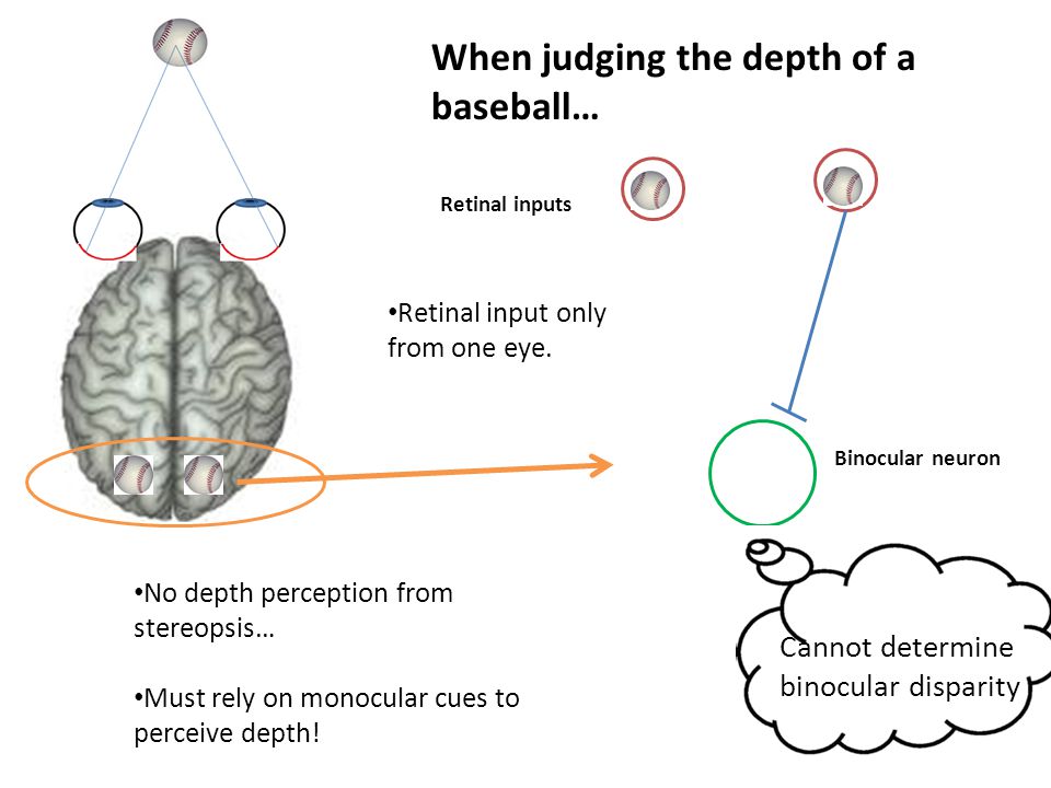
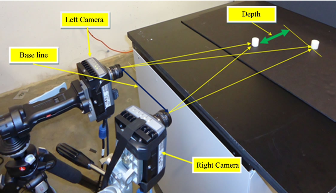
The distance between the two eyes on an adult is almost always 6.5 cm and that is the same distance in shift of an image when viewing with only one eye. Retinal disparity is the separation between objects as seen by the left eye and the right eye and helps to provide depth perception. Retinal disparity provides relative depth between two objects, but not exact or absolute depth. The closer objects are to each other, the retinal disparity will be small. If the objects are farther away from each other, then the retinal disparity will be larger. When objects are at equal distances, the two eyes view the objects as the same and there is zero disparity.[4]
Retinal disparity, sometimes called binocular disparity, is part of the process in visual perception that generates the depth and dimensionality. In the sequence of perception, this would occur at the surface/object stage. Specifically, retinal disparity is the space between the eyes that allows binocular vision to create depth perception.
The diagram below indicates a left and right eye. Both eyes converge on a box but due to retinal disparity, the angle of viewing is slightly different for each eye. The brain combines the two images to create the perception of a three-dimensional object.[13]
Retinal disparity is usually thought of as a 2D vector representing the deviation from retinal correspondence. It's assumed to decompose naturally into two orthogonal components, called horizontal and vertical disparity. Extensive literature has shown these components to be processed in fundamentally different ways. But when eye movements and non-identical correspondence patterns are taken into account, the simple definition of retinal disparity breaks down. In general, neither horizontal, nor vertical disparity, nor, indeed, the disparity vector itself, are well defined entities. Retinally, a binocular target is represented by one 2D position vector for each eye, or four dimensions. If disparity is assumed to be the difference between these projection vectors and a retinal correspondence pattern, the resulting entity has eight degrees of freedom - four more than a retinally located 2D disparity vector would have. Only when empirical retinal correspondence obeys certain constraints can disparity be reduced to such a vector. But even then it can not be simply split into retinal horizontal and vertical components, because moving eyes change the relationship between retinal locations and epipolar projection geometry. A practical consequence of these theoretical issues is demonstrated using the induced effect as an example.[14]

Stereoscopy creates the illusion of three-dimensional depth from given two-dimensional images. Human vision, including the perception of depth, is a complex process, which only begins with the acquisition of visual information taken in through the eyes; much processing ensues within the brain, as it strives to make sense of the raw information. One of the functions that occur within the brain as it interprets what the eyes see is assessing the relative distances of objects from the viewer, and the depth dimension of those objects.[6]
Stereo Vision Has Many Advantages
Stereo vision--or stereoscopic vision --probably evolved as a means of survival. With stereo vision, we can see WHERE objects are in relation to our own bodies with much greater precision--especially when those objects are moving toward or away from us in the depth dimension. We can see a little bit around solid objects without moving our heads and we can even perceive and measure "empty" space with our eyes and brains.[5]
Two Eyes = Three Dimensions (3D)
Each eye captures its own view and the two separate images are sent on to the brain for processing. When the two images arrive simultaneously in the back of the brain, they are united into one picture. The mind combines the two images by matching up the similarities and adding in the small differences. The small differences between the two images add up to a big difference in the final picture! The combined image is more than the sum of its parts. It is a three-dimensional stereo picture.[5]
Stereoblindness (also stereo blindness) is the inability to see in 3D using stereopsis, or stereo vision, resulting in an inability to perceive stereoscopic depth by combining and comparing images from the two eyes.

Reference:
- eyecarecontacts.com/binocular_vision_report.html
- ssc.education.ed.ac.uk/resources/vi&multi/eyeconds/binoc.html
- drsmilburn.com/eyenews.html
- en.wikipedia.org/wiki/Binocular_vision
- vision3d.com/stereo.html
- en.wikipedia.org/wiki/Stereoscopy
- en.wikipedia.org/wiki/Stereopsis
- ithacaeyecare.com/binocular-vision-dysfunction-
- northportwellnesscenter.com/blog/what-is-superior-oblique-palsy
- innerharbouroptometry.com/vertical-heterophoria
- .en.wikipedia.org/wiki/Strabismus
- opto.ca/health-library/binocular-vision
- artnet.nmu.edu/foundations/doku.php?id=retinal_disparity
- jov.arvojournals.org/article.aspx?articleid=2133633
- artisanoptics.com/artisan/the_eye_wire___artisan_optics_blog
- nvcofny.com/eye-care/what-are-the-symptoms-of-binocular-vision-dysfunction
- optometrists.org/childrens-vision/guide-to-childrens-eye-exams/what-is-a-binocular-vision-assessment
 وبلاگ تخصصی عینک شامل مجموعه مطالب پزشکی است که اطلاعات مفیدی در رابطه با عینک , چشم، لنز، سلامتی چشم و راه های پیشگیری از بیماریهای چشمی، کنترل و درمان آن را در اختیار شما کاربر محترم می گزارد.