دو تصور غلط در مورد معاينه چشم وجود دارد. يكي اين است كه اگر خوب مي بينيد نيازي به معاينه چشم نداريد و ديگري اينكه "تست بينايي" كه حدت بينايي را با استفاده از تابلو هاي مخصوص اندازه گيري مي كند (مشابه تست هاي بينايي كه در هنگام اخذ گواهينامه رانندگي انجام مي شود) همان معاينه چشمي است با اسمي ديگر. اما بايد توجه داشت كه چشم پزشك در واقع علاوه بر تست بينايي، چشم شما را از نظر بيماري هاي ديگري نيز كه ممكن است علائم زودرس نداشته باشند ولي نياز به درمان زودرس دارند معاينه مي كند. بنابراين معاينه كامل چشمي بسيار بيشتر از يك تست بينايي است.
چه كساني بايد مورد معاينه چشمي قرار گيرند؟
صرف نظر از سن و سلامت جسماني، هر شخصي بايد بصورت دوره اي و منظم مورد معاينه چشمي قرار گيرد. در بزرگسالان معاينه چشمي از جهت درست بودن شماره عينك و تشخيص زوردس بيماري ها اهميت دارد. در كودكان، معاينه چشمي نقش بسيار مهمي در تكامل بينايي كودك دارد.
از آنجاييكه بينايي نقش مهمي در فرايند يادگيري كودكان دارد، اهميت معاينات دوره اي در كودكان دو چندان است. مشكل بينايي كودك گاهي خود را بصورت افت تحصيلي و مشكل در انجام تكاليف مدرسه نشان مي دهد. در بسياري موارد، كودكان به اين دليل كه نمي دانند ديد "طبيعي" چگونه بايد باشد شكايتي از ديد خود ندارند. اگر كودك شما از نظر درسي در مدرسه مشكل دارد و يا در خواندن و يادگيري دچار مشكل است حتماً بايد جهت اطمينان از عدم مشكلات چشمي معاينه شود.
در معاينه چشم چه مشكلاتي مورد توجه قرار مي گيرند؟
مشكلاتي كه چشم پزشك در معاينه چشمي بدانها توجه مي كند
عبارتند از:
عيوب انكساري: شامل دوربيني، نزديك بيني، آستيگماتيسم، ...
تنبلي چشم (آمبليوپي): اين مشكل در موارد استرابيسم و يا اختلاف زياد بينايي دو چشم رخ مي دهد. در اين وضعيت مغز تصوير گرفته شده از چشم مشكل دار را ارسال نمي كند. آمبليوپي در صورت عدم درمان ممكن است باعث اختلال در تكامل بينايي شده و اختلال دائمي بينايي را بهمراه داشته باشد. اين اختلال معمولاً با بستن چشم بدون مشكل براي مدتي مشخص درمان مي شود.
استرابيسم (انحراف چشم): چشم پزشك چشم بيمار را از نظر هماهنگي حركتي و وضعيت قرار گيري نسبت به يكديگر بررسي مي كند. استرابيسم مي تواند سبب اختلال در درك عمق و آمبليوپي شود.
بيماريهاي چشمي: بسياري از بيماريهاي چشمي نظير گلوكوم و مشكلات ناشي از ديابت در مراحل اوليه علائم واضحي ندارند. چشم پزشك در معاينه چشمي به اين بيماري ها توجه كرده و در صورت برخورد با اين علائم درمان هاي اوليه را آغاز مي كند. در بسياري موارد تشخيص و درمان زودرس بيماريها سبب كاهش عوارض و از دست دادن دائمي ديد مي شود.
بيماريهاي ديگر: معاينه عروق ته چشم، پرده شبكيه و ديگر قسمت هاي چشم مي تواند ابتلا بيمار به بيماري هاي غير چشمي نظير فشار خون، ديابت، چربي بالا و بعضي بيماري هاي ديگر را نشان دهد.
معاينه چشمي به چه فواصلي بايد انجام شود؟ How Often Should You Get an Eye Exam

چشم پزشكان توصيه مي كنند كه هر فردي بسته به داشتن ريسك فاكتور و سلامت جسماني هر 1 تا 3 سال يكبار معاينه كامل چشمي شود.
كودكان: اين زمان در كودكان متفاوت است. تخمين زده مي شود كه از هر 20 كودك پيش دبستاني و هر 4 كودك دبستاني 1 كودك مشكل چشمي دارد كه در صورت عدم درمان مي تواند سبب كاهش دائمي بينايي شود. كودكاني كه علائمي نداشته و ريسك پاييني دارند بايد در 6 ماهگي، 3 سالگي و قبل از ورود به مدرسه معاينه كامل چشمي شوند. اين كودكان پس از آن بايد هر 2 سال مورد معاينه قرار گيرند.
اما كودكان داراي ريسك فاكتور مشكلات بينايي، نياز به معاينات بيشتري دارند.
بعضي از اين ريسك فاكتورها عبارتند از:
سابقه خانوادگي بيماري هاي چشمي family history of ocular diseases
سابقه صدمات چشمي the history of eye injuries
تأخير در تكامل developmental delay in children
انحراف چشم unusual ocular deviation
- تولد زودرس premature birth
كودكاني كه از عينك يا كنتاكت لنز استفاده مي كنند اغلب نياز به معاينات سالانه دارند تا در صورت تغيير در شماره چشم، عينك آنها اصلاح شود.

بزرگسالان: بطور كلي، بسته به ميزان تغييرات بينايي و سلامت جسماني، بزرگسالان بايد تا سن 40 سالگي هر 2 تا 3 سال تحت معاينه كامل چشمي قرار گيرند. در بيمارني كه به بيماري هايي نظير ديابت و فشار خون مبتلا هستند معاينات بيشتري توصيه مي شود زيرا اين بيماري ها تاثير سويي بر بينايي دارند.در افراد بالاي 40 سال بهتر است معاينه چشمي هر 1 تا 2 سال صورت گيرد. زيرا بعضي بيماري هاي نظير پير چشمي، كاتاراكت و دژنراسيون ماكولا با افزايش سن اتفاق مي افتند.از آنجا كه ريسك بيماري هاي چشم با افزايش سن بالا مي رود افراد بالاي 60 سال باز هر سال معاينه شوند.
Optometrists(OD) and ophthalmologists(eye MD) use a wide variety of tests and procedures to examine your eyes. These tests range from simple ones, like having you read an eye chart, to complex tests, such as using a high-powered lens to visualize the tiny structures inside of your eyes.

An eye examination is a series of tests performed by an ophthalmologist (medical doctor), optometrist, or orthoptist, optician (UK), assessing vision and ability to focus on and discern objects, as well as other tests and examinations pertaining to the eyes.
eye and vision tests that you are likely to encounter during a comprehensive eye exam:
- Ocular Motility (Eye Movements) Testing ; Stereopsis (Depth Perception) Test
- Autorefractors And Aberrometers ; Peripheral Visual Field Test
- Applanation Tonometry ; Contrast sensitivity Test
- Non-Contact Tonometry ; Fluorescein Angiogram
- Contact Lens Fittings ; Color Blindness Test
- Retinal Tomography ; The Glaucoma Test
- Visual Acuity Tests ; Keratometry Test
- Visual Field Test ; Slit Lamp Exam
- Pupil Dilation ; Retinoscopy
- Ultrasound ; Cover Test
- Refraction
Visual Ability
The basic mechanical skills of the visual system are: eye movements, which are important for following a moving object or looking from one object to another, as well as the ability to look at a single object, whether still or moving, for as long as is necessary; eye teaming, which refers to both eyes pointing at the same thing at the same time; without this ability there can be confusion and disorientation in processing visual information for meaning and response; and focusing, which is the ability to see clearly at any distance for any period of time with minimal effort. Other very important aspects of visual function are peripheral visual awareness and eye/hand coordination.
Eye health evaluation
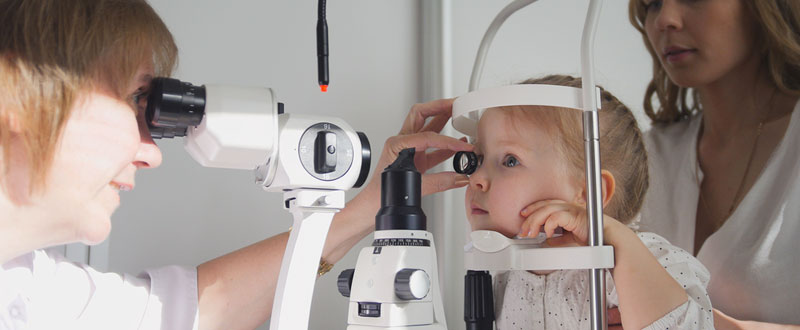
A wide variety of microscopes, lense, and digital technology will be used to assess the health of all the structures of the eye and the surrounding tissues. Dilating eye drops are often used to temporarily widen the pupil for better views of the structures inside the eye. In addition to measuring the pressure inside of the eye, this also is part of the eye exam where a doctor of optometry can detect otherwise unknown eye and systemic diseases.
Supplemental testing
Additional testing may be needed based on the results of the previous tests to confirm or rule out possible problems, to clarify uncertain findings, or to provide a more in-depth assessment.
At the completion of the examination, the doctor will assess and evaluate the results of the testing to determine a diagnosis and develop a treatment plan. He or she will discuss with you the nature of any visual or eye health problems found and explain available treatment options. In some cases, referral for consultation with, or treatment by, another doctor of optometry or other health care provider may be indicated. If you have questions about any diagnosed eye or vision conditions, or treatment recommendations, don't hesitate to ask your doctor for additional information or explanation. eResearch by Navid Ajamin -- spring 2012
The 8-Point Eye Exam

The key to any examination is to be systematic and always perform each element.
1. Visual acuity In the clinic, visual acuity is typically measured at distance. Otherwise, in a consult setting outside of the clinic, it’s measured at near. Don’t forget to have a near card with you. Make sure the patient is wearing his or her correction. Always have a pair of +3.00 readers with you, as many people in the emergency room won’t have their glasses with them. A pinhole occluder will also reduce the impact of uncorrected refractive error. If the patient is unable to see the biggest optotype on the card, the progression (from better to worse) is counting fingers (CF), hand motions (HM), light perception (LP) with projection, LP without projection and no light perception (NLP). For children who are too young to use Allen pictures, employ the “central, steady, maintain (CSM)” approach. Central: Is the corneal light reflex in the center of the pupil? Steady: Can the patient continue fixating when the light is slowly moved around? Maintain: Can the patient maintain fixation with the viewing eye when the previously covered eye is uncovered?
2. Pupils Look for anisocoria. If present, carefully check the pupil size in both well-lit and dark conditions. Check the reactivity of each pupil with a penlight or Finoff transilluminator. Use the swinging flashlight test to look for a relative afferent pupillary defect.

3. Extraocular motility and alignment Have the patient look in the six cardinal positions of gaze. Test with both eyes open to assess versions — repeat monocularly to test ductions. Use the cover/uncover test to assess for heterotropias. Use the alternate cover test to assess for the total amount of deviation. This amount minus any heterotropia is the amount of heterophoria.
4. Intraocular pressure Goldmann applanation tonometry is the gold standard and should be used in the clinic whenever possible. Outside of the clinic, Tono-Pen tonometry is much more practical. If you suspect a ruptured globe, skip this part of the exam.
5. Confrontation visual fields Assess each quadrant monocularly by having the patient count the number of fingers that you hold up. If acuity is particularly poor, have the patient note the presence of a light. Use the colored lid of an eyedrop bottle to define the position of a scotoma more accurately.
6. External examination Look for any ptosis by measuring the margin-to-reflex distance, which is the distance from the corneal light reflex to the margin of the upper lid. Look for lagophthalmos. Note any unusual growths or lesions that may require a biopsy. Palpate lymph nodes and the temporal artery if indicated by the history or exam. Measure proptosis or enophthalmos with an exophthalmometer. Perform a full cranial nerve exam for patients with diplopia or other neurologic symptoms.
7. Slit-lamp examination Lids/lashes/lacrimal system: Normal anatomy and contours? Any lesions? Conjunctiva/sclera: White and quiet? Injection? Lesions? Cornea: Clear? Epithelial disruptions? Stromal opacities? Endothelial lesions? Anterior chamber: Deep? Cell or flare? Iris: Round pupil? Transillumination defects? Nodules? Lens: Clear? Nuclear, cortical or subcapsular cataract? Anterior vitreous: Inflammation? Hemorrhage? Pigmented cells?
8. Fundoscopic examination Optic nerve: Cup-to-disc ratio? Focal thinning? Pallor? Symmetric? Macula: Foveal light reflex? Drusen, edema or exudates? Vessels: Contour and size? Intraretinal hemorrhage? Periphery: Tears or holes? Lesions? Pigmentary changes?
Understanding your vision and pinpointing the problems can be somewhat of a challenge at times.
What can sometimes be even more confusing is knowing what kind of eye exam to get: a comprehensive one or a regular one?
Knowing the difference between a comprehensive eye exam and a regular or routine eye exam is crucial in keeping your eyes healthy. It’s important to know which one to get done when it comes time to check your eyes.
The Difference Between a Comprehensive Eye Exam and a Regular One
A comprehensive eye exam normally takes about half an hour to an hour to complete, depending on how many exams you need to take. A comprehensive eye exam is a collection of a bunch of different tests used to diagnose disease and vision impairments.
Refraction
In physics, "refraction" is the mechanism that bends the path of light through the eye. In an eye exam, the term refraction is the determination of the ideal correction of refractive error. Refractive error is an optical abnormality in which the shape of the eye fails to bring light into sharp focus on the retina, resulting in blurred or distorted vision. Examples of refractive error are myopia, hyperopia, and astigmatism.
A refraction procedure consists of two parts: objective and subjective.
Objective refraction
An objective refraction is a refraction obtained without receiving any feedback from the patient, using a retinoscope or auto-refractor.
To perform a retinoscopy, the doctor projects a streak of light into a pupil. A series of lenses are flashed in front of the eye. By looking through the retinoscope, the doctor can study the light reflex of the pupil. Based on the movement and orientation of this retinal reflection, the refractive state of the eye is measured.
An auto-refractor is a computerized instrument that shines light into an eye. The light travels through the front of the eye, to the back and then forward through the front again. The information bounced back to the instrument gives an objective measurement of refractive error without asking the patients any questions.
Subjective refraction
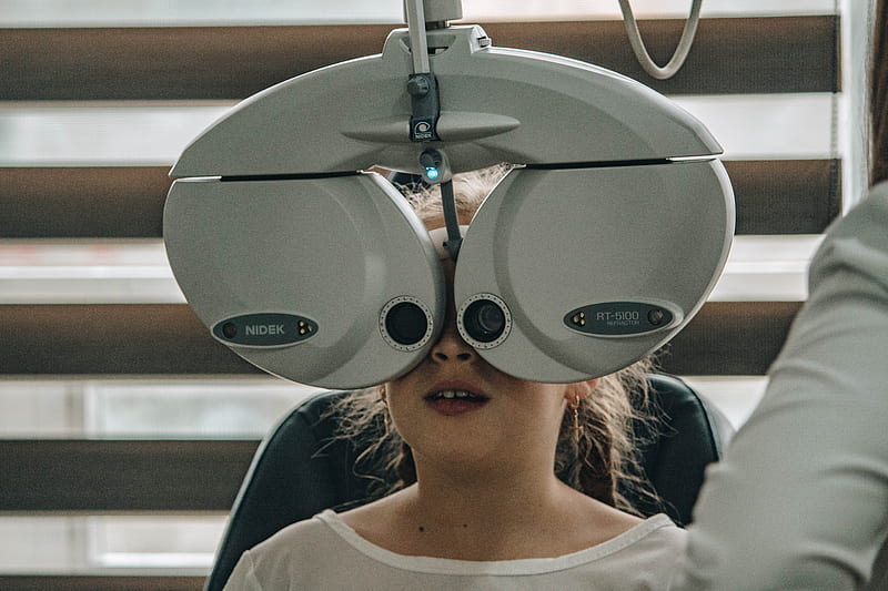
A subjective refraction requires responses from the patient. Typically, the patient will sit behind a phoropter or wear a trial frame and look at an eye chart. The eye care professional will change lenses and other settings while asking the patient for feedback on which set of lenses give the best vision.
Cycloplegic refraction
Sometimes, eye care professionals prefer to obtain a cycloplegic refraction, especially when trying to obtain an accurate refraction in young children who may skew refraction measurements by adjusting their eyes with accommodation. Cycloplegic eye drops are applied to the eye to temporarily paralyze the ciliary muscle of the eye.
Retinal examination
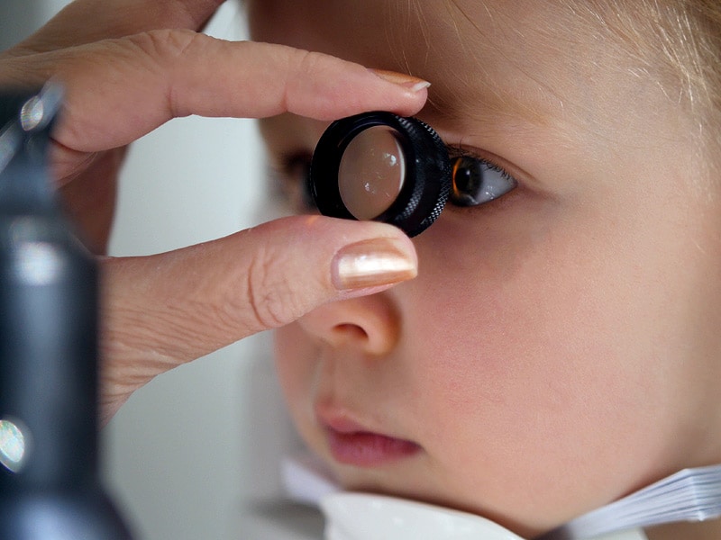
- Direct exam. Your eye doctor uses an ophthalmoscope to shine a beam of light through your pupil to see the back of the eye. Sometimes eyedrops aren't necessary to dilate your eyes before this exam.
- Indirect exam. During this exam, you might lie down, recline in a chair or sit up.
What Else Can Your Eye Exam Include?
Your ophthalmologist may suggest other tests to further examine your eye. This can include specialized imaging techniques such as:
- topography
- fundus photos
- fluorescein angiography (FA)
- optical coherence tomography (OCT)
Each part of the comprehensive eye exam provides important information about the health of your eyes. Make sure that you get a complete examination as part of your commitment to your overall health.
These tests can be crucial. They help your ophthalmologist detect problems in the back of the eye, on the eye's surface or inside the eye to diagnose diseases early.
Eye testing for infants
Babies should be able to see as well as adults in terms of focusing ability, color vision and depth perception by 6 months of age.
To assess whether your baby's eyes are developing normally, the doctor typically will use the following tests:
- Tests of pupil responses evaluate whether the eye's pupil opens and closes properly in the presence or absence of light.
- "Fixate and follow" testing determines whether your baby's eyes are able to fixate on and follow an object such as a light as it moves. (Infants should be able to fixate on an object soon after birth and follow an object by the time they are 3 months old.)
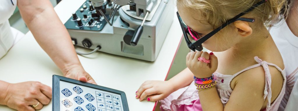
- Preferential looking involves using cards that are blank on one side with stripes on the other side to attract the gaze of an infant to the stripes. In this way, vision capabilities can be assessed without the use of a typical eye chart.
At-risk | Asymptomatic / low risk | Patient age (years) |
| At 6 to 12 months of age or as recommended | At 6 to 12 months of age | Birth through 2 |
| At least once between 3 and 5 years of age or as recommended | At least once between 3 and 5 | 3 through 5 |
| Before first grade and annually, or as recommended thereafter | Before first grade and annually thereafter | 6 through 17 |
The extent to which a child is at risk for the development of eye and vision problems determines the appropriate re-evaluation schedule. Children with ocular signs and symptoms require a prompt, comprehensive examination. Furthermore, the presence of certain risk factors may necessitate more frequent examinations based on professional judgment.

Factors placing an infant, toddler or child at significant risk for eye and vision problems include:
- Prematurity, low birth weight, prolonged supplemental oxygen at birth.
- Family history of myopia, amblyopia, strabismus, retinoblastoma, congenital cataracts, metabolic or genetic disease.
- Infection of mother during pregnancy (e.g., rubella, toxoplasmosis, venereal disease, herpes, cytomegalovirus or human immunodeficiency virus).
- Maternal smoking, use of alcohol or illicit drug use during pregnancy.
- Cortical visual impairment.
- Difficult or assisted labor, which may be associated with fetal distress.
- High or progressive refractive error.
- Strabismus.
- Anisometropia.
- Academic performance problems.
- Known or suspected neurodevelopmental disorders.
- Systemic health conditions with potential ocular manifestations.
- Wearing contact lenses.
- Functional vision in only one eye.
- Eye surgery or previous eye injury.
- Taking prescription or nonprescription drugs (e.g., over the counter medications, supplements, herbal remedies) with potential ocular side effects.

Don’t Do Anything Visually Stressful
It’s important that you don’t overexert your eyes in the hours before your eye exam. Using digital devices, reading, driving for prolonged periods, etc can all place considerable strain on your eyes, and this means that you are more likely to suffer from eye fatigue following your eye exam. For similar reasons, you should also try and get a good amount of sleep before your eye exam. Try and schedule your appointment for the morning to make sure that your eyes are as rested as possible.
Don’t Drink Coffee
Many people start the day with a cup of coffee, but what you might not realize is that drinking caffeine can affect your blood pressure, and the more you drink, the more significant this change is likely to be. This might not seem that important, but as part of your eye exam, your eye doctor will look at the blood vessels that are found at the back of the eye. These can reflect high blood pressure and potentially cause your eye doctor to be unnecessarily concerned.
Similarly, patients should also avoid drinking alcohol 24 hours before their appointment if possible. Alcohol also affects your blood pressure, as well as potentially making your eyes feel dry and irritated. And this could make your tests less comfortable.
Reference:
- aao.org
- allaboutvision.com
- mayoclinic.org
- en.wikipedia.org
- webmd.com
- precision-vision.com
- optometrists.org/vision-therapy
- urban-optics.com/blog/what-should-you-not-do-before-an-eye-exam.html
See also:
- Why do eye prescriptions differ between optometrists and ophthalmologists?
- Surprising Health Problems an Eye Exam Can Catch
- The Benefits Of An Enhanced Eye Examination
- What Are the Different Types of Eye Exams?
- What to expect from a DMV vision test
- 4 serious age-related eye problems
- Interesting Facts on Eye Chart
- Eyesight standards
 وبلاگ تخصصی عینک شامل مجموعه مطالب پزشکی است که اطلاعات مفیدی در رابطه با عینک , چشم، لنز، سلامتی چشم و راه های پیشگیری از بیماریهای چشمی، کنترل و درمان آن را در اختیار شما کاربر محترم می گزارد.