ابزار وبمستر

What is screen time defined as?

"Screen time" is a term used for activities done in front of a screen, such as watching TV, working on a computer, or playing video games.

What is a healthy screen time? [1]

Health experts say screen time at home should be limited to two hours or less a day. The time we spend in front of the screen, unless it's work- or homework-related, could be better spent being more physically active (increasing our energy out).

How much screen time by age?

Recommended time limits

  • Under 2 years old: Zero screen time, except for video chatting with family or friends.
  • 2-5 years old: No more than one hour per day co-viewing with a parent or sibling.
  • 5-17 years old: Generally no more than two hours per day, except for homework.

Screen time is a sedentary activity, meaning you are being physically inactive while sitting down. Very little energy is used during screen time.

Most American children spend about 3 hours a day watching TV. Added together, all types of screen time can total 5 to 7 hours a day.

Can mobile cause astigmatism?

For example, prolonged and frequent use of electronic devices such as smartphones, laptops, and tablets can cause eye strain and fatigue, leading to worsening astigmatism symptoms. Excessive exposure to UV rays from the sun or bright lights can also contribute to the development and progression of astigmatism.[2]

Too much screen time can:

  • Make it hard for your child to sleep at night
  • Raise your child's risk for attention problems, anxiety, and depression
  • Raise your child's risk for gaining too much weight (obesity)

Screen time increases your child's risk for obesity because:

  • Sitting and watching a screen is time that is not spent being physically active.
  • TV commercials and other screen ads can lead to unhealthy food choices. Most of the time, the foods in ads that are aimed at kids are high in sugar, salt, or fats.
  • Children eat more when they are watching TV, especially if they see ads for food.

Computers can help kids with their schoolwork. But surfing the internet, spending too much time on Facebook, or watching YouTube videos is considered unhealthy screen time.

How to Decrease Screen Time

Cutting down to 2 hours a day can be hard for some children because TV may be such a large part of their daily routines. But you can help your children by telling them how sedentary activities affect their overall health. Talk to them about things they can do to be healthier.

To decrease screen time:

  • Remove the TV or computer from your child's bedroom.
  • Do not allow TV watching during meals or homework.
  • Do not let your child eat while watching TV or using the computer.
  • Do not leave the TV on for background noise. Turn on the radio instead, or have no background noise.
  • Decide which programs to watch ahead of time. Turn off the TV when those programs are over.
  • Suggest other activities, such as family board games, puzzles, or going for a walk.
  • Keep a record of how much time is spent in front of a screen. Try to spend the same amount of time being active.
  • Be a good role model as a parent. Decrease your own screen time to 2 hours a day.
  • If it is hard not having the TV on, try using a sleep function so it turns off automatically.
  • Challenge your family to go 1 week without watching TV or doing other screen-time activities. Find things to do with your time that get you moving and burning energy.

Suicidal thoughts

The most recent study, published in the Journal of Youth and Adolescence, found that girls who had two to three hours of daily screen time beginning at age 13 were more likely to have suicidal thoughts in their later teen years. The same study showed that teenage boys, especially those who were the objects of cyberbullying, were also more likely to have suicidal thoughts in young adulthood.

Another study, published last month in the journal PLOS One, found that teens who spent more than three hours a day for reasons other than school are chronically stressed, sad, and think about suicide more often than those who spend less time with screens, the study showed. They also exhibited more emotional and physical problems.

“The results suggest the need for interventions that increase the awareness of the risks for adolescents who spend an excessive amount of time using the Internet,” the researchers concluded.

Critical thinking

Lead study author Sarah Coyne, associate director of the School of Family Life at Brigham Young University, tells parents not to panic, and not to forbid all screen time outright. Just set reasonable limits, she told the New York Post.

In addition, she encourages her own 13-year-old daughter to think critically and pay attention to the time she spends online.

“We say, ‘When you’re on TikTok, how does it make you feel? Who are you following?’ ” she told the paper. “If it ever feels like they’re bringing you down, or [making] you feel about yourself, you need to think, ‘Maybe I need to take a break,’ or ‘Maybe I need to not follow this person.”

Reference:

  1. medlineplus.gov/ency/patientinstructions/000355.htm
  2. nhlbi.nih.gov/health/educational/wecan/reduce-screen-time
  3. informedfamilies.org/catalyst/the-danger-of-too-much-screen-time-for-teens

See also:

  • Manage your child's screen time
  • What Causes Astigmatism to Worsen?
  • The Danger of Too Much Screen Time for Teens
  • Digital Eye Fatigue Can Lead to Lazy Eye in Children

What are the Benefits of Copper, Orange, Amber/Yellow and Brown/Bronze Tints [1]

Copper, orange, yellow/amber and brown/bronze lens tints make your environment appear brighter and are commonly used in low-light conditions. These lens tints block blue light and enhance contrast and depth perception making them helpful for overcast, hazy and foggy conditions.

While most people think that orange tinted glasses are only meant to be used during the daytime, there are actually many benefits to wearing them at night as well. For one, orange tinted glasses can help to reduce glare from oncoming headlights, making it easier to see while driving. Additionally, orange tinted glasses can also help to improve contrast and depth perception, making it easier to see in low-light conditions. And finally, orange tinted glasses can also help to reduce eye fatigue, making it easier to stay awake and alert when driving at night. So if you’re looking for a way to improve your night vision, consider investing in a pair of orange tinted glasses.

It’s suggested that bright orange prescription glasses are a good option for those who can’t avoid technology before bed. Light emitted by mobile devices, tablets, laptops, televisions, and e-readers, among other things, can be harmful to sleep. According to a Time/Qualcomm poll, nearly a quarter of 18- to 24-year-olds don’t get enough sleep because of technology. The pineal gland is particularly sensitive to blue light (460-480 nanometers). According to a 2006 study, blue light exposure prolonged melatonin production by more than twice as long as green light exposure. When the light is blue, there is also a decrease in feelings of sleepiness and activity in the brain during sleep. I wore a Fitbit Flex to collect objective sleep data, which showed me how long I sleep and how many times I sleep without sleep.

Blue light, with its shorter wavelength, scatters easier than other colors and makes focusing more difficult. Removing blue light therefore improves sharpness and depth perception and reduces fatigue. Note: These lens tints do cause some degree of color distortion, though brown/bronze lenses do so considerably less than do yellow/amber or orange lenses. eResearch by Navid Ajamin -- autumn 2024

Common users of copper, orange, yellow/amber and brown/bronze lens tints include baseball players, golfers, hunters and cyclists, as well as, those playing indoor sports and water sports. Individuals spending a considerable amount of time in front of a computer screen also find yellow/amber tints helpful because they reduce eye fatigue and strain by blocking blue light.

? What are orange lenses good for

The specific lens tint – copper, orange, yellow/amber or brown/bronze – depends on individual preference and situation.

Recent studies are showing new uses for lens tints that block blue light, and the potential applications would have significant impact for many individuals. Consider the following:

  • Sleep problems – Studies show that excessive light, especially blue light given off by computer screens, televisions and ambient light in most homes, suppresses melatonin. Melatonin, our natural sleep hormone, helps us get to sleep. For those struggling falling asleep, wearing lenses that block blue light for an hour before bed may prevent melatonin suppression, thereby allowing individuals to fall asleep more quickly and easily.
  • Bipolar disorder– Preliminary research shows that blocking blue light may help stabilize mood for individuals suffering from some forms of bipolar disorder. According to Dr. Jim Phelps, this “dark therapy” works basically in the opposite way as light therapy for depression.
  • Macular degeneration – Excessive blue light from sunlight may be one cause of age-related macular degeneration. This eye-disorder exists at the leading cause of blindness in the elderly.

    Based on this research, consider wearing copper, orange, yellow/amber or brown/bronze lens tints if you struggle falling asleep, have been diagnosed with bipolar disorder, or want to prevent age-related macular degeneration.

    While copper lenses block blue light better than the other lenses mentioned, they may be too dark for many to wear inside. Yellow/amber, orange and brown/bronze lenses still block enough blue light without the dimming effect to still produce some of the same benefits mentioned above.

    More research is needed, but exposure to blue light clearly has significant impact. In addition to the potential effects mentioned above, blue light may also increase cancer risk as well as have possible connections to diabetes and obesity.

    Because of its harmful potential, in addition to wearing lens tints that block blue light, consider also replacing night lights with dim, red lights to reduce exposure to blue light when trying to sleep, avoiding television and computer screens an hour or two before bed, and getting more natural light during the day to help regulate the body’s natural rhythms.

    Finding ways to regulate exposure to blue light may not only help you sleep better, preserve eyesight and stabilize mood, it may also go a long way in benefiting overall wellness and longevity. Take time today to assess your situation to determine if blue light may be having a significant impact on your health.

What does orange sunglasses mean? [2]

Yellow, orange or gold

They provide excellent depth perception and also work well for both outdoor and indoor sports sunglasses. They also improve the visibility of objects, make surroundings appear brighter and filter blue light. However, yellow, orange or gold sunglass lenses may distort color.

What are orange glasses lenses for? [3]

An orange filter provides 100 per cent blue light absorption for reduced glare and UV protection. This high-contrast filter is designed for improved definition. For indoor use and low intensity sun (53 percent absorption).

What are the benefits of yellow orange lenses? [4]

Enhanced Contrast and Clarity

One of the primary reasons shooters choose yellow or orange lenses is because they enhance contrast and clarity, especially in low-light conditions. These lenses filter out blue light, which is known to cause haze and reduce visual acuity.

Why wear orange tinted glasses? [5]

Wearing orange-tinted glasses is beneficial for those who spend a lot of time in front of screens or people who are exposed to bright artificial light sources. Studies show that wearing these glasses a few hours before bedtime can help you fall asleep and experience higher-quality rest.

What does orange light do to your eyes? [6]

Feeling dopey? Refresh your “circadian eye” with a burst of orange light. Light is a powerful wake-up call, enhancing alertness and activity. Its effect is controlled by a group of photoreceptor cells in the eyeball that make the light-sensing pigment melanopsin.

Reference:

  1. hi-techoptical.com/what-are-the-benefits-of-copper-orange-amber-yellow-and-brown-bronze-tints
  2. allaboutvision.com/sunglasses/color-lenses
  3. shop.rnib.org.uk/health-and-mobility/eyewear/orange-tint
  4. rx-safety.com/2024/08/why-people-prefer-yellow-or-orange-lenses-for-shooting-enhancing-vision-and-performance
  5. ocusleep.com/blogs/articles/how-do-orange-tinted-glasses-for-sleep-work
  6. newscientist.com/article/dn25195-a-burst-of-orange-light-wakes-up-our-circadian-eye
  7. learnglassblowing.com/3-benefits-of-wearing-orange-tinted-glasses-at-night

The Edinger–Westphal (EW) nucleus also called the accessory or visceral oculomotor nerve, is one of the two nuclei of the oculomotor nerve (CN III) located in the midbrain. It receives afferents from both pretectal nuclei (which have in turn received afferents from the optic tract). It contains parasympathetic pre-ganglionic neuron cell bodies that synapse in the ciliary ganglion. It contributes the autonomic, parasympathetic component to the oculomotor nerve (CN III), ultimately providing innervation to the iris sphincter muscle and ciliary muscle to mediate the pupillary light reflex and accommodation, respectively.

The Edinger-Westphal (EW) nucleus, which is part of the oculomotor nuclear complex (ONC), was first described in the literature in the 17th century. Although its most well known function is the control of pupil diameter, some controversy has arisen regarding the exact location of these preganglionic neurons. Currently, the EW is thought to consist of two different parts. The first part [termed the preganglionic EW-EWpg], which controls lens accommodation, choroidal blood flow and pupillary constriction, primarily consists of cholinergic cells that project to the ciliary ganglion. The second part [termed the centrally projecting EW-EWcp], which is involved in non-ocular functions such as feeding behavior, stress responses, addiction and pain, consists of peptidergic neurons that project to the brainstem, the spinal cord and prosencephalic regions.

Recently, it has been discovered that 2 different cell populations within the EW nucleus – subdivide into the EW preganglionic (EWpg) population and the EW nucleus centrally projecting (EWcp) population. However, the accepted nomenclature for these 2 groups varies.[9]

Schema of the oculomotor nerve nucleus and Edinger-Westphal nucleus (modified from the original figure by Wilson-Pauwels et al.). Oculomotor nerve nucleus consists of the lateral somatic cell column, caudal central nucleus, and medial cell column. Lateral somatic cell column consists of the dorsal subnucleus, intermediate column and ventral subnucleus, and regulates extraocular muscles on the ipsilateral side. The caudal central nucleus regulates levator palpebrae superioris muscles on both sides. The medial cell column regulates superior rectus muscles on the contralateral side. The Edinger-Westphal nucleus regulates sphincter pupillae muscles and ciliary muscles on the ipsilateral side.[8]

هستهٔ قرمز red nucleus از عناصر مهم سیستم حرکتی است. دسته‌ای از اکسون‌ها که از هستهٔ قرمز می‌آید، یکی از دو مجموعهٔ اصلی تارهای عصبی را درست می‌کند و پیام‌های حرکتی را از مغز به نخاع شوکی یاطناب نخاعی حمل می‌کند. هسته قرمز از بخش‌های مهم سیستم خارج هرمی است.

The pupillary reflex depends on the passage of light through eye structures, activation of the photoreceptors and retinal nerve fibers, and transmission along the optic nerve, which hemidecussates at the optic chiasm, to bilateral nuclei in pretectal areas of the rostral midbrain.[10]

The midbrain or mesencephalon is the uppermost portion of the brainstem connecting the diencephalon and cerebrum with the pons.

It consists of the cerebral peduncles, tegmentum, and tectum.[11]

It is functionally associated with vision, hearing, motor control, sleep and wakefulness, arousal (alertness), and temperature regulation.

The name mesencephalon comes from the Greek mesos, "middle", and enkephalos, "brain"

The Edinger–Westphal nucleus has two parts: [1]

  • The first is of preganglionic fibers (EWpg) that terminate in the ciliary ganglion.
  • The second is of centrally projecting cells (EWcp) that project to a number of brainstem structures.

The Edinger-Westphal nucleus, in the posterior midbrain, supplies parasympathetic fibers that terminate in the ciliary ganglion via cranial nerve III. It is mainly involved in pupillary constriction and the light accommodation reflex.[2]

Edinger-Westphal Nucleus. The optic nerve, afferent pathway, pretectal nucleus, optic tract, red nucleus, lateral geniculate nucleus, posterior commissure, and nerve with parasympathetic fibers are shown in the illustration. Illustration by Emma Gregory [4] eResearch by Navid Ajamin -- autumn 2024

The Edinger-Westphal nucleus is a small parasympathetic motor nucleus in the midbrain and one of the two nuclei for the oculomotor nerve. It is one of the cranial nerve nuclei.[5]

The Edinger–Westphal nucleus supplies preganglionic parasympathetic fibers to the eye, constricting the pupil, accommodating the lens, and convergence of the eyes.[1]

Cross-section of the midbrain at the level of the superior colliculus

Reference:

  1. en.wikipedia.org/wiki/EdingeWestphal_nucleus
  2. sciencedirect.com/topics/veterinary-science-and-veterinary-medicine/edinger-westphal-nucleus
  3. fa.wikipedia.org/wiki/هسته قرمز
  4. ncbi.nlm.nih.gov/books/NBK554555/figure/article-34491.image.f3
  5. radiopaedia.org/articles/edinger-westphal-nucleus-1
  6. medical-junction.com/light-reflex-pathway-and-defects
  7. tedmontgomery.com/the_eye/reflex.html
  8. researchgate.net/figure/Schema-of-the-oculomotor-nerve-nucleus-and-Edinger-Westphal-nucleus-modified-from-the_fig1_51904930
  9. pubmed.ncbi.nlm.nih.gov/26206178
  10. sciencedirect.com/topics/neuroscience/pupillary-reflex
  11. en.wikipedia.org/wiki/Midbrain

When we are severely stressed and anxious, high levels of adrenaline in the body can cause pressure on the eyes, resulting in blurred vision. People with long-term anxiety can suffer from eye strain throughout the day on a regular basis. Anxiety causes the body to become highly sensitised to any slight movement.

Stress impacts us mentally and physically, but did you know it can also affect our vision?

Stress is a common response to demands of daily life and is a normal part of everyday life, but when it becomes excessive or chronic, it can lead to some serious health issues and our eyes are certainly no exception.

When we are severely stressed and anxious, high levels of adrenaline in the body can cause pressure on the eyes, resulting in blurred vision. People with long-term anxiety can suffer from eye strain throughout the day on a regular basis.

Anxiety causes the body to become highly sensitised to any slight movement. Over time the strain from other senses can cause muscular tensions and headaches.

SYMPTOMS OF STRESS-RELATED VISION IMPAIRMENT: [1]

  • Sensitivity to light and movement; light may hurt your eyes or make it difficult for you to concentrate and focus.
  • Eye twitching; eyes can randomly spasm, with no pain but discomfort.
  • Very dry or very wet eyes; both can be a symptom, however, it can also be caused by other issues.
  • Blurry vision; finding it hard to concentrate, or focus.
  • Eye strain; discomfort or minor pain as a result of your eyes feeling tight and swollen.
  • Eye floaters; tiny spots that swim across your vision.

Stress can impair your vision in multiple ways. It may contribute to dry eyes, glaucoma, macular degeneration, and other eye diseases. It's always a good idea to speak with a doctor if you notice any changes to your vision.

Chronic stress may speed up the development of eye diseases like macular degeneration or glaucoma. In the short term, stress and behaviors related to stress may contribute to dry eyes.

Stress can also lead to psychogenic blindness. This is blindness with a psychological cause and no known structural problem.

The idea that stress may lead to vision loss isn’t new. It’s believed to date back at least 3,000 years.

Science has come a long way since then, but modern research also supports the idea that stress may contribute to vision loss.

Can stress cause blurry vision?

Stress can potentially contribute to the development of blurry vision in several ways, such as:

  • Drying out your eyes: In a 2022 review of studies, researchers found that dry eye disease was associated with depression and anxiety. Blurry vision is a potential symptom of dry eyes.
  • Medication side effects: Some medications used to treat anxiety or stress, such as tricyclic antidepressants, can lead to symptoms like pupil dilation and blurry vision.
  • Lead to retinal disease: Research suggests that perceived stress is a risk factor for central serous chorioretinopathy (CSCR). CSCR results in fluid collection in the macula (the central part of the retina), which can cause blurry vision.
  • Glaucoma: In a 2021 study, researchers found evidence that anxiety and depression contributed to the progression of glaucoma. Blurry vision is often one of the first symptoms of glaucoma.[2]

Here are a few ways anxiety can lead to visual symptoms:

  1. Pupil Dilation: Adrenaline causes the pupils to dilate, allowing more light to enter the eyes. This can lead to temporary blurred vision as the eyes struggle to adjust to the increased light.
  2. Eye Strain: Anxiety often leads to increased muscle tension, including in the muscles around the eyes. This tension can cause eye strain and, consequently, blurred vision.
  3. Hyperventilation: Mental stress can lead to rapid and shallow breathing, or hyperventilation. This can decrease carbon dioxide levels in the blood, affecting the oxygenation of the brain and eyes, and resulting in blurred vision.
  4. Dry Eyes: Anxiety can reduce the production of tears, leading to dry eyes, which can cause blurred vision and discomfort.

How to stop blurry vision from anxiety? Lifestyle Changes
Regular Exercise: Engage in regular physical activity to reduce overall anxiety levels and improve blood flow to the eyes. Healthy Diet: Consume a balanced diet rich in vitamins and minerals that support eye health, such as Vitamin A, C, and E.[3]

Reference:

  1. whitbyandco.com/stress-on-your-eyes
  2. healthline.com/health/stress/stress-blindness
  3. vision-specialists.com/binocular-vision-dysfunction/symptoms/anxiety

WHAT SPECIFIC PORTIONS OF THE ENVIRONMENTAL ELECTROMAGNETIC SPECTRUM CAUSE EYE DAMAGE?

With sufficient magnitude almost all portions of the electromagnetic spectrum can cause damage to the eye.
For example: Lasers of a wide variety of wavelengths from the short wavelength UV (Excimerlaser for LASIK) through the visible spectrum (Argon laser for diabetic retinopathy) to short wavelength IR (YAG laser for iridotomy and capsulectomy) are used to “damage” eye tissue in the treatment of various eye conditions.
However, in our “natural” environments with natural and man-made lights, the most offending portions of the EM spectrum are the UV-A (315 nm to 400 nm), UV-B (280 nm to 315 nm), and “blue-light” portion of the visible spectrum (380 nm to 500 nm). Our atmosphere generally protects us from UV radiation below 280 nm.
Additionally, as the cornea and crystalline lens absorbs almost all natural UV radiation, UV radiation is thought to cause damage to the anterior eye, while short visible light (“blue-light”) can cause damage to retinal structures. Also, as the damaging processes are thought to be at least partially photochemical in nature, the damaging effects can be cumulative in nature, which may compound across one’s lifetime.

can Bright Light Damage Your Vision?

In short, yes, staring at bright lights can damage your eyes. When the retina’s light-sensing cells become over-stimulated from looking at a bright light, they release massive amounts of signaling chemicals, injuring the back of the eye as a result.

The sun shines with such intensity that staring directly at it for just a few seconds can cause permanent retinal damage. Chronic exposure to UV rays over many weeks, months or years can also harm the macula, cornea and lens. A damaged macula leads to macular degeneration. A “sunburned” cornea can cause blurry vision and loss of eyesight. A damaged lens may develop a cataract, or clouding of the lens that blurs vision.

Blue light, even at moderate intensity levels, can damage your retinas slowly over time. Blue light has shorter wavelengths than warmer light, so it has more energy. Prolonged exposure may increase the risk of age-related macular degeneration (AMD). Sunlight is the most prominent source of blue light, but other examples include fluorescent lights, LEDs, TVs, computer monitors and Smartphone screens.

How to Treat and Prevent Eye Problems from Bright Light

Protect your vision with these tips:

  • Choose glasses with anti-reflective lenses to reduce glare from bright lights.
  • Wear dark-tinted sunglasses and a brimmed hat while outside. Polarized sunglasses with UV protection further shield your eyes from the effects of blue light and ultraviolet rays.
  • Decrease daily screen time and take frequent breaks to rest your eyes.
  • Turn on your computer’s “night light” feature to decrease the amount of blue light the screen emits.
  • Wear blue-blocking computer glasses with yellow-tinted lenses that ease digital eyestrain.
  • Choose CFLs and LEDs that emit “warm” light.
  • If you require cataract surgery, seek blue-blocking intraocular lens (IOL) implants to protect your retinas the same way sunglasses do.

Reference:

  • aoa.org/AOA/Documents/Education/Education_PDFs/Blue%20Light%20and%20Eye%20Damage.pdf
  • gersteineye.com/blog/2018/08/how-does-bright-light-affect-your-vision

The circle of Zinn is an arterial anastomotic ring surrounding the optic nerve head in the sclera formed by branches of the short posterior ciliary arteries. Multiple small branches from the circle of Zinn supply the anterior pia of the optic nerve, the optic disc and contribute to the blood supply of the posterior choroid.[1]

Schematic diagram: Blood Supply of the Border Tissue. Short posterior ciliary arteries form the circle of Zinn-Haller which supply the LC and border tissue of Elschnig. The central retinal artery is destined for the retina and does not contribute to the border tissue.[9]

What is the Circle of Zinn Haller?

The circle of Haller and Zinn comprises complete or incomplete anastomoses around the optic nerve between the medial and lateral short posterior ciliary arteries (SPCAs), which form a dense capillary plexus around the optic nerve.[2]

What is the function of the Circle of Zinn?

The para-optic branches have divided on each side to form the 'circle' of Haller and Zinn which provides pial branches to the retrolaminar optic nerve and recurrent choroidal branches to the peri-papillary choroid and peripheral vertical meridional choroid.[3]

What is The Circle of Zinn and Haller?

The circle of Zinn–Haller (CZH) is known to be an intrascleral arteriolar anastomosis derived from medial and lateral paraoptic short posterior ciliary arteries (SPCAs). The significance of this arterial circle in supplying the anterior optic nerve and peripapillary region has been the subject of controversy.[4] eResearch by Navid Ajamin -- autumn 2024

What is the circle of Zinn formed by?

The peripapillary arterial circle of Zinn-Haller (ZHAC) is an intrascleral arterial anastomosis derived from the paraoptic medial and lateral short posterior ciliary arteries. Arterial circle of Zinn-Haller provides the main vascular supply for the optic nerve head at the level of the lamina cribrosa.[5]

The ciliary arteries are divisible into three groups, the long posterior, short posterior, and the anterior.

  • The short posterior ciliary arteries from six to twelve in number, arise from the ophthalmic artery as it crosses the optic nerve.
  • The long posterior ciliary arteries, two for each eye, pierce the posterior part of the sclera at some little distance from the optic nerve.
  • The anterior ciliary arteries are derived from the muscular branches of the ophthalmic artery. [7]

Short posterior ciliary arteries

The short posterior ciliary arteries are a number of branches of the ophthalmic artery. They pass forward with the optic nerve to reach the eyeball, piercing the sclera around the entry of the optic nerve into the eyeball.

The number of short posterior ciliary arteries varies between individuals; one or more short posterior ciliary arteries initially branch off the ophthalmic artery, subsequently dividing to form up to 20 short posterior ciliary arteries.

The short posterior ciliary arteries branch off the ophthalmic artery as it crosses the optic nerve medially.

About 7 short posterior ciliary arteries accompany the optic nerve, passing anterior-ward to reach the posterior part of the eyeball, where they divide into 15-20 branches and pierce the sclera around the entrance of the optic nerve.

The short posterior ciliary arteries contribute arterial supply to the choroid, ciliary processes, optic disc, the outer retina, and Bruch's membrane.

Some branches of the short posterior ciliary arteries supply the optic disc by means of an anastomotic ring - the circle of Zinn-Haller or circle of Zinn - which is associated with the fibrous extension of the ocular tendons (common tendinous ring (also annulus of Zinn)).[6]

The peripapillary artery, also known as the circle of Haller and Zinn, is the vessel that provides most of the blood supply to the lamina cribrosa (LC) region of the optic nerve.[8]

Reference:

  1. radiopaedia.org/articles/circle-of-zinn
  2. pmc.ncbi.nlm.nih.gov/articles/PMC3308474
  3. nature.com/articles/eye199038.pdf
  4. bjo.bmj.com/content/83/7/862
  5. journals.lww.com/retinajournal/abstract/2017/02000/peripapillary_arterial_ring_of_zinn_haller_in.10.aspx
  6. en.wikipedia.org/wiki/Short_posterior_ciliary_arteries
  7. en.wikipedia.org/wiki/Ciliary_arteries
  8. pmc.ncbi.nlm.nih.gov/articles/PMC3308474
  9. researchgate.net/figure/Schematic-diagram-Blood-Supply-of-the-Border-Tissue-Short-posterior-ciliary-arteries_fig5_338955428
  10. link.springer.com/chapter/10.1007/978-1-4419-6724-4_7

See also:

  • Circle of Zinn vs annulus of Zinn

Leber congenital amaurosis (LCA) is a vision loss disorder that begins in infancy. Different genes are associated with LCA. They usually have autosomal recessive inheritance. In the present study, the genetic basis of congenital blindness in two Iranian families was examined.

Amaurosis (Greek meaning darkening, dark, or obscure) is vision loss or weakness that occurs without an apparent lesion affecting the eye. It may result from either a medical condition or excess acceleration, as in flight.

Theodor von Leber, a German ophthalmologist, described LCA in 1869 as a disorder characterized by profound visual loss present at or shortly after birth, nystagmus, sluggish pupillary reactions, and pigmentary retinopathy.

Theodor Karl Gustav von Leber

Leber congenital amaurosis, also known as LCA, is an eye disorder that is present from birth (congenital). This condition primarily affects the retina, which is the specialized tissue at the back of the eye that detects light and color. People with this disorder typically have severe visual impairment beginning at birth or shortly afterward. The visual impairment tends to be severe and may worsen over time.

Leber congenital amaurosis is also associated with other vision problems, including an increased sensitivity to light (photophobia), involuntary movements of the eyes (nystagmus), and extreme farsightedness (hyperopia). The pupils, which usually expand and contract in response to the amount of light entering the eye, do not react normally to light. Instead, they expand and contract more slowly than normal, or they may not respond to light at all.

A specific behavior called Franceschetti's oculo-digital sign is characteristic of Leber congenital amaurosis. This sign consists of affected individuals poking, pressing, and rubbing their eyes with a knuckle or finger. Poking their eyes often results in the sensation of flashes of light called phosphenes. Researchers suspect that this behavior may contribute to deep-set eyes in affected children.

Various retinal phenotypes among LCA patients

In very rare cases, delayed development and intellectual disability have been reported in people with the features of Leber congenital amaurosis. Because of the visual loss, affected children may become isolated. Providing children with opportunities to play, hear, touch, understand and other early educational interventions may prevent developmental delays in children with Leber congenital amaurosis.

At least 20 genetic types of Leber congenital amaurosis have been described. The types are distinguished by their genetic cause, patterns of vision loss, and related eye abnormalities.

Leber's congenital amaurosis (LCA) is the most severe and earliest of the inherited retinal dystrophies that cause congenital blindness. It has an incidence of 2-3 per 100,000 births and accounts for 10-18% of cases of congenital blindness among children in blind institutes and 5% of all retinal dystrophies, a figure that is likely to be greater in countries with a greater percentage of consanguinity.

In most cases, LCA is inherited in an autosomal recessive pattern, as established by Alström and Olson in 1957. Some patients only have retinal blindness, whereas others have multi-systemic involvement that includes renal, cardiac, skeletal, and, most notably, central nervous system anomalies.

What are the types of congenital blindness?

Congenital anomalies such as anophthalmos, microphthalmos, coloboma, congenital cataract, infantile glaucoma, and neuro-ophthalmic lesions are causes of impairment present at birth.


a Focal perimacular atrophy and pigment accumulation. b Fundus hypopigmentation with diffuse RPE granularity. c Macular atrophy and bone spicule pigmentation. d Macular RPE clumping and atrophy accompanying peripheral bone spicule changes

Reference:

  • medlineplus.gov/genetics/condition/leber-congenital-amaurosis
  • pmc.ncbi.nlm.nih.gov/articles/PMC3119278
  • en.wikipedia.org/wiki/Amaurosis

Retinoblastoma is a kind of eye cancer that starts as a growth of cells in the retina. The retina is the light-sensitive lining on the inside of the eye. The retina is made up of nerve tissue that senses light as it comes in through the front of the eye. The light causes the retina to send signals to the brain.

Retinoblastoma happens most often in young children. It's usually diagnosed before age 2. It most often affects one eye. Sometimes it happens in both eyes.

There are several treatments for retinoblastoma. For most children, treatment doesn't require removing the eye to get rid of the cancer. The outlook for children diagnosed with retinoblastoma is quite good.

Symptoms

Retinoblastoma signs and symptoms include:

  • A white color in the center circle of the eye when light is shone in the eye. It might show up in flash photos.
  • Eye redness.
  • Eye swelling.
  • Eyes that seem to be looking in different directions.
  • Vision loss.

When to see a doctor

Make an appointment with a doctor or other healthcare professional if you notice any changes to your child's eyes that concern you.

Request an appointment

Causes

Retinoblastoma is caused by changes inside the cells in the eye. It's not always clear what causes the changes that lead to this eye cancer.

Retinoblastoma starts when cells in the eye get changes in their DNA. A cell's DNA holds the instructions that tell the cell what to do. In healthy cells, the DNA gives instructions to grow and multiply at a set rate. The instructions also tell the cells to die at a set time. In cancer cells, the DNA changes give different instructions. The changes tell the cancer cells to make many more cells quickly. Cancer cells can keep living when healthy cells would die. This causes too many cells.

In retinoblastoma, this growth of cells happens in the retina. The retina is the light-sensitive lining on the inside of the eye. The retina is made up of nerve tissue that senses light as it comes in through the front of the eye. The light causes the retina to send signals to the brain. The brain interprets the signals as images.

As the cancer cells build up in the retina, they can form a mass, called a tumor. The tumor can grow to invade and destroy healthy body tissue. In time, cancer cells can break away and spread to other parts of the body. When cancer spreads, it's called metastatic cancer. Retinoblastoma rarely spreads, especially if it's found early.

For most instances of retinoblastoma, it's not clear what causes the DNA changes that lead to cancer. However, it's possible for children to inherit DNA changes from their parents. These changes can increase the risk of retinoblastoma.

Risk factors

Risk factors for retinoblastoma include:

  • Young age. Retinoblastoma is most common in very young children. It's typically diagnosed by age 2. Retinoblastoma that happens later in life is very rare.
  • DNA changes that run in families. DNA variations that increase the risk of retinoblastoma can be passed from parents to children. Children with these inherited DNA changes tend to get retinoblastoma at a younger age. They also tend to have retinoblastoma in both eyes.

Complications

Children with retinoblastoma can develop complications.

Cancer that comes back

After treatment, there is a risk that the cancer might come back in the eye or near it. For this reason, your child's healthcare team will create a plan of follow-up appointments. Your child's follow-up plan will depend on the treatments your child received. A typical plan might involve eye exams every few months for the first few years after treatment.

Increased risk of other cancers

Children with the form of retinoblastoma that can run in families may have a higher risk of getting other kinds of cancer.

The risk of these cancers is increased:

  • Bone cancer.
  • Bladder cancer.
  • Breast cancer.
  • Hodgkin lymphoma.
  • Lung cancer.
  • Melanoma.
  • Pineoblastoma.
  • Soft tissue sarcoma.

Your child's healthcare team might recommend tests to screen for these other types of cancers.

Prevention

There's no way to prevent retinoblastoma.

Some retinoblastomas are caused by DNA changes that run in families. If retinoblastoma runs in your family, tell your healthcare professional. Together you might consider genetic testing to look for variations in your DNA that increase the risk of retinoblastoma. Your health professional might refer you to a genetic counselor or other healthcare professional trained in genetics. This person can help you decide whether to undergo genetic testing.

If your children have an increased risk of retinoblastoma, care can be planned to manage that risk. For instance, eye exams may begin soon after birth. That way, retinoblastoma may be diagnosed very early. These screening tests could find the cancer when it is small and has a greater chance of being cured.

If you haven't had children, but are planning to, talk with your healthcare team about your family history of retinoblastoma. Genetic testing might help you and your partner understand whether there is a risk of passing DNA variations to your future children. Your healthcare team may have options to help you manage this risk.

Reference:

  • mayoclinic.org/diseases-conditions/retinoblastoma/symptoms-causes/syc-20351008
  • together.stjude.org/en-us/about-pediatric-cancer/types/retinoblastoma.html
  • step2.medbullets.com/oncology/120405/retinoblastoma

Did you know that an error of only 2mm can lead to a loss of more than 40% of your clear vision field?

What happens if you get your PD wrong?

Reduced Depth Perception: An incorrect PD can affect your depth perception, making it difficult to judge distances accurately. Adjustment Issues: You might find it hard to adjust to the glasses, leading to frustration and possibly abandoning their use altogether.

Ensuring the accuracy of your pupillary distance (PD) is crucial when it comes to ordering prescription glasses. If you enter incorrect prescription information, including an inaccurate pupillary distance measurement, the consequences could affect your vision with the new glasses.

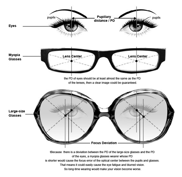
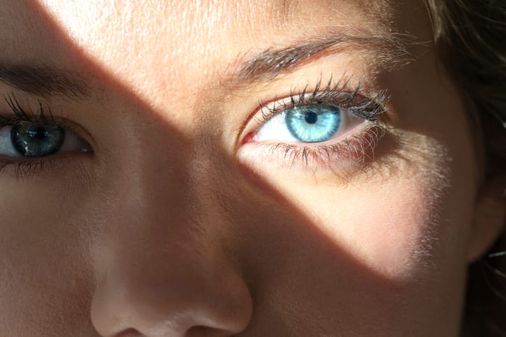
PD, or pupillary distance, refers to the distance in millimeters between the center of one pupil to the center of the other. It is used to determine where the optical center of the lens should be placed on your glasses that need to line up over the center of the pupil. Having a correct measurement of your PD on your glasses prescription ensures that you’re looking through the ideal precise placement spot (Optical Center) in your lenses.

Mean IPD in adult subjects was 61.1±3.5 mm in women and 63.6±3.9 mm in men (p<0.001). Mean IPD increased 4.8 mm during the second decade, 1.7 mm during the third decade, and 0.6 mm during the fourth and fifth decades of life.

There are two types of pupillary distance measurements:

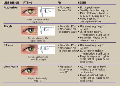
Monocular PD. This is the distance between the center of your pupil and the middle of the bridge of your nose. Monocular PDs are required for accurate fitting of progressive lenses.

Binocular PD. This is the distance between the center of the pupil of one eye and the center of the pupil of the other eye.

There are two ways to measure the distance: single PD and dual PD.

Single PD will measure the distance between the center of one pupil to the other while dual PD consists of two numbers and is the distance between the centers of each pupil to the bridge of the nose.

For Example, The average adult's single PD is between 54-74 mm; kids' are between 43-58 mm but a fun fact is that most of us are not symmetrical and therefore your dual PD will be most likely not even 32/30.

What is the tolerance for PD?

While the PD tool is consistent up to ±1 mm for 95% of the scans, the PD tolerance in the industry is up to ±2.5 mm, which means you are good to go with any single result from the PD scan. ** No results should be obtained within ±2.5 mm or higher from each other, if the card was held properly flat against the forehead.

What is standard PD?

What is a normal pupillary distance? The average pupillary distance of a typical adult ranges from 54 to 74 mm without much difference between the average female and male PD. A child's distance ranges from 43 to 58 mm. During an eye checkup, your optometrist will typically measure your PD.

Misaligned glasses put extra load on your eye muscles, forcing them to work harder to bring objects into focus. This may cause migraines, eyestrain, and discomfort. Prolonged eye muscle strain may cause more serious eye defects like asthenopiaeye fatigue.

Pupillary Distance (PD), also known as interpupillary distance, is the horizontal distance between the centres of your pupils—the dark circle in your eye that shrinks to focus near objects and widens to accommodate light from far objects. PD is measured in millimetres.

The PD value is used to properly align your glasses with your eyes. Wearing misaligned glasses may cause side effects like eye strain, nausea, and distorted vision. Your PD value doesn’t appear on your prescription; as a result, you have to take this measurement yourself to be able to order your glasses from us.

Benefits of Accurate PD Values

An accurate PD measurement helps us align your lenses to fit your eyes perfectly. Here are some of the benefits of wearing properly aligned glasses:

  • + Properly aligned lenses give you the best possible clarity and definition.
  • + They provide you with comfort and reduce headaches and eyestrain by making sure that your eye muscles do not do more work than necessary.
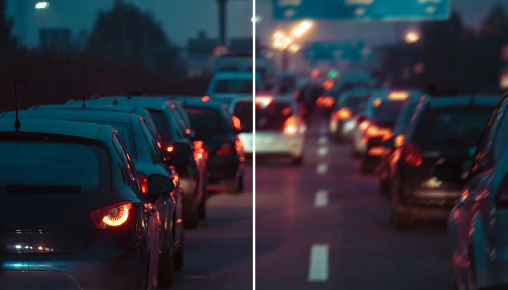
  • + Properly aligned glasses give you an accurate perception of depth and distance, helping you perform tasks like driving and walking safely.
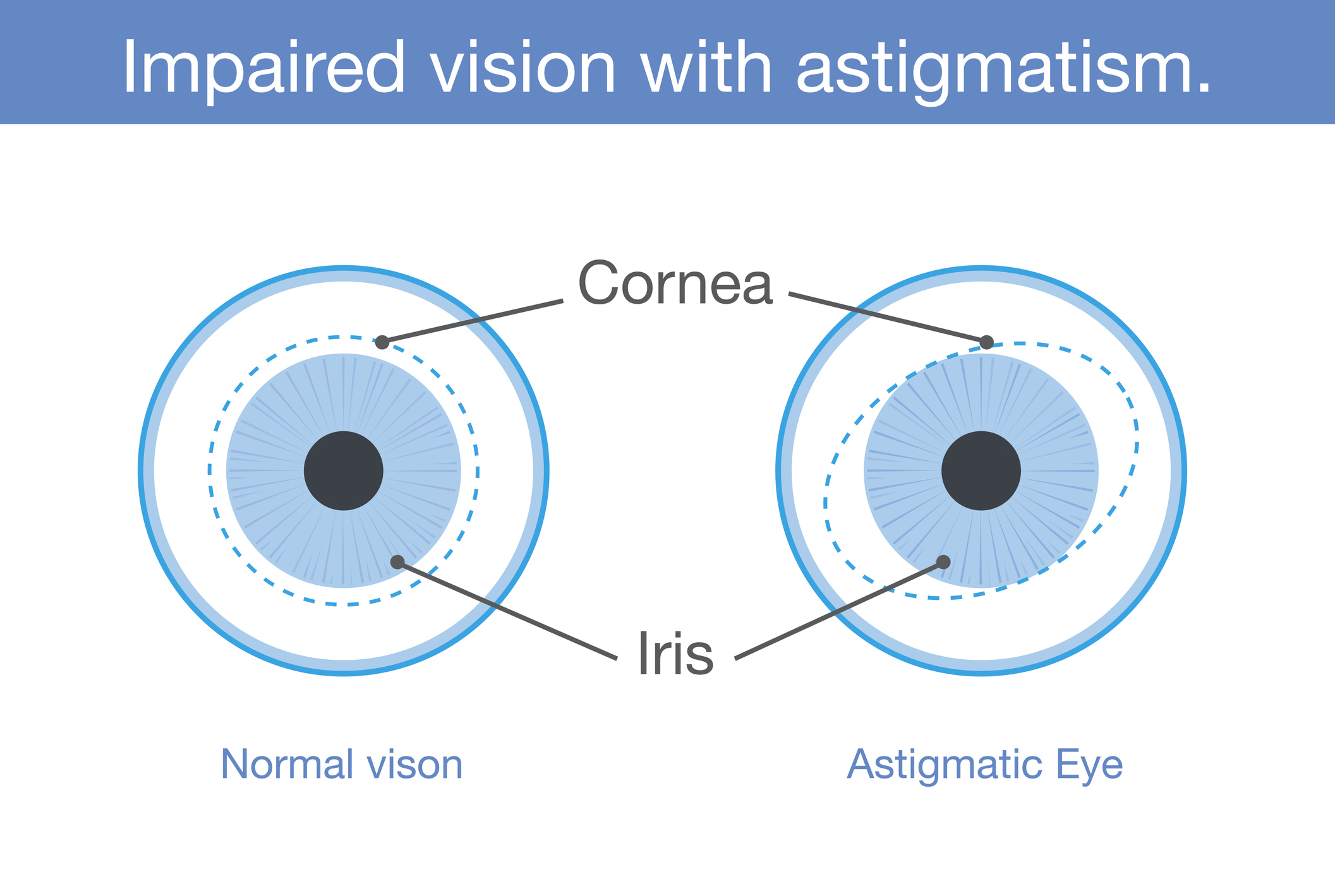
  • + Properly aligned glasses make sure that the corrective power of your lens is applied accurately, with the right power and at the right angle/axis. This is particularly important for cylindrical lenses and astigmatism correction.

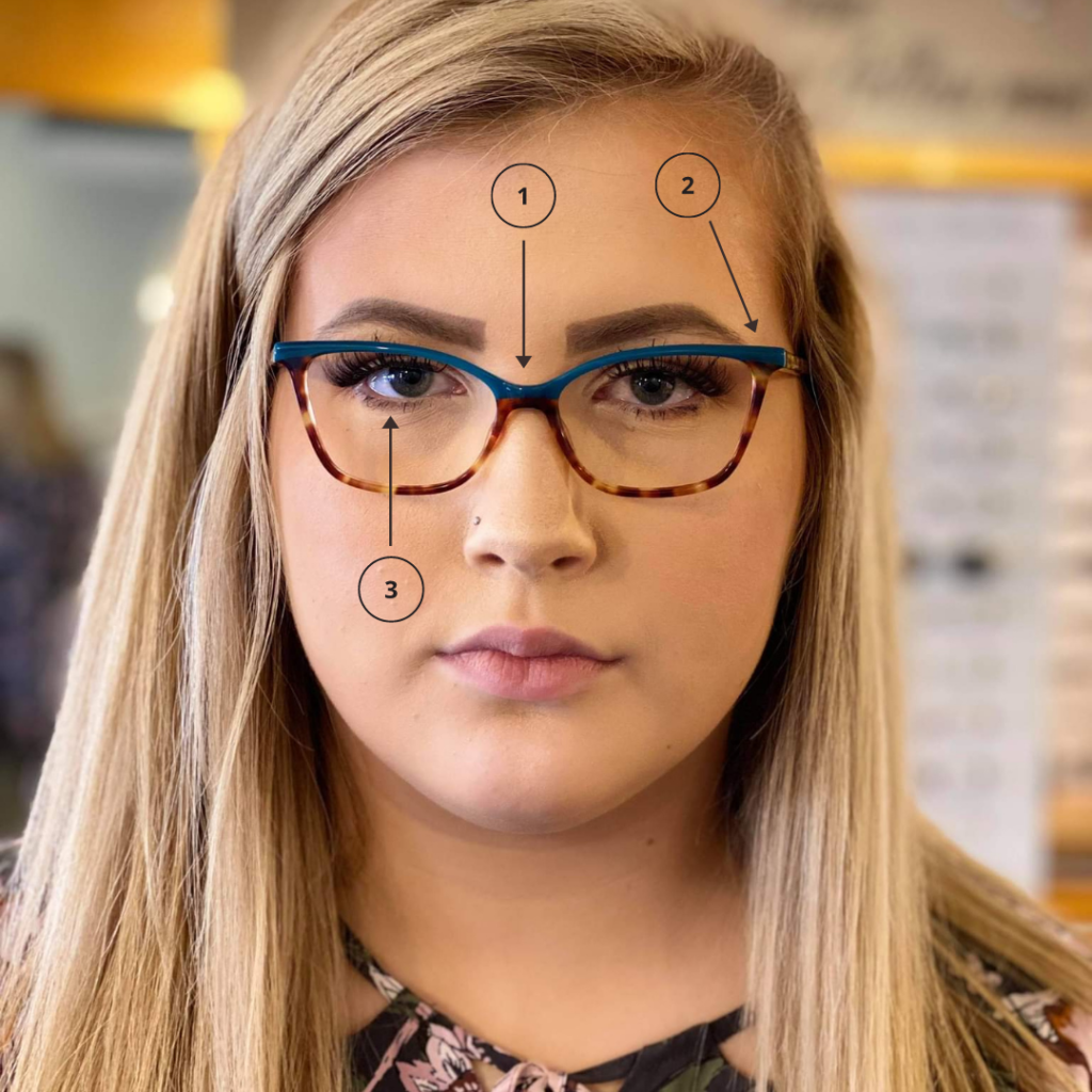
  1. The bridge is narrow
  2. The frame ends before the wearer’s temples – and sits too close to the side of the wearer’s head
  3. The eyes are not centered in the frame – they are sitting too high

Side Effects of Wrong PD Values

Entering the wrong PD value while ordering your eyeglasses will unfortunately lead to you receiving misaligned glasses. The list below includes a few of the side effects of wearing misaligned glasses:

  • - Misaligned glasses put extra load on your eye muscles, forcing them to work harder to bring objects into focus. This may cause migraines, eyestrain, and discomfort. Prolonged eye muscle strain may cause more serious eye defects like asthenopia—eye fatigue
  • - Improper lens alignment may cause blurred or double vision, making basic tasks, like reading more difficult.
  • - Misaligned lenses also give a false sense of depth and distance, making it harder to drive or walk. This may even lead to an increased risk of accidents.
  • - They may further weaken the ability of the eyes to focus, worsening eye defects in the process. This is why we often advise not to wear other people’s prescribed glasses.

How do you calculate near PD for reading glasses?

Subtract 3 mm from your distance PD. For example, if your distance PD is 63mm, then your near PD is 60mm. If you use dual PD to calculate near PD, then subtract 1.5mm from each eye's measurement.

Do I use dist PD or near PD when ordering progressive glasses?

If you need progressive or bifocal lenses, you'll also need to measure your near focus PD. You will need someone else to measure this for you to get the most accurate reading. The person measuring should follow the same instructions as they would to get your regular PD. eResearch by Navid Ajamin -- autumn 2024

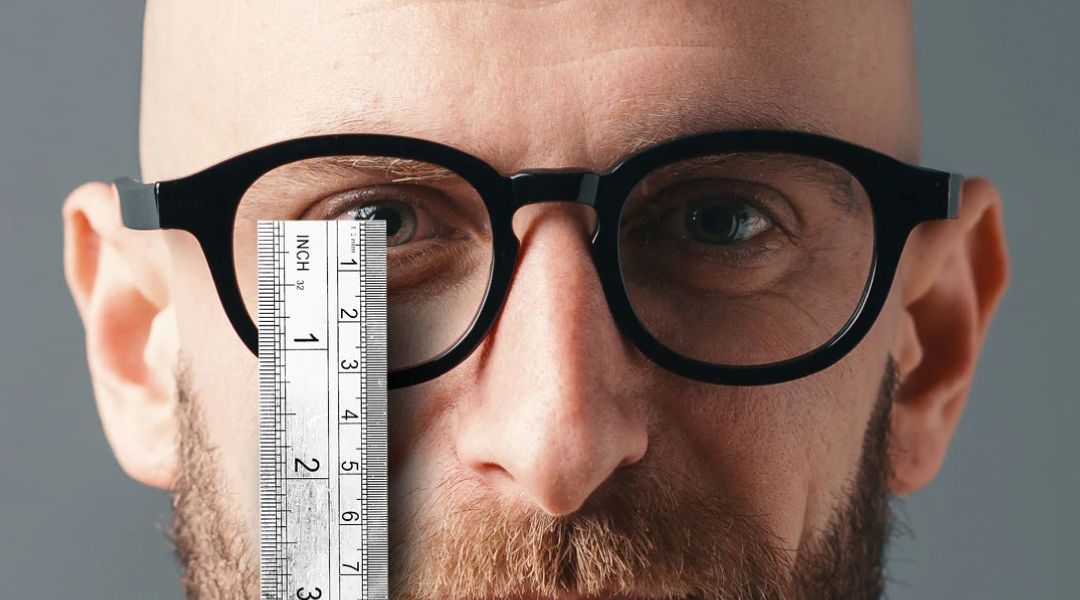
Fitting glasses. For the best viewing experience, you want the center of your lens to sit right in front of your pupil. The center has the clearest part of your prescription, so lining it up with your pupil helps you see well and lowers eye strain. The measurement will change, though, depending on the type of glasses you buy.

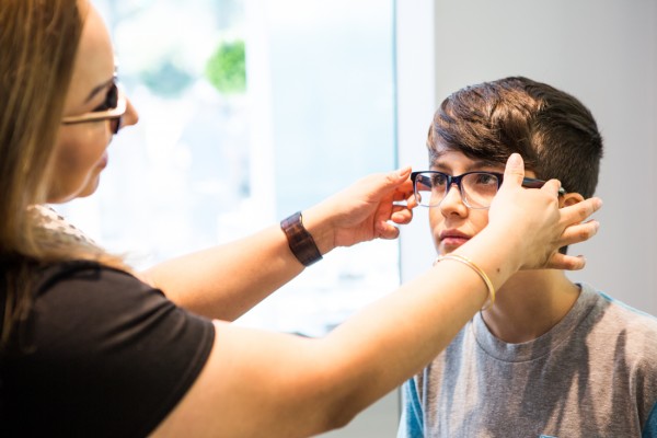
An optician might use a handheld tool called a pupillometer or a digital PD meter. They hold this up to your face and adjust it to get the right distance. They might also use a special ruler to measure, or they might make marks on a pair of glasses and then measure the distance between them.

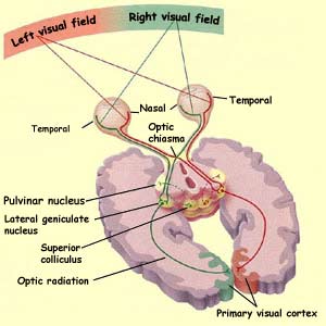
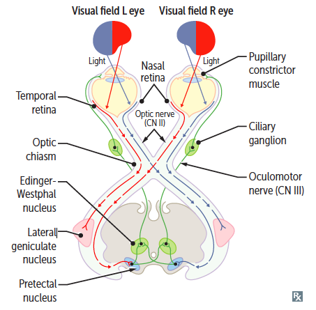
Testing stereo acuity. Your eye doctor will use PD to understand your stereo acuity, which is your depth perception. Both eyes see images and objects individually, but your brain puts those left and right visions together to form a three-dimensional image. The pupillary distance helps your doctor understand how much your field of vision differs between your left and right eyes.

Testing near-point convergence. Near-point convergence occurs when your eyes focus together on a nearby object. Your doctor might test this by bringing a pencil or other object toward your nose and watching how well your eyes come together to focus.

Your pupillary distance gets shorter as your eyes converge, or come together, so your doctor might check to see that your PD is changing.

The pupillary distance is especially important for fitting reading glasses.

How The Wrong PD Will Give You Headaches

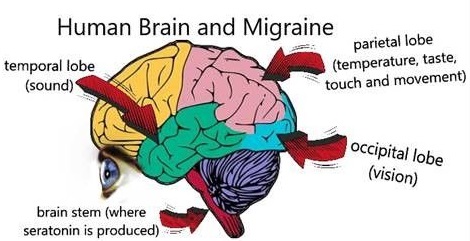
With the light bent “wrong” when arriving at your retinas while your PD isn’t right, things aren’t going well. The visual cortex in your brain is working overtime, trying to to “fix” the incorrect visual signal. Like your computer overheating when you tax the CPU with lots of computing, your brain is going to tell you that it’s not dealing well with this work – via fatigue and headaches.

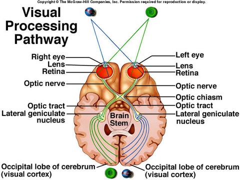
It’s important to remember that vision happens in your brain. So when things go wrong with the visual signal, it’s your headaches that are the most common result.

If you're wearing glasses made using an incorrect PD measurement, you'll probably notice symptoms related to the misalignment. Those include eyestrain, headaches and distortions in your vision. These symptoms aren't dangerous, but they also aren't pleasant.

What is the most common type of eye disorder?

Refractive errors are some of the most common eye conditions. They're also usually easier to treat, with corrective lenses like eyeglasses or contact lenses helping to adjust eyesight and improve visual acuity.

The top 25 common eye problems and their definitions are listed below.

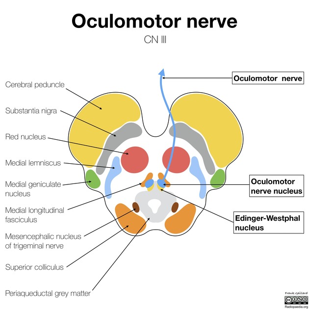
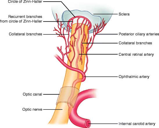
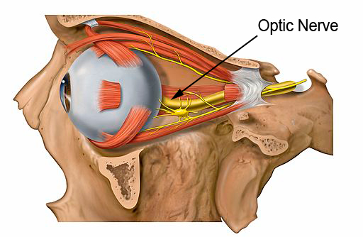
  1. Glaucoma: Glaucoma is an eye condition that causes a loss of peripheral vision (visual field loss) through damage to the optic nerve.
  2. Cataracts: Cataracts form in the eye lens as a result of clumping of protein, leading to a decline in clear vision which commonly occurs naturally with advancing age.
  3. Uveitis: Uveitis refers to a group of conditions that causes eye inflammation and damages the uvea which is the middle layer of the eye.
  4. Dry eyes: Dry eye syndrome is caused by either the tears in the eyes evaporating too quickly or the eyes not producing enough tears.
  5. Presbyopia: Presbyopia is an age-related eye condition that occurs when the lens inside the eye becomes more rigid, becoming less able to focus clearly at near distances.
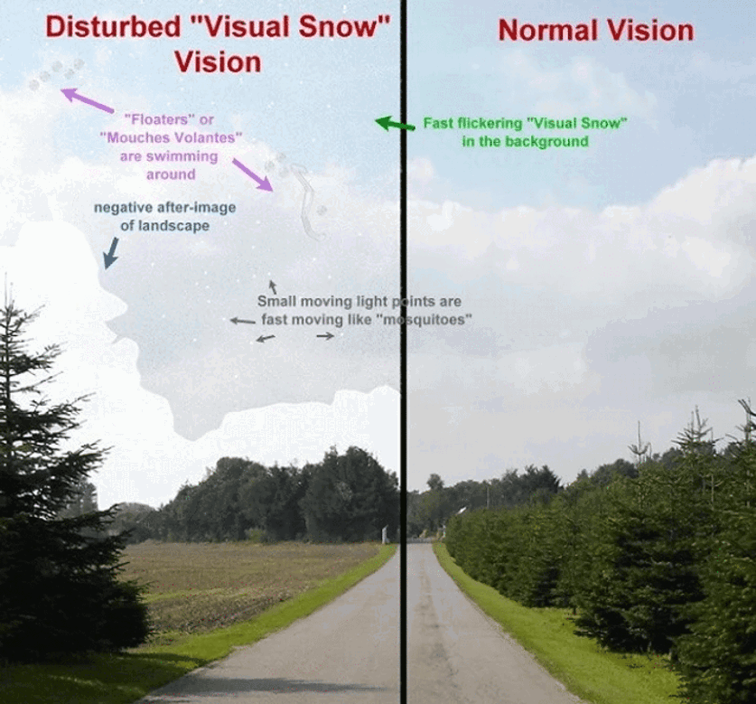
  6. Floaters: Floaters in the eye is a very common condition that usually occurs naturally with age and causes spots and lines to drift and float through a person’s vision.
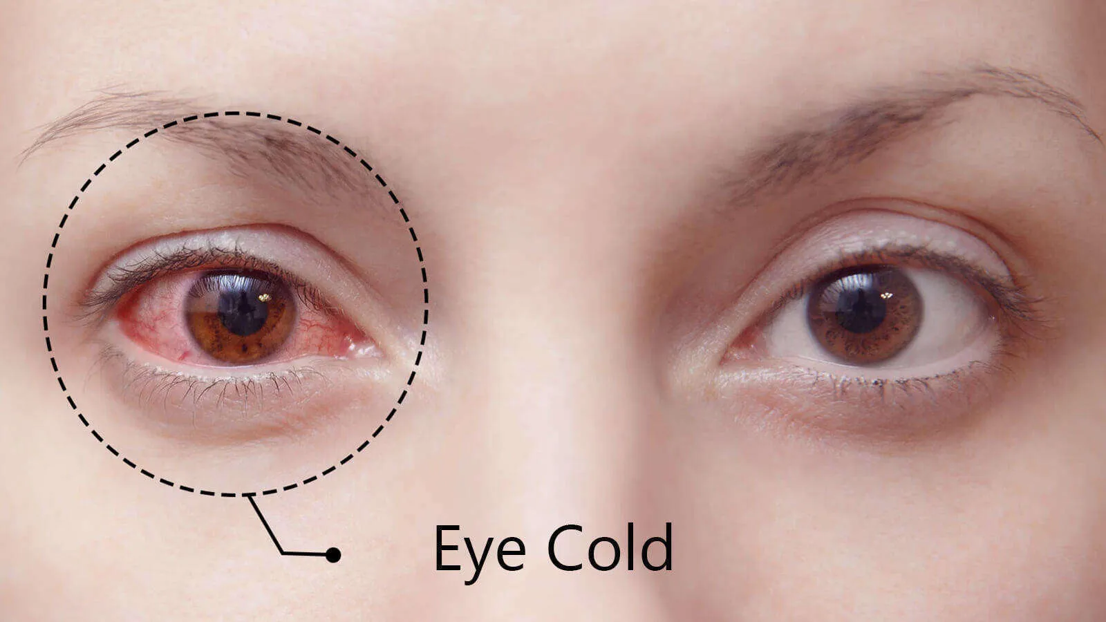
  7. Red eyes: Red eyes occur when the tiny blood vessels on the white part of the eye dilate, making the eye look more red than usual.
  8. Night blindness: Night blindness is an eye problem that impairs a person’s vision meaning they struggle to see in dim light or at night.
  9. Lazy eyes: Lazy eyes, also known as amblyopia, is an eye condition that describes a person with one eye that has weaker vision than the other.
  10. Colour blindness: Colour blindness is an eye condition that means a person is able to perceive colour but has trouble telling the difference between certain colours.
  11. Eyestrain: Eyestrain is a very common eye problem and is caused by the eyes concentrating on tasks or objects for a long period of time.
  12. Vision changes: Vision changes are a common sign of many eye conditions and are characterised by a sudden difference in your ability to see normally.
  13. Excess tearing: Excess tearing causes watery eyes due to the eyes producing too many tears or there being an issue with the film drainage of the eyes.
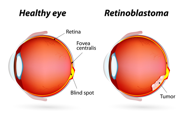
  14. Retinal disorders: Retinal disorders are a term for a group of conditions that affect the retina. Common retinal disorders and diseases include age-related macular degeneration and retinal tear and detachment.
  15. Eyelid problems: Eyelid problems are eye conditions that affect the eyelids, potentially making them swollen, painful and irritated.
  16. Corneal diseases: Corneal diseases are diseases that mainly affect the cornea. Some common corneal diseases include keratitis, eye herpes and pterygium.
  17. Problems with contact lenses: People may experience problems with contact lenses that can lead to certain eye conditions and symptoms.
  18. Cross eyes (strabismus) and nystagmus: Crossed eyes is an eye condition that occurs when one eye turns in a different direction than the other eye. Nystagmus causes involuntary movement of the eyes which can be side-to-side, circular or up and down.
  19. Conjunctivitis (pink eye): Conjunctivitis is an inflammation of the conjunctiva, a lining covering the white part of the eye and underneath the eyelids.
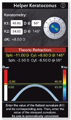
  20. Keratoconus: Keratoconus is an eye disease that affects the cornea, making it thin and bulge outwards.
  21. Chalazion (eyelid cyst): A chalazion is an eyelid cyst that is a sign of a blocked oil gland, usually underneath the upper eyelid, resulting in swelling of the affected area.
  22. Pterygium (noncancerous growth on the conjunctiva): Pterygium, also known as surfer’s eye, is a benign, noncancerous eye growth on the conjunctiva.
  23. Pinguecula (yellowish growth on the conjunctiva): A pinguecula is a yellow, raised growth on the conjunctiva that usually contains calcium, fat or protein.
  24. Diabetic retinopathy: Diabetic retinopathy is a complication of diabetes where the small blood vessels in the back of the eye are damaged.
  25. Age-related macular degeneration: Age-related macular degeneration is a common eye condition that causes a painless loss of central vision.

Reference:

  • glasses.com
  • docs.luna.io/docs/pd-reade
  • webmd.com/eye-health/pupillary-distance
  • zennioptical.com/measuring-pd-infographic
  • lowcostglasses.co.uk/distance-pd-vs-near-pd
  • oscarwylee.com.au/glasses/eye/common-problems
  • eyebuydirect.com/guides/how-to-measure-your-pd
  • endmyopia.org/how-to-measure-pd-pupillary-distance
  • allaboutvision.com/eye-care/measure-pupillary-distance
  • rx-safety.com/2014/02/does-pupillary-distance-have-to-be-exact
  • researchgate.net/figure/Mean-interpupillary-distance-mm-values-in-various-age-groups
  • finestglasses.com/landingpage/would-your-vision-become-worse-if-you-wear-large-sized-glasses-often.html
  • visionsource-meadville.com/2021/09/03/answers-to-4-faqs-about-eyeglasses-to-help-you-find-the-perfect-fit
  • eyecareleaders.net/blog/2023/6/9/what-is-pupillary-distance-pd-and-why-is-it-important-to-have-an-accurate-measurement-of-it

See also: Normal Interpupillary Distance Values in an Iranian Population

What do k readings mean?

Recording accurate corneal curvatures (K-readings) before a lens is placed on the eye is a critical baseline measure. in contact lens practice. Subtle changes in corneal contour. induced by contact lenses or pathology may be leading edge.

What is normal K reading range? K readings.

The normal maximum keratometry (K) reading (K max) is less than 49.00 D. The normal difference between K max and the steep simulated K (sim K) is less than 1.00 D. The K max should be used in the calculation of photorefractive treatments of hyperopia.

What is K1 and K2 in keratometry?

Corneal power (keratometry – K) was measured in both the flat (K1) and steep K (K2). The final K value was taken as the mean of K1 and K2. For calculation of astigmatism, difference between K1 and K2 were taken and for the axis of astigmatism the axis of the steeper power, i.e., K2 was taken.

What is K1 K2 in keratometry readings?

K1: flat meridian of the anterior corneal surface, K2: steep meridian of the anterior corneal surface.

What is K value in optometry?

In this case, keratometry is normally measured along these two orthogonal meridians, giving the maximum and minimum corneal power. These extrema are known as K-values or corneal Ks. The difference between the K-values is a measure of corneal astigmatism.

Keratometry (K) is the measurement of corneal curvature, which is used to evaluate the cornea's power. Astigmatism is caused by differences in power across the cornea (opposite meridians); thus, keratometry quantifies astigmatism. It can be obtained manually or through automated ways using a variety of devices.

What is the K index of the eye?

The corneal keratometric index is a crucial parameter for ophthalmic procedures and calculations, particularly for IOL power calculation. Notably, the estimated corneal keratometric index value of 1.329/1.331 in this study is lower than the commonly used 1.3375 index.

Keratometric readings are also important when fitting contact lenses, especially gas permeable lenses, to monitor corneal disease and detect keratoconus. Keratometry is frequently used to identify keratocones.

How to calculate base curve using k-readings?

For example, if the post-keratorefractive surgery measured Ks are found to be 35.88 x 180 and 33.75 x 090, a good choice for the over-refraction contact lens base curve would be calculated as follows: Base curve = 0.95 * (0.50 * (K1+K2))Base curve = 0.95 * (0.50 * (35.88 D + 33.75 D))

Reference:

  • toddzarwell.com/journal/2012/9/21/documenting-keratometry
  • latamoptical.com/what-is-the-significance-of-keratometry
  • onlinelibrary.wiley.com/doi/10.1155/2016/3437125

Flickers are sparkles that shimmer in vision ("scintillations") Flickers usually come from activated visual cortex in migraine, but importantly also in transient ischemic attack, seizure, damaged retina, and damaged optic nerve.

  • Flashes are bright sparks or streaks of light that appear suddenly and briefly in vision
  • Flashes usually come from tugging on retinal photoreceptors, which may signal impending or actual vitreous detachment, retinal hole, or retinal detachment
  • Flickers are sparkles that shimmer in vision ("scintillations")
  • Flickers usually come from activated visual cortex in migraine, but importantly also in transient ischemic attack, seizure, damaged retina, and damaged optic nerve
    Ocular migraines vs. migraine auras
  • Flashes appear abruptly like lightning bolts in outer edge of visual field
  • Flashes may be provoked by eye movement
  • Flickers may be transient or persistent
  • Flickers that are part of visual aura of migraine often expand across hemifield in 20-30 minutes and disappear
  • Flickers of migraine usually precede headache and other manifestations
  • Flickers of damaged retina or optic nerve are often persistent

In visual perception, flicker is a human-visible change in luminance of an illuminated surface or light source which can be due to fluctuations of the light source itself, or due to external causes such as due to rapid fluctuations in the voltage of the power supply (power-line flicker) or incompatibility with an external dimmer. eResearch by Navid Ajamin -- summer 2024

What is a migraine?

Migraine is a neurological condition that is characterized by often incapacitating symptoms including severe, throbbing and recurring pain that’s usually centered on one side of the head.

Other disabling symptoms of a migraine are nausea, vomiting, dizziness, tingling or numbness in the extremities or face, and extreme sensitivity to sound, light, touch and smell.

Migraine affects about 1 billion men, women and children worldwide and approximately 40 million Americans.

Migraine headaches can last up to three days and produce moderate to severe pain.

Migraines are most common between the ages of 18 and 44, and they affect women disproportionately. Eighteen percent of American women suffer from migraines, compared to 6 percent of men and 10 percent of school-age children.

There is a strong genetic link for migraines, with about 90 percent of sufferers reporting a family history of these headaches.

Migraine is a "diagnosis of exclusion," which means it is reached by a process of elimination since there is no test or biomarker to confirm its presence.

Just as every person is unique, so are migraine headaches. Migraines differ from person to person, and migraines also can present in different ways and with different symptoms in the same person.

Retinal migraine is a retinal disease often accompanied by migraine headache and typically affects only one eye. It is caused by ischaemia or vascular spasm in or behind the affected eye.

The terms "retinal migraine" and "ocular migraine" are often confused with "visual migraine", which is a far-more-common symptom of vision loss, resulting from the aura phase of migraine with aura. The aura phase of migraine can occur with or without a headache. Ocular or retinal migraines happen in the eye, so only affect the vision in that eye, while visual migraines occur in the brain, so affect the vision in both eyes together. Visual migraines result from cortical spreading depression and are also commonly termed scintillating scotoma.

Northern lights, aurora, borealis, scenic

Migraine aura

A migraine aura starts in your brain, not your eye. The aura is one or more symptoms that can happen right before a headache starts. These visual symptoms happen in both eyes.

Migraine with aura (also called classic migraine) is a recurring headache that strikes after or at the same time as sensory disturbances called aura. These disturbances can include flashes of light, blind spots, and other vision changes or tingling in your hand or face.

Treatments for migraine with aura and migraine without aura (also called common migraine) are usually the same. You can try to prevent migraine with aura with the same medications and self-care measures used to prevent migraine.

Symptoms

Migraine aura symptoms include temporary visual or other disturbances that usually strike before other migraine symptoms — such as intense head pain, nausea, and sensitivity to light and sound.

Migraine aura usually occurs within an hour before head pain begins and generally lasts less than 60 minutes. Sometimes migraine aura occurs without headache, especially in people age 50 and older.

Visual signs and symptoms

Most people who have migraine with aura develop temporary visual signs and symptoms, which tend to start in the center of the field of vision and spread outward. These might include:

  • Blind spots (scotomas), which are sometimes outlined by simple geometric designs
  • Zigzag lines that gradually float across your field of vision
  • Shimmering spots or stars
  • Changes in vision or vision loss
  • Flashes of light

Some people experience an aura without any pain at all. Doctors call this an “acephalgic migraine” or a “migraine aura without headache.”

It's also common for people to call them "visual migraines." This may be why they get mixed up with ocular migraines so often. Here is an easy way to remember the difference: "visual migraines" happen in your vision, but "ocular migraines" happen in your eye.

About 8% of the population gets migraines with aura.

One in every four people who get migraines sees an aura beforehand. Others will only experience symptoms like headache, nausea and vomiting.

  • Refer patient with flashes urgently to ophthalmologist because they suggest intraocular disorder (vitreous, retina, optic nerve)
  • Refer patient with flickers urgently to ophthalmologist, neuro-ophthalmologist or neurologist unless diagnosis of migraine is obvious because they could also suggest transient ischemic attack or seizure
  • Vitreous detachment may rarely cause retinal tear and detachment which must be repaired promptly to protect vision
  • Visual aura of migraine is usually harmless, but transient ischemic attack and seizure have health consequences
?Does Migraine Cause Hallucinations

How do I tell the difference between aura and stroke?

Strokes produce visual, sensory and/or speech symptoms almost instantaneously, and most frequently they are “negative” phenomena—that is, a loss of vision, numbness or weakness. Also with strokes, there is no sense of movement of the phenomena to other parts of the body. They don’t progress but are maximal at onset. Strokes usually are continuous and do not remit in one hour. A headache may or may not occur with a stroke. In addition, an aura occurring for the first time after the age of 40, with numerous vascular risk factors such as hypertension, diabetes and hyperlipidemia, should be investigated for transient ischemic attacks. If the visual symptom is ONLY negative (that is, a hemianopic scotoma), the person should be further investigated.

A migraine is a recurrent and severe headache which a whopping one in ten New Zealanders suffer from.

There are a few different types of migraines which people usually experience:

  1. A classic migraine occurs in about 40% of migraine sufferers and has an aura (visual, auditory, olfactory or tactile).
  2. A common migraine occurs in about 60% of migraine sufferers and has no aura.
  3. A silent migraine is when a person experiences the aura, but no headache or pain afterwards.
  4. A visual migraine is a silent migraine when the aura is visual.
  5. A retinal migraine is very rare and its visual symptoms are the partial or total loss of vision, temporarily in one eye. In recurrent cases, it is most likely to affect the same eye each time. The vision fades out over five minutes, and can be described as a dimming of vision, flashes of light or patches of blank spots (scotomas) that enlarge to block out all sight in that eye. The vision returns to normal within an hour. This loss of vision is caused by reduced blood flow or spasms of the blood vessels in the retina or behind the eye, not in the brain.

Migraines might occur rarely, once or twice a year, or they could strike several times a month. The frequency differs drastically between sufferers and is often associated with an underlying cause. Migraines usually run in families as they have a genetic component. People with two parents who suffer from migraines have a 75% chance of having migraines themselves. Women are also three times more likely than men to get them.

Reference:

  • denarend.com/miscellaneous
  • en.wikipedia.org/wiki/Retinal_migraine
  • allaboutvision.com/conditions/migraine-aura
  • allaboutvision.com/conditions/ocular-migraine.htm
  • kellogg.umich.edu/theeyeshaveit/common/flashes_flickers.html
  • mayoclinic.org/diseases-conditions/migraine-with-aura/symptoms-causes
  • wefixeyes.co.nz/articles/the-relationship-between-eyesight-and-migraines
  • americanheadachesociety.org/wp-content/uploads/2020/08/AHS-First-Contact-Migraine-w-Aura.pdf

Astigmatism is a refractive error that occurs when the cornea’s shape affects how light reaches your eye. However, astigmatism differs from other refractive errors like myopia (nearsightedness) and hyperopia (farsightedness) because it can affect your vision at near and far distances.

Most people have some degree of astigmatism, but it can become more apparent if you develop another refractive error alongside it.

Aside from blurry vision, astigmatism may also cause eye strain and headaches.

Astigmatism Symptoms

Symptoms of astigmatism may include:

  • Blurry or distorted vision
  • Eyestrain
  • Headaches
  • Trouble seeing at night

Symptoms of astigmatism are squinting to see clearly, seeing glares or halos around lights at night, headaches and eye fatigue. The possible symptoms of astigmatism are listed below.

  • Squinting to see clearly: Squinting to see clearly is a symptom of astigmatism because the eyes are attempting to focus harder to overcome the vision disruption caused by astigmatism.
  • Seeing glares or halos around lights at night: Seeing glares or halos around lights at night is a symptom of astigmatism because the misshapen cornea or lens disrupts the eye’s ability to perceive light, causing visible halos and glares.
  • Headaches: Headaches are a symptom of astigmatism as this condition may cause eye strain and eye fatigue, leading to headaches.
  • Eye fatigue: Eye fatigue is a symptom of astigmatism because as the eyes squint and increase in focus in order to overcompensate for the compromises in vision caused by astigmatism, they become more exhausted and fatigued, causing eye strain and eye pain.
  • Blurry vision: Blurry vision is a common symptom of astigmatism, caused by the misshapen cornea or lens not bending light correctly.

What improves astigmatism?

Glasses or contacts can correct almost all cases of astigmatism. But if you have only a slight astigmatism and no other vision problems, you may not need them. If you have a common level of astigmatism, you'll probably have corrective lenses, like glasses or contacts, or surgery.

Can vision therapy improve astigmatism?

Even though vision therapy can't help correct astigmatism and how it affects your visual acuity, it can help address some of its more uncomfortable symptoms while also strengthening the relationship between your eyes and brain share.

Vision therapy is a doctor-led treatment program specifically designed to help strengthen the relationship between your eyes and brain. We typically recommend vision therapy to patients with amblyopia (lazy eye), strabismus (crossed eyes), or sports vision problems, but we can recommend this treatment to anyone who may want to enhance the visual skills they use in everyday life.

Some of the most common visual skills vision therapy can help support include:

  • Hand-eye coordination
  • Eye-tracking
  • Binocular movements
  • Visual memory
  • Focusing

What causes astigmatism?

Astigmatism occurs when the front surface of the eye (the cornea) or the lens inside the eye is more oval or cylindrical than round. The cornea and lens are mostly responsible for properly focusing light entering your eyes. This allows you to see things clearly. Astigmatism is caused by small differences in the growth and alignment of the components of the eye. Genetics may play a role in the development of refractive error. Astigmatism may also result from such factors as pressure of the eyelids on the cornea.

Sometimes astigmatism develops following an eye injury or eye surgery. In rare cases there is also a condition called keratoconus that may occur. Keratoconus is a condition in which the cornea becomes progressively thinner and cone shaped. This causes a large amount of astigmatism resulting in poor vision that cannot be effectively corrected with glasses. In these cases, the clearest vision is achieved with contact lens wear. Corneal transplants or other corneal treatments may be considered, depending on the case

Astigmatism often occurs early in life, so it is important to schedule children for a comprehensive eye exam with an optometrist to avoid vision problems in school from uncorrected astigmatism. eResearch by Navid Ajamin -- winter 2024


No.1) Here are our top 5 eye exercises for astigmatism:

1.Rectus muscle relaxation

The rectus eye muscle can come into considerable strain over time, and astigmatism might be the result of a weak or tight rectus muscle. The rectus muscle relaxation exercise is done by gradually and gently relaxing the eye rectus muscles. The exercise should be done in the following steps:

  • Put your thumb in front of your face just above the nose (about 10cm)
  • Move your thumb as if it is running on a clock. First slowly move your thumb to 12 o’clock where it will disappear and leave it there for 2-3 seconds.
  • If you experience tension here, that’s okay. Return to the initial nose spot and move back to 1 o’clock then again to 3,5,6,7,9, 11 and back to noon.
  • From the center out, don’t forget to breathe. Exhaling slowly will help relax your muscles.

Duration: 2 minutes

Repetitions: 2-4 times per day

2.Eye Massage

Eye massages are an effective and relaxing way to reduce strain for those suffering from astigmatism. This exercise is effective because it helps restore the shape of the lens as astigmatism is when the lens has been distorted.

  • Close your eyelids and place two fingers on each lid
  • With gentle pressure move your fingers from right to left, top to bottom in a circular motion
  • Move clockwise and counterclockwise 10-15 times repeating both the right to left and circular motions

Duration: 1 minute

Repetitions: 2-4 times per day

3.Reading

Now we don’t mean pull out just any book and start reading chapter after chapter, this is to be done a certain way to improve the pressure and strain caused by astigmatism.

  • Use corrective lenses and pull out a book
  • Place a playing card or another object to the side and focus on that after you’ve read one paragraph
  • Switch back and forth from the book to the object
  • Continue off and on until your eyes start to feel tired

Duration: 10 minutes

Repetitions: 2-4 times per day

4.See a chiropractor

People who have astigmatism tend to tilt or position their neck and head in one position more than another. Because of eye strain, most people have an incorrect stature or tilted head. It is suggested to get an adjustment to relax the neck and spine while also correcting posture.

5.Paper and peripheral vision

Take a piece of paper that is long and wide enough to cover both of your eyes.

  • Make sure the side is exposed so you are able to peek out, strengthening your peripheral vision.
  • Grab your corrective lenses and attach the paper to your face at the bridge of your nose.
  • Move your hand to the peripheral side of the paper one after another.
  • Without moving the head, look to see the finger on either side
  • Repeat

Duration: 5 minutes

Repetitions: 2-4 times per day

Each exercise is meant to improve the strain put on the eyes by astigmatism. Daily practice will absolutely help the damage that has been caused by astigmatism while giving your eyes new pathways of practice to improve your traction and span and reduce pressure. Consult with your eye doctor and ask if these exercises will help your case.

Astigmatism in Children

No.2) Eye Exercises for Astigmatism:

Astigmatism is a common eye disorder that affects your vision. The muscles around your eyes are affected which causes undue stress on the cornea which causes the cornea to lose its shape, which in turn causes blurry vision. Some of the other symptoms of astigmatism include double vision, eyestrain, eye irritation, and headaches. It can be prevalent at birth or could be a result of trauma, congenital conditions or eye surgery. It can be very annoying as it makes a simple task like reading a book complicated.

However, there are several natural ways to treat astigmatism and one of them is eye exercises. Benefits of eye exercises for Astigmatism It’s true that there are eye exercises to treat astigmatism. Just like the other muscles in our body, our eye muscles also work on a simple logic of keeping them in use them or else you lose them. Therefore, it is important that you keep your eye muscles active throughout the day. Other than by staring straight at the computer screen or at the road ahead while driving doesn’t exercise the muscles to their full potential, additional eye exercises are must. Here are some benefits of eye exercises:

  • Help to reduce the stress.
  • Strengthen the eyes and relaxes the eye muscles.
  • Improve vision over time or in between 1 to 4 weeks.

6 Eye Exercises to Treat Astigmatism

1. Eye Massage This exercise restores the shape of the cornea

  • Close your eyes and keep your two fingers on each of your eyelids.
  • By applying gentle pressure, slowly move your fingers in a circular motion from top to bottom and right to left.
  • Move your fingers clockwise as well as anti-clockwise and repeat it for 10–15 times, 2 to 4 times a day.

2. Reading It helps to release the strain and pressure caused by astigmatism.

  • Pull out a book.
  • Place an object on the side. Focus on the side object after reading a paragraph from the book.
  • It is advised to continue this until your eyes start to feel tired, 2-4 times a day.

3. Vision Breaks It relieves eye pressure and strain.

? What Causes Astigmatism to Worsen
  • Take a short break from writing, reading, or staring at the computer.
  • Focus for 20 seconds on other objects that are kept in the distance
  • Repeat the exercise as many times as possible in a day.

4. Head Tilting It helps the extraocular muscles to regulate the force they exert on the eyeball.

  • After looking in the mirror, find out if you tilt your head to one side.
  • Spend time every day to tilt your head in the opposite direction.

5. Eye Yoga It strengthens eye muscles, sharpens focus and improves vision.

  • Stand, sit in the chair or on the floor and keep your posture straight.
  • Close your eyes and breathe while concentrating.
  • Slowly and start moving your eyeballs from side to side.
  • Do this exercise several times a day.

6. Rectus muscle relaxation: It relaxes the rectus muscles and can be done in the following steps

  • Place your thumb just above the nose, move it clockwise and leave it there for 2-3 seconds.
  • Move your thumb back to the original position. Now, move it to 1 o’clock position, then to 3,5, so on and back to 12. You can do this exercise 2-4 times per day.
  • Don’t forget to breathe while you are moving the thumb from the center out and slowly exhale to relax your muscles.

Daily performing these exercises will reduce the symptoms and eventually treat astigmatism. But make sure to consult your eye doctor to find out if these exercises will work for you.

Reference:

  • Astigmatism | The Canadian Association of Optometrists opto.ca/eye-health-library/astigmatism
  • Astigmatism: Definition, Causes, Symptoms, Risk Factors and Treatment (oscarwylee.com.au)
  • 5 Vision Therapy Exercises To Reduce Astigmatism Symptoms (urbanoptique.ca)
  • Astigmatism: Causes, Symptoms, Diagnosis, and Treatment (webmd.com)
  • centreforsight.net/blog/treat-astigmatism-with-daily-eye-exercises

See Also: Exercise benefits your eyes

What is screen addiction?

Screen addiction is a group of behaviors that are negative, some negative outcomes that can happen when we use too much technology during our day. So prolonged use of watching TV, video games, scrolling through social media all of that use acts like a digital drug for our brain.

How many hours of screen time is considered an addiction?

How Much Screen Time Is Too Much? The American Optometric Association (AOA) found that people who use screens continuously for two or more hours a day are at the greatest risk of developing computer vision syndrome, the most prevalent side-effect of screen addiction.

Are screens bad for your brain?

Screen time may affect its growth. A study called Adolescent Brain Cognitive Development (ABCD) revealed that some kids who use screens more than seven hours a day had a thinner cortex than those who used screens less. Future studies hope to show how this will affect kids' brains over time.

How much screen time is healthy?

What's a healthy amount of screen time for adults? Experts say adults should limit screen time outside of work to less than two hours per day. Any time beyond that which you would typically spend on screens should instead be spent participating in physical activity.

How much screen time per age?

Yousuf said pediatricians generally recommend the following guidelines: Under 2 years old: Zero screen time, except for video chatting with family or friends.

  • 2-5 years old: No more than one hour per day co-viewing with a parent or sibling.
  • 5-17 years old: Generally no more than two hours per day, except for homework.

Physical Effects

The most prevalent physical effect of screen use is the great demand it puts on the eyes. Digital eye strain and computer vision syndrome are reported in 50% to 90% of people who work at computers daily.

Symptoms of digital eye strain and computer vision syndrome include:

  • Dry eyes
  • Blurred vision
  • Double vision
  • Headaches
  • Neck and shoulder pain

While many of these physical symptoms will resolve after screen use has stopped, long-term eye strain can lead to permanent vision problems.

Physiological And Mental Effects

Screen addiction can contribute to physiological and mental effects like insomnia, stress, and depression.

Depression

Depression is the most common mental health condition associated with screen addiction. A study from Preventative Medicine Reports found a correlation between higher screen time usage and moderate to severe depression levels, with higher rates found among females. The study found that four or more hours of daily screen time was the tipping point for heightened risk of depression.

Insomnia And Poor Sleep

Using screens before bed can lead to many issues that affect a person’s sleep quality. Blue light from devices can delay melatonin production, leading to a lack of sleepiness. Likewise, engaging in stimulating games or watching exciting shows can also make a person feel more alert. Screen addiction heightens the risk for these factors, as a person spends excessive time on these devices and puts off sleep.

Stress

Some people claim to use screen time to zone out and take a break from the day’s stressors, but the reality may be that this time is a source of stress. Fear of missing out (FOMO), lowered self-esteem, and loneliness are some of the stressors associated with screen addiction. The Journal of Medical Internet Research found that people who used screen time for entertainment and social networking experienced 19% more emotional stress than those who used screen time for professional purposes.

How To Cut Back Screen Time

If you feel like you are spending too much time looking at a screen, there are methods for cutting back.

20-20-20 Rule

For temporary eye strain relief, the AOA suggests using the 20-20-20 method, which directs a person to take a 20-second break every 20 minutes to focus on an object at least 20 feet away. This gives the eye a break from the blue light and allows it to relax by focusing on something further away.

Time Limits

Many studies recommend limiting personal screen time to 30 minutes per day for optimal effects. Since this may be a drastic change for some, limiting screen use to one hour a day is also beneficial.

Likewise, taking a three-to-four-hour break from screens can create an overall healthier relationship, as the perception changes from looking at screen time as a necessity to an activity of choice.

Digital Detox

A digital detox may be the best option for people who feel like their screen addiction has gotten out of control. This does not necessarily mean giving up all screen use but encourages users to cut back on non-work-related screen time.

Taking scheduled breaks or putting the phone away before bedtime can be the first step in resetting and creating new, healthier relationships with screens.

Treatment Options For Screen Addiction

Screen addiction treatment often involves a mixture of behavioral therapy, support groups, and lifestyle changes.

Lifestyle Changes

Lifestyle changes often include limiting screen time and searching for alternative ways to fill free time, specifically ones that do not involve technology. Finding new hobbies and interests can help reduce dependence on technology. Engaging in social interactions in person is also a great alternative to using smartphones for social interactions. In some cases, prescribed medication may also help address underlying mental health conditions, such as anxiety or depression, that may contribute to screen addiction.

More formal treatment options used to address screen addiction may include:

Cognitive-Behavioral Therapy (CBT)

Cognitive-Behavioral Therapy encourages people to change their thoughts and behaviors related to screen use and addiction. CBT can help individuals develop new and healthy coping skills and habits, identify and address triggers related to screen use, and learn how to utilize their time and energy more appropriately.

Mindfulness-Based Therapy

This type of therapy refers to mindfulness strategies that can help an individual learn to manage stress and anxiety, which are often common triggers for screen use. Practicing mindfulness can teach a person to be more present and aware of their thoughts and feelings. This increased awareness can help individuals reduce their desire for screen time.

Family Therapy

Screen addiction can significantly impact relationships with loved ones. Family therapy can help address these issues. Family therapy can help family members develop healthier communication patterns, boundary setting around screen use, and other ways to support each other in reducing screen time and spending more time with each other.

Group Therapy

Group therapy can offer a supportive and non-judgmental atmosphere for individuals struggling with screen addiction. Individuals can share their coping strategies for managing screen use and gain support from others experiencing the same challenges.

Individual Counseling

Individual counseling can help people get to the root of their problems. There may be a bigger reason why someone has immersed themselves in technology. Individual counseling can help a person address many deep-rooted issues from trauma and relationship problems to grief and loss.

Reference:

  • premierhealth.com/your-health/articles/health-topics/screen-addiction-affects-physical-and-mental-health#
  • How to Encourage Technology Literacy in Kids Without Screen Addiction? (narayanaschools.in)
  • Screen Addiction Explained: Effects and Coping Strategies (addictioncenter.com)

Low temperatures can make the blood vessels in and around the eye constrict, which can cause blurriness and double vision. However, this vision change is usually temporary and your vision should return to normal once you move into a warmer environment and out of the cold.

1. Can extreme cold damage eyes?

What extreme cold does to your eyes. Excessively cold temperatures can constrict your eye's blood vessels or even freeze your cornea. This can cause blurred vision, double vision, or even a loss of sight.

2. What temperature should your eyes be?

The growing importance of thermography and temperature in understanding the ocular surface has also increased the importance of establishing normal values in healthy eyes. The single time-point OST of healthy eyes typically ranges from 31 to 37°C in older literature.

3. Does temperature affect eyesight?

Higher temperatures in low humidity conditions are associated with a higher risk of dry eye diagnosis. Excessive heat in low humidity conditions has also been shown to worsen dry eye symptoms. This occurs because high heat causes tears to evaporate faster. Lower temperatures can be detrimental to the eye as well.

4. Can weather affect eyes?

Here are some ways weather can affect your eyes: Dryness: In dry weather conditions, such as during winter or in arid climates, the air can be lacking in moisture, leading to dry eyes. Dry eyes can cause discomfort, redness, itching, and a gritty feeling in your eyes.

5. Can vision be affected by a cold?

There are chances of developing swelling in your eyelids when you have a common cold. With a condition like this, the vision may get narrowed and you may feel discomfort and pain while blinking. The swelling usually reduces after 24 hours. It is recommended to get a check-up at the earliest. eResearch by Navid Ajamin -- winter 2024

6. Can common cold cause blurry vision in one eye?

Symptoms include red/watery eyes, discharge, and swollen eyelids. Blurred vision is uncommon but can certainly happen because of these factors. When the infection spreads into the eye, blurry vision may result, as well as seeing halos around lights in some cases.

7. Does sunlight help dry eyes?

The sun: The sun's rays can cause a person's tears to evaporate more quickly. If their eyes cannot produce tears quickly enough to replace the evaporated moisture, they will become dry. Windy environments: The wind blowing in a person's eyes can also cause them to become dry before they can replenish the moisture.

8. Can blocked sinuses cause blurred vision?

Sinus problems such as chronic sinusitis can also cause blurry vision, vision loss and other problems due to optic nerve damage caused by chronic inflammation, although this is rare.

9. Why is heat good for your eyes?

Warm compresses can help dry eyes by providing moisture and heat. These compresses can help you produce more tears, unclog the meibomian glands, and relieve pain and swelling. Research shows that heating devices like warm compresses on the eyes can help relieve dry eye symptoms.

10. Is Hot or cold good for eyes?

Warm compresses help to reduce the sticky buildup of discharge on the eyelids or crust that forms on your eyelashes, while cold compresses help to relieve itching and inflammation. If you have allergic conjunctivitis, it's important to avoid rubbing the eye, since this can worsen your symptoms.

Why do my eyes run in the cold?

When cold air causes more evaporation, our eyes are left with a thinner cushion of tears that protect the sensitive surface cells. This condition can trigger a reflex that tells the lacrimal gland (the eye gland that secretes an aqueous layer of tear film) to produce extra tears.

Can a cold affect your eyes?

It may sound dramatic, but when you have a cold your body is essentially under attack, and vulnerable to infection as a result. Whilst a cold can see your eyes produce too much mucus as mentioned above, it can also do the opposite, which as a result can cause dry eyes. This can make your eyes feel irritated and itchy.

An eye cold occurs when you have viral conjunctivitis, commonly called “pink eye.” Viruses affect mucous membranes in your body, including those in the eyes, lungs, and nose. Viral conjunctivitis is highly contagious and can be spread by person-to-person contact or by touching an object contaminated by the virus.

How long does a cold in your eye last?

In most cases, an eye cold simply runs its course and clears up on its own in 7 to 10 days or less. But it can take as long as two or three weeks to clear up for some people. An eye cold is very contagious, especially while you still have symptoms.

What does a cold in the eye look like?

If your eyes are bothering you, there are a few symptoms to look out for that an eye cold presents. First is an increase in tearing or redness of the whites of your eyes. You might also experience some inflammation. If inflammation increases, your eyelids will get swollen and more sensitive.

To avoid temporary winter vision changes:

  • Wear sunglasses with UV protection
  • Wear goggles with UV protection during winter sports
  • Close your eyes in the winter wind
  • Use eye drops on your ophthalmologist’s recommendation
  • Drink lots of water to stay hydrated and avoid blurry vision

Reference:

  • nyulangone.org
  • ealthline.com/health/eye-cold-conjunctivitis
  • focusclinics.com/blog/can-a-cold-affect-your-eyes
  • lowvisionmd.org/3-ways-cold-weather-can-affect-your-vision
  • georgiacenterforsight.com/blog/why-your-eyes-water-when-its-cold-outside
  • flvisioninstitute.com/eye-disorders/symptoms-causes-and-treatments-of-an-eye-cold

See also:

  • Can A Cold Affect Your Eyes & Vision?
  • Hot vs. Cold Compresses for Eyes: Which is Best and When?
nicetoview.blogfa.com
عینک eyewear وبلاگ تخصصی عینک شامل مجموعه مطالب پزشکی است که اطلاعات مفیدی در رابطه با عینک , چشم، لنز، سلامتی چشم و راه های پیشگیری از بیماریهای چشمی، کنترل و درمان آن را در اختیار شما کاربر محترم می گزارد.

Always Be Healthy
همیشه سلامت باشید

Navid Ajamin نوید عجمین
eMail: navid.aj@outlook.com
موضوعات وب
پیوندها

unicef for every child

شمارنده