The Edinger–Westphal (EW) nucleus also called the accessory or visceral oculomotor nerve, is one of the two nuclei of the oculomotor nerve (CN III) located in the midbrain. It receives afferents from both pretectal nuclei (which have in turn received afferents from the optic tract). It contains parasympathetic pre-ganglionic neuron cell bodies that synapse in the ciliary ganglion. It contributes the autonomic, parasympathetic component to the oculomotor nerve (CN III), ultimately providing innervation to the iris sphincter muscle and ciliary muscle to mediate the pupillary light reflex and accommodation, respectively.
The Edinger-Westphal (EW) nucleus, which is part of the oculomotor nuclear complex (ONC), was first described in the literature in the 17th century. Although its most well known function is the control of pupil diameter, some controversy has arisen regarding the exact location of these preganglionic neurons. Currently, the EW is thought to consist of two different parts. The first part [termed the preganglionic EW-EWpg], which controls lens accommodation, choroidal blood flow and pupillary constriction, primarily consists of cholinergic cells that project to the ciliary ganglion. The second part [termed the centrally projecting EW-EWcp], which is involved in non-ocular functions such as feeding behavior, stress responses, addiction and pain, consists of peptidergic neurons that project to the brainstem, the spinal cord and prosencephalic regions.
Recently, it has been discovered that 2 different cell populations within the EW nucleus – subdivide into the EW preganglionic (EWpg) population and the EW nucleus centrally projecting (EWcp) population. However, the accepted nomenclature for these 2 groups varies.[9]

Schema of the oculomotor nerve nucleus and Edinger-Westphal nucleus (modified from the original figure by Wilson-Pauwels et al.). Oculomotor nerve nucleus consists of the lateral somatic cell column, caudal central nucleus, and medial cell column. Lateral somatic cell column consists of the dorsal subnucleus, intermediate column and ventral subnucleus, and regulates extraocular muscles on the ipsilateral side. The caudal central nucleus regulates levator palpebrae superioris muscles on both sides. The medial cell column regulates superior rectus muscles on the contralateral side. The Edinger-Westphal nucleus regulates sphincter pupillae muscles and ciliary muscles on the ipsilateral side.[8]
هستهٔ قرمز red nucleus از عناصر مهم سیستم حرکتی است. دستهای از اکسونها که از هستهٔ قرمز میآید، یکی از دو مجموعهٔ اصلی تارهای عصبی را درست میکند و پیامهای حرکتی را از مغز به نخاع شوکی یاطناب نخاعی حمل میکند. هسته قرمز از بخشهای مهم سیستم خارج هرمی است.
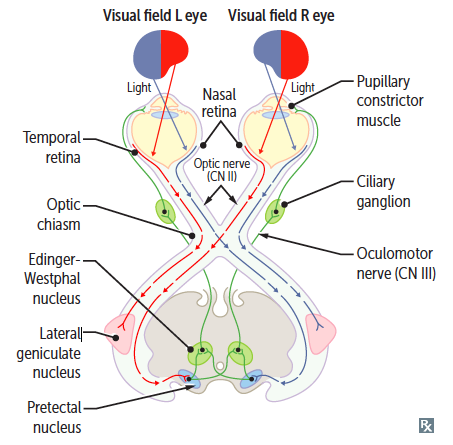
The pupillary reflex depends on the passage of light through eye structures, activation of the photoreceptors and retinal nerve fibers, and transmission along the optic nerve, which hemidecussates at the optic chiasm, to bilateral nuclei in pretectal areas of the rostral midbrain.[10]
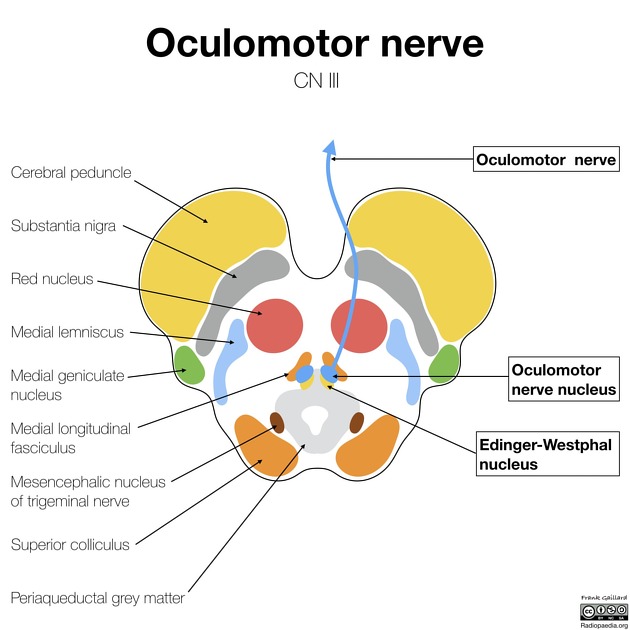
The midbrain or mesencephalon is the uppermost portion of the brainstem connecting the diencephalon and cerebrum with the pons.
It consists of the cerebral peduncles, tegmentum, and tectum.[11]
It is functionally associated with vision, hearing, motor control, sleep and wakefulness, arousal (alertness), and temperature regulation.
The name mesencephalon comes from the Greek mesos, "middle", and enkephalos, "brain"
The Edinger–Westphal nucleus has two parts: [1]
- The first is of preganglionic fibers (EWpg) that terminate in the ciliary ganglion.
- The second is of centrally projecting cells (EWcp) that project to a number of brainstem structures.
The Edinger-Westphal nucleus, in the posterior midbrain, supplies parasympathetic fibers that terminate in the ciliary ganglion via cranial nerve III. It is mainly involved in pupillary constriction and the light accommodation reflex.[2]

Edinger-Westphal Nucleus. The optic nerve, afferent pathway, pretectal nucleus, optic tract, red nucleus, lateral geniculate nucleus, posterior commissure, and nerve with parasympathetic fibers are shown in the illustration. Illustration by Emma Gregory [4] eResearch by Navid Ajamin -- autumn 2024
The Edinger-Westphal nucleus is a small parasympathetic motor nucleus in the midbrain and one of the two nuclei for the oculomotor nerve. It is one of the cranial nerve nuclei.[5]
The Edinger–Westphal nucleus supplies preganglionic parasympathetic fibers to the eye, constricting the pupil, accommodating the lens, and convergence of the eyes.[1]

Cross-section of the midbrain at the level of the superior colliculus
Reference:
- en.wikipedia.org/wiki/EdingeWestphal_nucleus
- sciencedirect.com/topics/veterinary-science-and-veterinary-medicine/edinger-westphal-nucleus
- fa.wikipedia.org/wiki/هسته قرمز
- ncbi.nlm.nih.gov/books/NBK554555/figure/article-34491.image.f3
- radiopaedia.org/articles/edinger-westphal-nucleus-1
- medical-junction.com/light-reflex-pathway-and-defects
- tedmontgomery.com/the_eye/reflex.html
- researchgate.net/figure/Schema-of-the-oculomotor-nerve-nucleus-and-Edinger-Westphal-nucleus-modified-from-the_fig1_51904930
- pubmed.ncbi.nlm.nih.gov/26206178
- sciencedirect.com/topics/neuroscience/pupillary-reflex
- en.wikipedia.org/wiki/Midbrain
 وبلاگ تخصصی عینک شامل مجموعه مطالب پزشکی است که اطلاعات مفیدی در رابطه با عینک , چشم، لنز، سلامتی چشم و راه های پیشگیری از بیماریهای چشمی، کنترل و درمان آن را در اختیار شما کاربر محترم می گزارد.