There are several types of specialists for you to contact for help with questions and problems regarding your eyesight. The following definitions were provided by each corresponding professional organization.
Ophthalmologists
An ophthalmologist is a physician (doctor of medicine or doctor of osteopathy) who specializes in the comprehensive care of the eyes and visual system in the prevention of eye disease and injury. The ophthalmologist has completed four or more years of college premedical education, four or more years of medical school, one year of internship, and three or more years of specialized medical and surgical training and experience in eye care. The ophthalmologist is a physician who is qualified by lengthy medical education, training and experience to diagnose, treat and manage all eye and visual system problems, and is licensed by a state regulatory board to practice medicine and surgery. The ophthalmologist is the medically trained specialist who can deliver total eye care: primary, secondary and tertiary care services (i.e., vision services, contact lenses, eye examinations, medical eye care and surgical eye care), and diagnose general diseases of the body.
Optometrists
The optometrist is a health care professional trained and state licensed to provide primary eye care services. These services include comprehensive eye health and vision examinations; diagnosis and treatment of eye diseases and vision disorders; the detection of general health problems; the prescribing of glasses, contact lenses, low vision rehabilitation, vision therapy and medications; the performing of certain surgical procedures; and the counseling of patients regarding their surgical alternatives and vision needs as related to their occupations, avocations and lifestyle. The optometrist has completed pre-professional undergraduate education in a college or university and four years of professional education at a college of optometry, leading to the doctor of optometry (O.D.) degree. Some optometrists complete a residency.
Opticians
Opticians are professionals in the field of designing, finishing, fitting and dispensing of eyeglasses and contact lenses, based on an eye doctor's prescription. The optician may also dispense colored and specialty lenses for particular needs as well as low-vision aids and artificial eyes.
Certified Ophthalmic Registered Nurses
A certified ophthalmic registered nurse is a registered nurse who has a specialized body of knowledge, skills and experience. Ophthalmic nurses perform ophthalmic examinations, patient assessments based on human responses to ophthalmic diseases, triage, teach patients about their ophthalmic conditions and prevention, assist in eye surgeries and provide emotional support to patients and their families. Ophthalmic registered nurses work in operating rooms, ambulatory clinics, private offices and hospitals. The goal of ophthalmic nursing is to assist patients in preserving and maximizing the vision that they have, prevent disabling eye disease through education, promote independence, and enhance the patient's quality of life. Eligibility for certification (CRNO) requires two years of practice in ophthalmology before taking the written examination.
Certified Orthoptists
The orthoptist, an allied health professional in ophthalmology, works in an adjunctive capacity with an ophthalmologist in the diagnostic and therapeutic assessment of children and adults with strabismus, amblyopia, diplopia and disturbances of binocular function. Expert in the visual assessment of nonverbal patients and in the performance of diagnostic tests used to evaluate visual function, the orthoptist may also be skilled in refraction, visual field testing, electrophysiologic testing, contact lens evaluation and low vision assessment.
Certified Ophthalmic Personnel
These individuals, such as ophthalmic assistants, ophthalmic technicians and ophthalmic medical technologists, are qualified to assist the ophthalmologist in a variety of procedures, from history taking and basic tonometry to visual field testing and ophthalmic photography, depending on the level of certification. Certification in the subspecialty areas of Ophthalmic Surgical Assisting and Assisting in Low Vision are also available. The Joint Commission on Allied Health Personnel in Ophthalmology is the certifying agency.
Paraoptometric
A person who works under the direct supervision of a licensed doctor of optometry, collects patient data, administers routine yet technical tests of the patient's visual capabilities and assists in office management. The paraoptometric may assist the optometrist in providing primary patient care examination and treatment services, including contact lenses, low vision, vision therapy and optical dispensing and office management. State laws may limit, restrict or otherwise affect the duties that may be performed by the paraoptometric.
Optometric Assistant
A paraoptometric who is primarily involved in front office procedures, optical dispensing and contact lens patient education. The optometric assistant may be trained on the job or may have completed a formal education program that is less than one academic year in length, and successful completed the National Optometric Assistant Registry Examination. A registered optometric assistant will be designed by Opt. A., R.
Optometric Technician
A paraoptometric who is prepared for widely diversified job duties through academic and clinical experience. Technicians work directly with optometrists in the areas of patient examination and treatment, including contact lenses, low vision, vision therapy and optical dispensing and office management. The optometric technician may have completed a college program in optometric technology that is a minimum of one academic year in length, or career ladder to the position by successfully completing the Optometric Technician Registry Examination. A registered optometric technician will have the Opt. T., R. designation.
Reference: www.preventblindness.org (The Vision Learning Center)
Have you ever had a migraine? Do you know someone who has? Chances are, whether it is you or someone you know, the first thing you want to do if you have a migraine is to get out of the light and into a dark room.
Migraine glasses are specialized eyewear. They filter out specific light wavelengths, like blue light, to reduce the frequency and intensity of migraine episodes. Over 90% of people with migraine episodes experience light sensitivity, or photophobia, which can trigger episodes.
Migraine glasses target a crucial element in the eye known as intrinsically photosensitive retinal ganglion cells (IpRGCs). These cells are light-sensitive and are involved in regulating our body’s circadian rhythm and pupil responses. They contain melanopsin, a pigment that’s most sensitive to blue-green light.
Migraine glasses function by reducing the stimulation of these IpRGCs. By blocking certain wavelengths of light, such as blue-green light, these glasses can effectively decrease the frequency and intensity of migraine episodes triggered by light sensitivity.
Light, any type of light, is a migraineurs enemy once the pain has begun. With all the research that is going in to trying to find causes and cures for migraines, one angle has been to look at how light affects migraines as opposed to “regular” headaches, no matter how severe the headaches are.
Researchers from Beth Israel Deaconess Medical Center have published study findings in the advance online issue of NatureNeuroscience that indicate they have identified a new visual pathway that underlies sensitivity to light during migraine in both blind people and in those with normal eyesight. eResearch by Navid Ajamin -- winter 2009
By studying both groups, those with sight and those without, the researchers were able to determine that photophobia (avoiding light because of the pain it causes) somehow is affected by the optic nerve.
Blindness may be caused by many problems and it also varies on the type of blindness. For example, there are people with no vision who can detect light and, therefore, have somewhat of a day/night cycle that their body is accustomed to. Others who are blind have no perception of light whatsoever, meaning they have no idea if there is any sun or light around them at any time.
By examining people who fall into either group of blindness, the researchers found that patients who were blind with no sense of light but who were experiencing a migraine were unaffected when they were exposed to light. However, those who were blind but who could perceive light did experience intensified pain when exposed to light.
The researchers then took these findings into the labs where they looked at animal eyes and the path between the eyes and the brain, through the optic nerve.
The researchers discovered a group of neurons that become electrically active during migraine.
The take-away message for this is that sunglasses, even at night, may be helpful for people who are experiencing a migraine. Even the more subtle lighting can intensify the migraine pain.
کراتوکونوس Keratoconus یا قوز قرنیه، بیماری پیشروندهای است که معمولا" در سنین نوجوانی یا اوایل دهه 20 زندگی بروز میکند. در این بیماری قرنیه نازک شده و شکل آن تغییر میکند. قرنیه به طور طبیعی شکلی گرد یا کروی دارد ولی در کراتوکونوس قرنیه برآمده و مخروطی شکل میشود. این مسئله بر روی انکسار نور هنگام ورود به چشم تأثیر گذاشته و سبب کاهش وضوح بینایی و تاری دید میشود. کراتوکونوس ممکن است در یک یا هر دو چشم رخ دهد ولی در 90% موارد در هر دو چشم دیده میشود.
علائم و نشانهها
Keratoconus (KC) is a disorder of the eye that results in progressive thinning of the cornea. This may result in blurry vision, double vision, nearsightedness, irregular astigmatism, and light sensitivity leading to poor quality-of-life. Usually both eyes are affected. In more severe cases a scarring or a circle may be seen within the cornea.
Keratoconus is a vision disorder that occurs when the normally round cornea (the front part of the eye) becomes thin and irregular (cone) shaped. This abnormal shape prevents the light entering the eye from being focused correctly on the retina and causes distortion of vision.
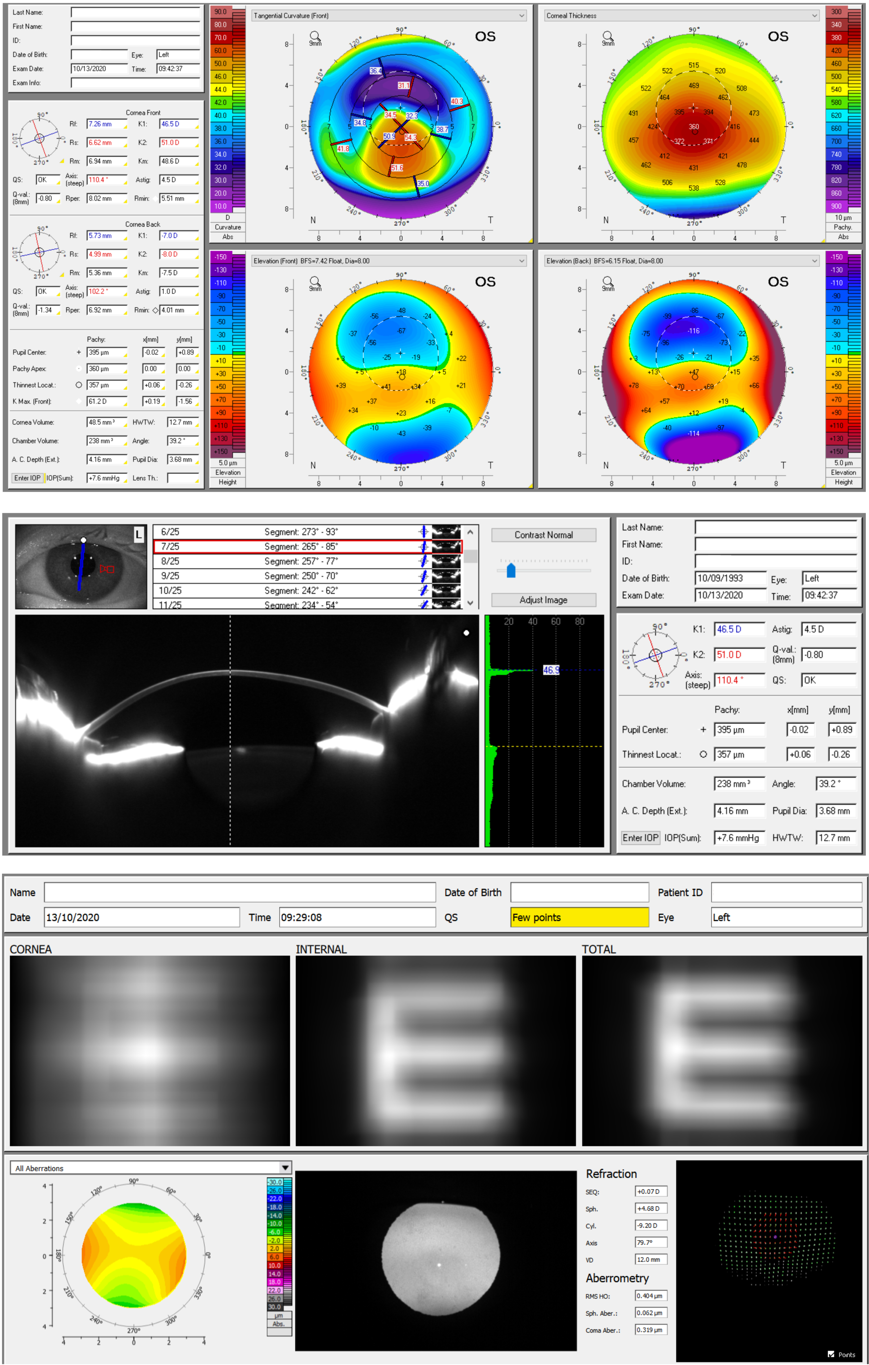
While the cause is unknown, it is believed to occur due to a combination of genetic, environmental, and hormonal factors. Patients with a parent, sibling, or child who has keratoconus have 15 to 67 times higher risk in developing corneal ectasia compared to patients with no affected relatives. Proposed environmental factors include rubbing the eyes and allergies. The underlying mechanism involves changes of the cornea to a cone shape. Diagnosis is most often by topography. Topography measures the curvature of the cornea and creates a colored "map" of the cornea. Keratoconus causes very distinctive changes in the appearance of these maps, which allows doctors to make the diagnosis.
تشخیص کراتوکونوس ممکن است به دلیل بروز و پیشرفت آهسته آن مشکل باشد. این بیماری ممکن است با نزدیک بینی و آستیگماتیسم همراه باشد. در نتیجه ممکن است موجب عدم وضوح و تاری دید شود. بیمار همچنین ممکن است دچار هاله بینایی و حساسیت به نور باشد. بیماران معمولا" در هر بار مراجعه به چشم پزشک نسخهشان تغییر میکند.
عللکراتوکونوس
علت کراتوکونوس مشخص نیست. این بیماری ممکن است ژنتیکی باشد زیرا بیماران متعددی در یک خانواده دیده میشوند. و ممکن است بصورت اتوزوم مغلوب یا غالب به ارث برسد.
Treatment
Eyeglasses or soft contact lenses may be used to correct the mild nearsightedness and astigmatism that is caused by the early stages for keratoconus. Corneal cross-linking surgery is indicated early after diagnosis to stabilize the structure of the cornea and slow progression. As the disorder progresses and cornea continues to thin and change shape, rigid gas permeable contact lenses can be prescribed to correct vision adequately. In most cases, this is adequate. The contact lenses must be carefully fitted, and frequent checkups and lens changes may be needed to achieve and maintain good vision. In a few cases, a corneal transplant is necessary. However, even after a corneal transplant, eyeglasses or contact lenses are often still needed to correct vision.
Corneal cross-linking surgery halts or slows progression and is indicated early after diagnosis, or if later in the course of the disease there is still an increase in the glasses prescription. Intacs intracorneal removable inserts can stretch the cornea to stabilize astigmatism and near-sighted refractive errors. Once the progression has stabilized from life or cross-linking, an implantable Collamer lens surgery can correct higher prescriptions so that glasses or conventional soft contact lenses can be utilized.
درمان
در انواع خفیف بیماری ، عینک یا لنز نرم (Soft Contact Lenses) ممکن است کمک کننده باشد ولی با پیشرفت بیماری و نازکتر شدن و تغییر شکل بیشتر قرنیه این درمانها دیگر چندان کارساز نخواهد بود.
درمان بعدی بیماری استفاده از لنزهای سخت دارای قابلیت نفوذ گاز (Rigid gas permeable contact lenses) است. این لنزها قابلیت بیشتری برای اصلاح آستیگماتیسم نامنظم ناشی از کراتوکونوس دارند. قرار گیری مناسب لنز بر روی قرنیه مبتلا به کراتوکونوس کاری ظریف و زمان گیر است. به همین دلیل مراجعات مکرر برای تنظیم لنز و اصلاح نسخه امری قابل انتظار است. این فرایند با نازکتر شدن و تغییر شکل بیشتر قرنیه نیاز به تکرار داشته و نسخه بیمار باید مجددا" تغییر داده شود.
در صورتی که قرنیه قادر به تحمل لنز سخت نبوده و یا لنز توانایی اصلاح دید بیمار را نداشته باشد قدم بعدی پیوند قرنیه است. بیمار ممکن است حتی بعد از پیوند قرنیه برای دید بهتر به عینک و یا لنز نیاز داشته باشد.
در حال حاضر در بیمارانی که قادر به تحمل لنزهای سخت نیستند و از طرفی انجام پیوند قرنیه بدلیل عوارض آن در آنها توصیه نمیشود از روشهای جدیدتری نظیر قرار دادن حلقههای داخل قرنیه مثل Intacs و Ferrara استفاده میشود. مطالعه بر روی این روش در چند سال اخیر در جریان بوده و نتایج نشان دهنده بهبود نسبی بیماران است. هر چند نشان داده نشده است که این روش مانع پیشرفت بیماری شود.
The classic symptom of keratoconus is the perception of multiple "ghost" images, known as monocular polyopia. This effect is most clearly seen with a high contrast field, such as a point of light on a dark background. Instead of seeing just one point, a person with keratoconus sees many images of the point, spread out in a chaotic pattern. This pattern does not typically change from day to day, but over time, it often takes on new forms. People also commonly notice streaking and flaring distortion around light sources. Some even notice the images moving relative to one another in time with their heartbeat.
The predominant optical aberration of the eye in keratoconus is coma. The visual distortion experienced by the person comes from two sources, one being the irregular deformation of the surface of the cornea, and the other being scarring that occurs on its exposed highpoints. These factors act to form regions on the cornea that map an image to different locations on the retina. The effect can worsen in low light conditions, as the dark-adapted pupil dilates to expose more of the irregular surface of the cornea.
کراتوکونوس و جراحی عیوب انکساری
انجام هیچکدام از اعمال جراحی اصلاح عیوب انکساری نظیر لیزیک (LASIK) و لیزر (PRK) در بیماران مبتلا به کراتوکونوس بدلیل نازکی پیشرونده قرنیه امکان پذیر نیست.
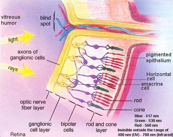
In the normal eye, light rays pass through the cornea, pupil, and lens and focus directly on the retina. When the cornea fails to focus light rays directly on the retina, refractive errors such as myopia, hyperopia, and astigmatism occur.
What should a normal eye look like?
The white part of the eye, the sclera, with the overlying conjunctiva, is not red and inflamed. The cornea is bright and clear. The pupil is black and round. If the pupil and iris are well seen, this confirms that the cornea must be clear.
Myopia
Myopia, or nearsightedness, occurs when the curvature of the cornea is too steep or the eyeball is too long. Therefore, light rays entering the eye focus in front of the retina. This results in blurred vision at distance.
Hyperopia
Hyperopia, or farsightedness, occurs when the curvature of the cornea is too flat or the eyeball is too short. Therefore, light rays entering the eye focus in back of the retina. This results in blurred vision at near.
Astigmatism
Astigmatism occurs when the curvature of the cornea is irregularly shaped, like the shape of a football. Therefore, light rays entering the eye focus at two different foci, causing blurred or distorted vision at distance and near. Astigmatism can occur alone or in conjunction with myopia and hyperopia.
Presbyopia
Presbyopia is a vision condition in which the crystalline lens loses its flexibility or elasticity, making it difficult to focus on near objects. Generally, presbyopia becomes noticeable in the early to mid-40s. Presbyopia is an unpreventable age-related process. To help alleviate symptoms of presbyopia, reading glasses, bifocals, progressive lenses, and multifocal contact lenses can be prescribed.
Dry Eye Syndrome
Dry eye syndrome is a physical condition in which the front of the eye becomes dry. Symptoms of dry eyes will affect the majority of people at some time. Patients with dry eyes often report feelings of sandy, gritty, burning, tearing, and itchiness in the eye. Other symptoms include fluctuation in vision, contact lens intolerance and recurrent infections. Symptoms may worsen in hot and dry climates and can become more irritated by smoke, wind, and air conditioned environments.
Achieving relief from the symptoms of dry eye is easy with dry eye therapy. Therapy may be as simple as using over the counter artificial tears, but may be complex to involve the use of prescription medications and/or punctal plugs. A dry eye therapy plan is covered by most major medical insurances and can be easily implemented to help relieve the pesky ailments of dry eyes.
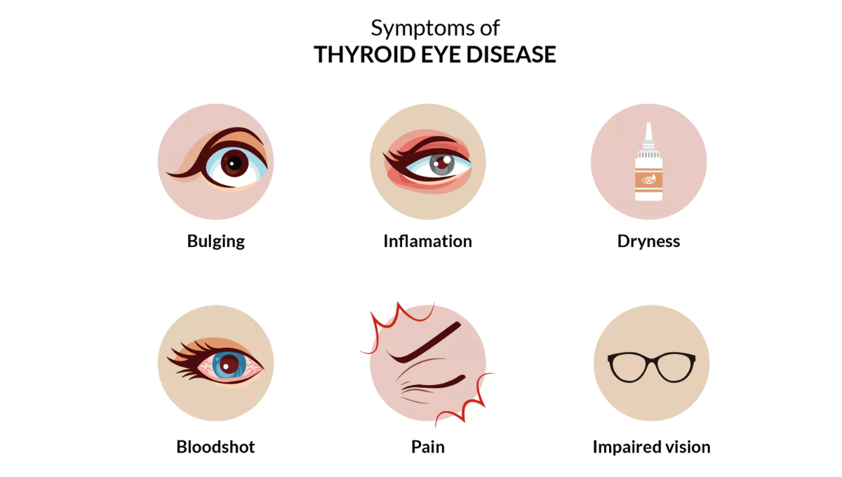
What is thyroid eye disease?
Also known as TED (thyroid eye disease) or Graves’ eye disease, thyroid eye disease is an autoimmune disorder involving an imbalance or recognition problem with your immune system.
Most commonly, this happens as part of thyroid disease from an overactive thyroid gland, affecting the skin and eyes. This autoimmune disease causes inflammation and swelling, stimulating the production of muscle tissue and fat behind the eye. Up to one-half of people with Graves’ disease develop these eye symptoms.
Stages of thyroid eye disease
There are two phases of thyroid eye disease. The first is the inflammatory phase which can last from six months to two years, while the second is the stable phase, during which the active inflammation is dormant.
Following the inflammatory phase, many individuals are left with eye protrusion, eyelid retraction, or double vision, which can be treated in various ways. If you believe you have thyroid eye disease, you must talk to your doctor immediately.
What does thyroid eye disease look like?
Thyroid eye disease symptoms can vary significantly from one person to another. For some individuals, symptoms can lead to pain, disfigurement, or threatened eyesight. For others, the disorder remains unchanged for many years but for others it can either worsen or slightly improve.
Allergy and Infections
Red eyes that itch and burn are commonly related to an allergy or in worse cases, an infection. Allergies are common and often present symptoms similar to those of dry eyes, but will definitely become more prevalent during hay fever season. Irritations caused by allergies can be alleviated with prescription eye drops.
When allergy-like symptoms are accompanied with pain or discomfort, eye secretions, vision loss, and light sensitivity, it may be due to an eye infection. Infections of the eyes may result from many scenarios, such as eye abrasions from injuries, foreign bodies, or contact lens wear. Symptoms of an emerging infection can be easily dismissed for an allergy or dry eye, hence it is important that all patients seek immediate care for any suspicious redness, pain, or sudden loss of vision.
Cataract
Cataract is the clouding of the eye’s natural crystalline lens. This loss of transparency decreases the amount of light that can reach the retina, resulting in overall blurriness of images. Signs and symptoms of cataract include: blurry, hazy vision, reduced intensity of colors, increased sensitivity to glare, increased difficulty with night vision, and changes in the eye’s refractive error. Cataracts are typically due to age-related changes in the natural lens. Other precipitating factors may include: ultraviolet radiation exposure, diabetes, corticosteroid use, smoking, high alcohol consumption, and certain nutrient deficiency. Recent studies have shown that antioxidants (e.g. vitamin C, vitamin E, carotenoids) may decrease cataract formation.
Glaucoma
Glaucoma is an eye disease caused by an increased pressure in the eye. This can damage the optic nerve that transmits visual information to the brain, resulting in the loss of vision. Glaucoma is the second leading cause of blindness in the U.S. Individuals over the age of 40, individuals with a family history of glaucoma, and African Americans are at an increased risk of developing glaucoma. Other risk factors for the development of glaucoma include: thinner corneas, systemic vascular conditions (e.g. diabetes, hypertension, heart disease), prolong corticosteriod use, high myopia, chronic ocular inflammation, and ocular trauma.
What is Retinal Vein Occlusion?
Retinal Vein Occlusion (RVO) is a condition where the veins carrying blood away from the retina, located at the back of the eye, become blocked. This blockage causes blood and fluid to leak into the retina, leading to visual problems.
Macular Degeneration
Macular degeneration (MD) is the leading cause of blindness in Americans over the age of 55. MD causes a deterioration and loss of photoreceptors and other cells in the macula, the part of the retina responsible for sharp, clear central vision. Because only central vision is usually affected, people rarely go blind from the disease. However, MD can sometimes make it difficult to read, drive, or perform other daily activities that require fine, central vision. Most people with MD have the dry form, for which there is no known treatment. The less common wet form may respond to laser procedures, if diagnosed and treated early. A major National Eye Institute study (AREDS) indicates that certain nutrients such as beta carotene (vitamin A) and vitamins C and E may help prevent or slow progression of MD. eResearch by Navid Ajamin -- winter 2010
Retinal Detachment
The retina is the light-sensitive tissue that lines the inside back wall of the eye. In retinal detachment, the retina is separated from its underlying supportive tissue, depriving it from nutrients and oxygen. The longer the retina is detached, the greater the risk of permanent vision loss. Retinal detachment, thus, is a medical emergency requiring prompt surgical treatment to preserve vision. Warning signs of retinal detachment include: floaters, flashes of light, a sudden decrease in vision, and a shadow or curtain over the vision. Risk factors for retinal detachment include: high myopia, previous severe eye injury/trauma, family history of retinal detachment, and previous history of retinal detachment in the other eye.
Unusual Eye Conditions You Didn’t Know About
3Meter (10ft) HOTV Translucent Eye Chart
In any medical field, there are always going to be certain medical conditions that we see on a daily basis. For ophthalmologists, common conditions include glaucoma, which per the Glaucoma Research Foundation afflicts over 3 million people, and cataracts, which according to the National Eye Institute, half of individuals will have a cataract or cataract surgery by the age of 80. However, there are also eye-related medical conditions that people may never encounter. The following rare eye conditions appear in less than .01% of United States citizens, per the National Eye Institute.
Anophthalmia and Microphthalmia
While these two rare eye conditions are commonly used in substitution for each other, they are actually two separate but related conditions. Anophthalmia is a birth defect that results in the absence of one or both eyes. Micropthalmia is when one or both eyes is noticeably too small.
In the cases of both conditions, genetic mutations and abnormal chromosomes are believed to be at fault. Environmental factors are a challenge to pinpoint, put researchers have suggested X-rays, chemicals, drugs, pesticides, toxins, radiation and viruses may also be to blame. In other words, more research is still needed to determine what causes this defects.
Bietti’s Crystalline Dystrophy
Back in 1937, Italian ophthalmologist Dr. G. B. Bietti had three different patients with similar symptoms. Crystals were in the cornea and yellow shiny deposits were on the retina. Eventually, the back layers of the eye – the retina, choriocapillaries and choroid – would begin to atrophy. In patients since, crystals have also been found in white blood cells.
Currently, there is no treatment for BCD, though some believe that treatment will arise out of more genetic research.
Retinitis Pigmentosa
Retinitis Pigmentosa refers to a group of rare genetic disorders, all of which lead to the breaking down of cells in the retina. The retina is responsible for processing light and hosts rods and cones that interpret color and allow us to see at night. In RP, over 50 different genes can be afflicted. When the genetic mutations are severe enough, the cells in the retina aren’t provided with enough protein to function. In some cases, the protein is toxic. Over time, RP will cause the rods and cones in the eyes to die, impeding night and peripheral vision. Patients with the condition will also often find bright lights uncomfortable. Treatments for Retinitis Pigmentosa includes low-vision improvement aids for children and vitamin A drops for adults.
Retinoblastoma
Perhaps the most life-threatening and most rare eye condition on the list, retinoblastoma is a cancer that affects the retina. Unfortunately, it is most commonly found in children under the age of five. The good news is that if the cancer is diagnosed early enough and treatment is delivered promptly, the vision and life of the child are possible to save.
Usher Syndrome
Unlike some of the other rare eye conditions on the list, Usher’s syndrome can also affect the hearing capacities of patients. While quite rare, it is curiously enough the most common condition that affects both hearing and vision. Usher syndrome is related to Retinitis Pigmentosa, but in addition to experiencing the symptoms of RP, people with Usher syndrome often have severe balance issues and hearing loss. Severity of the condition is typically broken down into three tiers, depending on the severity of vision, hearing, and balance impairment.
Uveal Coloboma
Because it is one of the most-rare eye conditions, it is not always properly diagnosed. For this reason, the National Eye Institute estimates that Uveal Coloboma occurs in between 0.5 to 2.2 cases per 10,000 births. Coloboma is used to help describe the absence of normal tissue in or around the eye. Consequently, the coloboma can affect the eyelid, lens, macula (handles daylight, fine and color vision), and the optic nerve. Because Uveal Coloboma patients are missing a component of the eye, it is responsible for a significant portion of blindness in newborns. However, not all patients are blind. Depending on the part of the eye affected, people with UC may suffer from mere light sensitivity or a more limited field of vision.
Uveal Coloboma has no cure, but corrective treatments are available for some patients.
Photophobiais a symptom of abnormal intolerance to visual perception of light.As a medical symptom, photophobia is not a morbid fear or phobia, but an experience of discomfort or pain to the eyes due to light exposure or by presence of actual physical sensitivity of the eyes, though the term is sometimes additionally applied to abnormal or irrational fear of light such as heliophobia.The term photophobia comes from the Greek φῶς (phōs), meaning "light", and φόβος (phóbos), meaning "fear". Photophobia is a common symptom of visual snow.
Light sensitivity, or photophobia, is an intolerance of light. Some only feel discomfort from bright lights, while others in extreme cases can not stand any type of light. Sources can range from sunlight, fluorescentlight, incandescent light or flames of candles or fires. Some people tend to squint or close their eyes if their sensitivity is too strong. There are many different reasons why someone could have a sensitivity to lights, but the biggest issue is the underlying cause, as photophobia is a symptom, not a condition or disease.Photophobia is known to happen to all ages, young and old.
Photophobia is classified as an extreme sensitivity to light. Photophobia is not a disease on its own. It is usually a symptom caused by another condition. It can be extremely painful, frustrating and debilitating at times.
When exposed to bright light, look for the following:
the inability to be in the sun without squinting
searching out dark or shady areas for relief
nausea or dizziness
headaches or migraines
eye pain
Some of these symptoms can be normal if they are mild and are not associated with pain. If they become significant enough that you avoid the sun or alter your habits, it is time to see a doctor.
Symptoms of Photophobia
There are a few obvious symptoms to recognize your sensitivity to light has increased, such as:
Discomfort
Need to close eyes
Need to squint
Burning
Excessive tearing
In some cases, there might not be any sort of symptoms except the sensitivity to light itself. People have reported nothing one day, then sensitivity the next day. Each individual is unique and experiences different symptoms. Again, it depends on the underlying cause. In other cases, people will suffer many other types of symptoms, depending on the condition or disease that is causing the light sensitivity.
Glare-control sunglasses, like these by Corning, are helpful if you are sensitive to sunlight or even strong indoor lighting.
Causes of Photophobia eResearch by Navid Ajamin -- winter 2010
There are several different reasons why someone might be suffering from photophobia or sensitivity to light. It’s not a disease, disorder, problem or condition. In fact, it’s a symptom of many different diseases, disorders, problems and conditions. For example, an infection or inflammation that irritates the eyes can cause photophobia. Also, it can be a symptom of an underlying disease such as a viral illness or a severe headache or migraine.
People can be sensitive to light for many different reasons. It doesn’t always occur because of an eye condition, and sometimes there isn’t a cause at all – some people are just more sensitive to light than others.
IPRGC cells or melanopsin cells transduce light into pain.
Some eye diseases cause this symptom, including:
Dry eye
Uveitis (swelling of the inside of your eye)
Keratitis (swelling of your cornea, the clear layer that covers the colored part of your eye)
Iritis (swelling of the colored ring around your pupil)
Cataracts (cloudy coverings over the lenses of your eyes) Corneal abrasion (a scratch on your cornea)
Conjunctivitis (inflammation of the conjunctiva, the clear tissue that sits over the white part of your eye)
Damage to your retina, the light-sensitive layer in the back of your eye Blepharospasm (a condition that makes your eyelids close uncontrollably)
Photophobia may also affect some people who have thesementalhealth conditions:
Agoraphobia(a fear of being in public places)
Anxiety
Bipolar disorder
Depression
Panic disorder
Some things which can make you light sensitive include:
Medicationstaken for other conditions – for example tetracycline (an antibiotic), and digitalis (a drug used for heart problems).
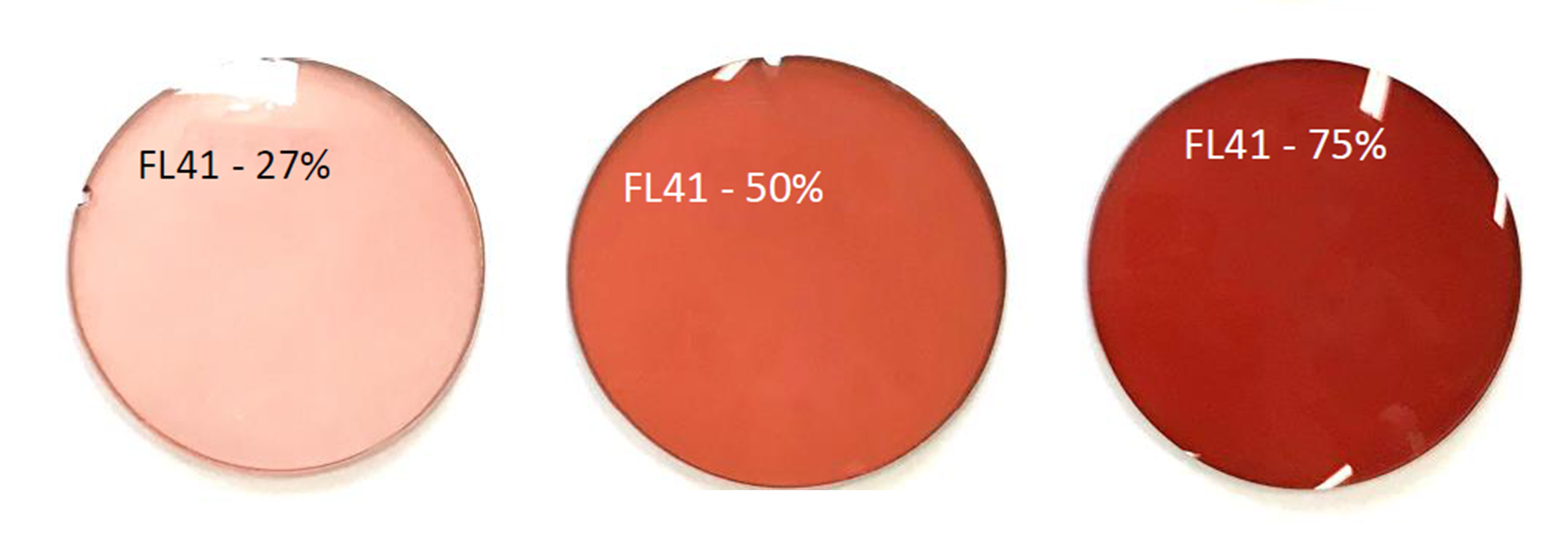
People who suffer or have suffered with migrainestend to be more sensitive to light. Some find that special coloured tinted lenses can help. These coloured lenses are individually prescribed by some optometrists and can also be used to help people who experience glare from pages of text, such as people with dyslexia.
Meningitiscan cause light to become painful quite quickly. If your light sensitivity comes on very suddenly or a child that you’re caring for becomes very light sensitive very quickly, this should be checked straight away by a medical professional in case it is the first sign of this more serious condition.
If you begin to experience light sensitivity, have your eyes checked by an optometrist (optician) – they can check that there is no underlying eye condition which may be causing this and may be able to suggest ways to help you cope.
Photophobia preventionat home
Once the underlying cause has been determined, a few practical tips can help minimize photophobia.- Use polarized sunglasses when outdoors
Wear a hat or cap when outdoors
Avoid bright fluorescent lights
Utilize natural light where possible for indoor settings
Control indoor lighting with dimmers and consider replacing any fluorescent or cool white LED light bulbs with a warm white LED light bulb or an incandescent light bulb.
Control the brightness on your screen by adjusting the settings on your TV, computer, phone and other devices
Wear light-filtering lenses or tinted lenses indoors At home is where we have the greatest ability to control our light environment.
Photophobia Treatment
The best treatment for light sensitivity is to address the underlying cause. Once the triggering factor is treated, photophobia disappears in many cases.
If you are taking a medication that causes light sensitivity, talk to your prescribing physician about discontinuing or replacing the drug.
If you're naturally sensitive to light, avoid bright sunlight and other harsh lighting sources. Wear wide-brimmed hats and sunglasses with ultraviolet (UV) protection when outdoors in daylight. Also, consider wearing eyeglasses with photochromic lenses. These lenses darken automatically outdoors and block 100 percent of the sun's UV rays.
For bright sunlight, considerpolarized sunglasses. These sun lenses provide extra protection against glare-causing reflections of light from water, sand, snow, concreteroadways and other reflective surfaces.
In an extreme case, you may consider wearing prosthetic contact lenses that are specially colored to look like your own eyes. Prosthetic contact lenses can reduce the amount of light that enters the eye and make your eyes more comfortable.
Reference:
rnib.org.uk
webmd.com
axonoptics.com
en.wikipedia.org
sciencedirect.com/science/article
allaboutvision.com
michiganheadandneck.com
See also:
GUIDE TO PHOTOPHOBIA / LIGHT SENSITIVITY-- axonoptics.com
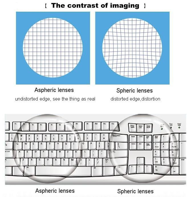
You might feel self-conscious about your glasses and how they make you look, but aspheric lenses can help. With a flatter curve, there's less central thickness and less eye magnification. They also correct distortion and create a higher-quality image. Aspheric lenses can also improve your peripheral vision.
What Are Aspheric Lenses Used For?
You might want to choose aspheric lenses when you have a strong prescription or you experience dramatic refractive errors. Dramatic refractive errors mean you have significant problems with the way light focuses on your retina, which means you’ll need stronger corrective measures.
Relevant problems include conditions like:
Myopia, where objects in the distance are blurry
Hypermetropia, where objects close to you are blurry
Astigmatism, where objects near and far are blurry or distorted
Presbyopia, where you can’t see things close up as you get older
Your eye doctor might recommend lenses that are both aspheric and high-index. Where aspheric refers to the lens profile, high-index refers to lens material and thickness. The higher the index number, the thinner the lens.
It’s called a high-index lens because it has a high refractive index, which means light travels quickly through the material. The material bends light more efficiently, making it better at correcting high refractive errors.
An aspheric and high-index option means your lenses will be easier to wear with a strong prescription.
What is an Aspheric Lens ?
An aspheric lens or asphere(often labeled ASPHon eye pieces) is a lens whose surface profiles are not portions of a sphere or cylinder. In photography, a lens assembly that includes an aspheric element is often called an aspherical lens.
Aspheric lenses are also sometimes used for eyeglasses. Aspheric eyeglass lenses allow for crisper vision than standard "best form" lenses, mostly when looking in other directions than the lens optical center. Moreover, the reduction of the magnification effect of a lens may help with prescriptions that have different powers in the 2 eyes (anisometropia). Not related to the optical quality, they may give a thinner lens, and also distort the viewer's eyes less as seen by other people, producing better aesthetic appearance.[4]
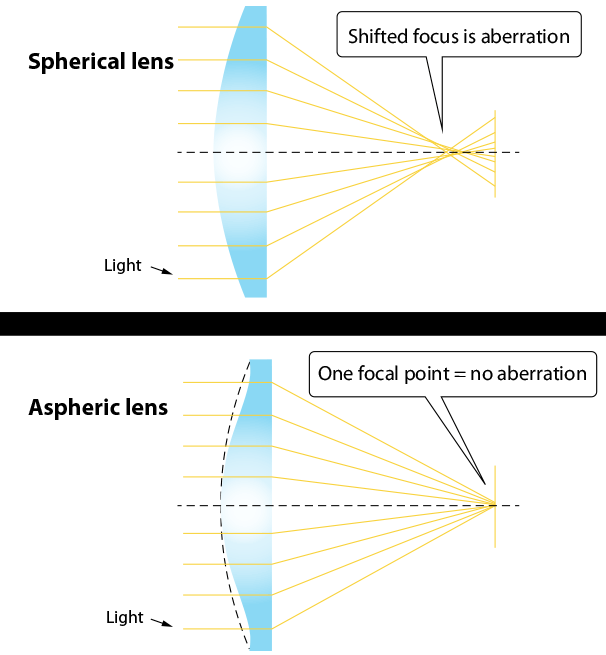
An aspheric lens is designed for aberration correction. With the help of aspheric lens, the image formed is distortion free.
An aspheric lens is a lens whose surfaces profile neither a portion of a sphere nor of a circular cylinder. Since it is not spherical, the conventional processes no longer apply to making aspheric surfaces. This is why aspheric cost many times what spherical surfaces do.
In optics, a lens assembly that includes an aspheric element is often called an aspheric lens.
Benefit
The asphere's more complex surface profile can eliminate spherical aberration and reduce other optical aberrations compared to a simple lens. A single aspheric lens can often replace a much more complex multi-lens system. These lenses are small, lighter and in general, better than similar lenses which only employ spherical elements.
As magnifier, aspheric lens enhances image quality and minimizes distortion throughout the viewing area, it reduces distortion at wide angles, improves corner resolution. You can get more detail and high-resolution of the image.[1]
Asphericity allows lens designers to flatten a lens form in order to improve cosmesis, without sacrificing opical performance. The lens aberrations produced by using flattened lens forms are simply eliminated using the surface astigmatism of the aspheric design. While aspheric lenses do not provide better vision than best form lenses, they do provide equivalent vision in a flatter, thinner, and lighter lens.
Aspheric lenses allow lens designers to produce lenses that are considerably flatter, thinner, and lighter in weight than conventional best form lenses.
It is interesting to note that aspheric surfaces produce thinner lenses for two reasons:
Aspheric lenses generally use flatter front curves, which reduce the center thickness in plus lenses and the edge thickness in minus lenses.
The geometry of an aspheric surface also provides additional thickness reduction. Some aspheric lenses are even designed solely for cosmesis, and actually use more asphericitythan what is optically required. This produces a thinner lens at the expense of reduced optical performance.
As with the base curveof a best form lens, the amount or degree of asphericity will depend upon the focal power of the lens.
Additionally, the surface (that is, front or back) upon which the asphericity has been applied will also make a difference:
Plus lenses. If asphericity is applied to the front surface of a plus lens, the surface will become flatter away from the center. If it is applied to the back surface, the surface will become steeper away from the center.
Minus lenses. If asphericity is applied to the front surface of a minus lens, the surface will become steeper away from the center. If it is applied to the back surface, the surface will become flatter away from the center.
Ideally, aspheric lenses should be optimized for each individual focal power. In practice, however, small ranges of powers are grouped upon common aspheric base curves—just like with best form lenses. Nevertheless, asphericity gives lens designers the freedom to optimize just about any base (front) curve for the chosen focal power—or range of powers. (Generally, flatter base curves are chosen for cosmesis.) [2]
An aspheric lens or asphere is a lens whose surfaces have a profile that is neither a portion of a sphere nor of a circular cylinder. In photography, a lens assembly that includes an aspheric element is often called an aspherical lens.
The asphere's more complex surface profile can eliminate spherical aberration and reduce other optical aberrations compared to a simple lens. A single aspheric lens can often replace a much more complex multi-lens system. The resulting device is smaller and lighter, and possibly cheaper than the multi-lens design.[3]eResearch by Navid Ajamin -- winter 2008
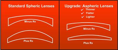
Traditional lenses have a bulgy, curved shape. Imagine the spherical surface of a ball. Aspheric lenses are designed with less curvature than their traditional counterparts. Think flatter and thinner. In both far and nearsighted prescriptions, aspheric lenses provide a slimmer profile and minimize eye distortion without compromising optical quality. Let’s take a look at how conventional lenses are designed for far and nearsighted prescriptions:
Lenses for farsightedness have a convex curve, meaning they are thicker in the center and thinner at the edges. The stronger the prescription, the more the middle of the lens bulges outwards.
Lenses for nearsighted prescriptions have a concave curve, meaning they are thinnest at the center of the lens and thickest at the edge.
For both types of prescriptions, aspheric lenses reduce the curve of the surface, either by minimizing the thickness of the center or the edges of the lens.
Benefits of Aspheric Lenses
Less bulging of the lens, giving you a sleeker profile
More frame options for individuals with strong prescriptions
More natural appearance of the eye (reduces the eye magnification that occurs with farsightedness and the smaller appearance of the eye that occurs with nearsightedness)
Lightweight (less material is used to make the lens)
Better peripheral vision
Higher image quality (more consistent magnification throughout the lens)
The surface radian of aspherical lens is different from that of ordinary spherical lens. In order to pursue the thinness of the lens, it needs to change the surface of the lens. Aspheric lens should be designed as flat as possible. However, the optical properties of flattened lenses, even if they are designed with aspheric surfaces, will decline rapidly. At the same time, the lens is lighter, thinner and flatter, and it still maintains excellent impact resistance, so that the wearer can use it safely.
Advantages of aspherical lenses:
Optical advantages: reduce the aberration of the lens and make the vision clearer.
Clear images can also be obtained at high luminosity: Although the spherical point focus lens is designed by the best base arc, the luminosity beyond + 7.00D-22.00D is not within the Cherning ellipse, and the aberration can not be eliminated. Only aspheric design can achieve better quality.
It can make the lens flatter, thinner and more beautiful. The higher the lenticity of spherical lens, the worse the appearance. Aspheric lens can be designed with flat base arc, which not only makes the appearance beautiful, but also reduces the peripheral magnification. Let others see the wearer's eyes, will not change a lot in size.
Defects of aspherical lenses:
Aspheric lens has a relatively small light area. When the eyeball rotates around, it will blur a little when looking at the outside through the lens edge, that is, the visual range of the line of sight becomes smaller.
Human's eyeball is spherical. The eyeball rotates to the edge. Through aspheric lens, the object near the eye appears protruding.
Age-related macular degeneration (AMD) results in damaged sharp and central vision. Central vision is needed for seeing objects clearly and for reading and driving. AMD affects the macula, the central part of the retina that allows the eye to see fine details.
Age-related macular degeneration
Vision loss with AMD
Vision loss with cataract
Vision loss with glaucoma
Amblyopia, also referred to as "lazy eye," is the most common cause of vision impairment in children. With amblyopia, the vision in one eye is reduced because the eye and the brain are not working together properly. The eye itself looks normal, but it is not being used normally because the brain is favoring the other eye.
Amblyopia
Strabismus involves an imbalance in the positioning of the two eyes. Strabismus can cause the eyes to cross in (esotropia) or turn out (exotropia).
CDC is the nation's leading science-based, data-driven, service organization that protects the public's health. CDC puts science into action to help children stay healthy so they can grow and learn; to help families, businesses, and communities fight disease and stay strong; and to protect the public's health.
nicetoview.blogfa.com
وبلاگ تخصصی عینک شامل مجموعه مطالب پزشکی است که اطلاعات مفیدی در رابطه با عینک , چشم، لنز، سلامتی چشم و راه های پیشگیری از بیماریهای چشمی، کنترل و درمان آن را در اختیار شما کاربر محترم می گزارد.


 وبلاگ تخصصی عینک شامل مجموعه مطالب پزشکی است که اطلاعات مفیدی در رابطه با عینک , چشم، لنز، سلامتی چشم و راه های پیشگیری از بیماریهای چشمی، کنترل و درمان آن را در اختیار شما کاربر محترم می گزارد.