Blind spot test
To see or not to see.
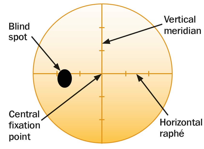
The eye’s retina receives and reacts to incoming light and sends signals to the brain, allowing you to see. One part of the retina, however, doesn't give you visual information—this is your eye’s “blind spot.”
At the back of your eye is the retina. Your retina is made up of light-sensitive cells which send messages to your brain about what you see. Everyone has a spot in their retina where the optic nerve connects. In this area there are no light-sensitive cells so this part of your retina can’t see. We call this the blind spot.
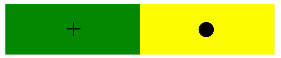
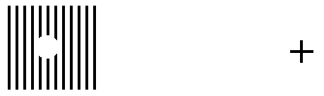
Most of the time you don’t notice your blind spot because the spot in one eye doesn’t match the spot in the other eye. Each eye supplies information to the brain, filling in what’s missing. Also, sometimes the brain will fill in the missing information with what it thinks should be there. That causes one kind of optical illusion. eResearch by Navid Ajamin -- Autumn 2025

Instructions:
R ... L
Place your eye a distance from the screen approximately equal to three times the distance between the R and the L. Move your eye towards or away from the screen until you notice the other letter disappear. For example, close your right eye, look at the "L" with your left eye, and the "R" will disappear.

(Upper row) A cross section of the eye showing the blind spot and retinal veins, as well as the fact that light goes through all the retinal layers before hitting the photoreceptors. (Lower row) Top-down view of the retina, showing how big the blind spot and retinal veins are relative to the fovea, which is the high-resolution region of the retina. (Images adapted with permission from Webvision-University of Utah).
Night blind spot
It is estimated that once fully adapted to darkness, the rods are 10,000 times more sensitive to light than the cones, making them the primary receptors for night vision. Since the cones are concentrated near the fovea, the rods are also responsible for much of the peripheral vision. The concentration of cones in the fovea can make a night blind spot in the center of the field of vision.
Description: Students will make a simple prop and use it to find their blind spot
Purpose: To locate and identify the blind spot
Length of Activity: 20 minutes
Materials:
- One 3 x 5 inch card (or other stiff paper) per student.
- Black markers.
- 1 ruler per student.
Steps:
1. Students should be instructed to make a dot and an X on the white side of the index card as pictured.
2. They should then hold the card so the X is on the right side and raise it to eye level about an arm's length away.

3. Have students close their right eye.
4. Student should look directly at the X with their left eye only. They should note that they can also see the dot, but should not focus on it.
5. While looking at the X, and keeping an awareness of the dot, have students bring the cards slowly towards their faces. At some point they should be aware that the dot has disappeared and then reappeared.
6. Now have students repeat but this time close their left eyes. They should use their right eyes to look at the dot while keeping aware of (but not looking directly at) the X. This time the X will disappear and then reappear as the card is slowly brought towards their faces.
7. Now have students take their markers and ruler to draw a straight line through the center of both the dot and the X.
8. Repeat the activity. Note that this time the line seems to be continuous, with no gap, even as the X or dot disappears.
What’s Going On?
At the back of your eye is the retina. Your retina is made up of light-sensitive cells which send messages to your brain about what you see. Everyone has a spot in their retina where the optic nerve connects. In this area there are no light-sensitive cells so this part of your retina can’t see. We call this the blind spot. The point at which the mark on the card disappears is where your blind spot is.
When you draw a line through the dot and X you set up an optical illusion. The brain knows that a line is there and fills in the gap, even as it loses sight of the dot or X.

Reference:
- researchgate.net/figure/Upper-row-A-cross-section-of-the-eye-showing-the-blind-spot-and-retinal-veins
- aao.org/museum-eye-openers/experiment-blind-spot
- en.wikipedia.org/wiki/Blind_spot_(vision)
- exploratorium.edu/snacks/blind-spot
 وبلاگ تخصصی عینک شامل مجموعه مطالب پزشکی است که اطلاعات مفیدی در رابطه با عینک , چشم، لنز، سلامتی چشم و راه های پیشگیری از بیماریهای چشمی، کنترل و درمان آن را در اختیار شما کاربر محترم می گزارد.