The cornea is the transparent front part of the eye that covers the iris, pupil, and anterior chamber. The cornea, with the anterior chamber and lens, refracts light, with the cornea accounting for approximately two-thirds of the eye's total optical power. In humans, the refractive power of the cornea is approximately 43 dioptres. While the cornea contributes most of the eye's focusing power, its focus is fixed. The curvature of the lens, on the other hand, can be adjusted to "tune" the focus depending upon the object's distance. Medical terms related to the cornea often start with the prefix "kerat-" from the Greek word κέρας, horn. [1]
Although the cornea is clear and seems to lack substance, it is actually a highly organized group of cells and proteins. Unlike most tissues in the body, the cornea contains no blood vessels to nourish or protect it against infection. Instead, the cornea receives its nourishment from the tears and aqueous humor (a fluid in the anterior portion of the eye) that fills the chamber behind it. The cornea must remain transparent to refract light properly, and the presence of even the tiniest blood vessels can interfere with this process. To see well, all layers of the cornea must be free of any cloudy or opaque areas.
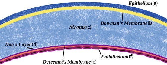
The corneal tissue is arranged in five basic layers, each having an important function. These five layers are:
- Epithelium The epithelium is the cornea's outermost region, comprising about 10 percent of the tissue's thickness. The epithelium functions primarily to: (1) block the passage of foreign material, such as dust, water, and bacteria, into the eye and other layers of the cornea; and (2) provide a smooth surface that absorbs oxygen and cell nutrients from tears, then distributes these nutrients to the rest of the cornea. The epithelium is filled with thousands of tiny nerve endings that make the cornea extremely sensitive to pain when rubbed or scratched. The part of the epithelium that serves as the foundation on which the epithelial cells anchor and organize themselves is called the basement membrane.
- Bowman's Layer Lying directly below the basement membrane of the epithelium is a transparent sheet of tissue known as Bowman's layer. It is composed of strong layered protein fibers called collagen. Once injured, Bowman's layer can form a scar as it heals. If these scars are large and centrally located, some vision loss can occur.
- Stroma Beneath Bowman's layer is the stroma, which comprises about 90 percent of the cornea's thickness. It consists primarily of water (78 percent) and collagen (16 percent), and does not contain any blood vessels. Collagen gives the cornea its strength, elasticity, and form. The collagen's unique shape, arrangement, and spacing are essential in producing the cornea's light-conducting transparency.
- Descemet's Membrane Under the stroma is Descemet's membrane, a thin but strong sheet of tissue that serves as a protective barrier against infection and injuries. Descemet's membrane is composed of collagen fibers (different from those of the stroma) and is made by the endothelial cells that lie below it. Descemet's membrane is regenerated readily after injury.
- Endothelium The endothelium is the extremely thin, innermost layer of the cornea. Endothelial cells are essential in keeping the cornea clear. Normally, fluid leaks slowly from inside the eye into the middle corneal layer (stroma). The endothelium's primary task is to pump this excess fluid out of the stroma. Without this pumping action, the stroma would swell with water, become hazy, and ultimately opaque. In a healthy eye, a perfect balance is maintained between the fluid moving into the cornea and fluid being pumped out of the cornea. Once endothelium cells are destroyed by disease or trauma, they are lost forever. If too many endothelial cells are destroyed, corneal edema and blindness ensue, with corneal transplantation the only available therapy.
What is the function of the cornea?
Because the cornea is as smooth and clear as glass, but is strong and durable, it helps the eye in two ways:
- It helps to shield the rest of the eye from germs, dust, and other harmful matter. The cornea shares this protective task with the eyelids, the eye socket, tears, and the white part of the eye (sclera).
- The cornea acts as the eye's outermost lens. It functions like a window that controls and focuses the entry of light into the eye. The cornea contributes between 65-75 percent of the eye's total focusing power.
When light strikes the cornea, it bends--or refracts--the incoming light onto the lens. The lens further refocuses that light onto the retina, a layer of light sensing cells lining the back of the eye that starts the translation of light into vision. For you to see clearly, light rays must be focused by the cornea and lens to fall precisely on the retina. The retina converts the light rays into impulses that are sent through the optic nerve to the brain, which interprets them as images.
The refractive process is similar to the way a camera takes a picture. The cornea and lens in the eye act as the camera lens. The retina is similar to the film. If the image is not focused properly, the film (or retina) receives a blurry image. The cornea also serves as a filter, screening out some of the most damaging ultraviolet (UV) wavelengths in sunlight. Without this protection, the lens and the retina would be highly susceptible to injury from UV rays.[2]

Researchers at The University of Nottingham have discovered a new layer of the human cornea located at the back of the cornea between the corneal stroma and Descemet’s membrane.
Scientists have discovered a previously undetected layer in the cornea, the clear window at the front of the human eye.

The breakthrough, announced in a study published in the academic journal Ophthalmology, could help surgeons to dramatically improve outcomes for patients undergoing corneal grafts and transplants.
he new layer has been dubbed the Dua’s Layer after the academic Professor Harminder Dua who discovered it.
 Professor Dua, Professor of Ophthalmology and Visual Sciences at The University of Nottingham, said: “This is a major discovery that will mean that ophthalmology textbooks will literally need to be re-written. Having identified this new and distinct layer deep in the tissue of the cornea, we can now exploit its presence to make operations much safer and simpler for patients. [3]
Although the layer is just 15 microns thick —the entire cornea is around 550 microns thick or 0.5mm — it is incredibly tough and is strong enough to be able to withstand one and a half to two bars of pressure. eResearch by Navid Ajamin -- summer 2013
Dua's layer, according to a 2013 paper by Harminder Singh Dua's group at the University of Nottingham, is a layer of the cornea that had not been detected previously. It is hypothetically 15 micrometres (0.59 mils) thick, the fourth caudal layer, and located between the corneal stroma and Descemet's membrane. Despite its thinness, the layer is very strong and impervious to air. It is strong enough to withstand up to 2 bars (200 kPa) of pressure. While some scientists welcomed the announcement, other scientists cautioned that time was needed for other researchers to confirm the discovery and its significance. Others have met the claim "with incredulity".[5]
The scientists now believe that corneal hydrops, a bulging of the cornea caused by fluid build up that occurs in patients with keratoconus (conical deformity of the cornea), is caused by a tear in the Dua’s layer, through which water from inside the eye rushes in and causes waterlogging.

The discovery will have an impact on advancing understanding of a number of diseases of the cornea, including acute hydrops, Descematocele and pre-Descemet’s dystrophies. [4]
Reference:
- en.wikipedia.org/wiki/Cornea
- nei.nih.gov/health/cornealdisease
- scitechdaily.com/new-layer-of-the-human-cornea-discovered
- sci-news.com/othersciences/anthropology/article01151-human-eye-duas-layer.htm
- en.wikipedia.org/wiki/Dua%27s_layer
See Also:
Cornea Structure and Function - Washington University Physicians
Limbal epithelial stem cells of the cornea -- stembook.org
 وبلاگ تخصصی عینک شامل مجموعه مطالب پزشکی است که اطلاعات مفیدی در رابطه با عینک , چشم، لنز، سلامتی چشم و راه های پیشگیری از بیماریهای چشمی، کنترل و درمان آن را در اختیار شما کاربر محترم می گزارد.