The choroid, also known as the choroidea or choroid coat, is the vascular layer of the eye, containing connective tissue, and lying between the retina and the sclera. The human choroid is thickest at the far extreme rear of the eye (at 0.2 mm), while in the outlying areas it narrows to 0.1 mm. The choroid provides oxygen and nourishment to the outer layers of the retina. Along with the ciliary body and iris, the choroid forms the uveal tract.
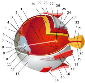
1.posterior compartment 2.ora serrata 3.ciliary muscle 4.ciliary zonules 5.canal of Schlemm 6.pupil 7.anterior chamber 8.cornea 9.iris 10.lens cortex 11.lens nucleus 12.ciliary process 13.conjunctiva 14.inferior oblique muscule 15.inferior rectus muscule 16. medial rectus muscle 17.retinal arteries and veins 18.optic disc 19.dura mater 20.central retinal artery 21.central retinal vein 22.optical nerve 23.vorticose vein 24.bulbar sheath 25.macula 26.fovea 27.sclera 28.choroid 29.superior rectus muscule 30.retina

قسمتهای مختلف کره چشم انسان: ۱. زجاجیه ۲. حاشیه دندانهدار ۳. ماهیچه مژکی ۴. گردالکهای مژگانی ۵. مجرای اشلک ۶. مردمک ۷. اتاق جلویی ۸. قرنیه ۹.عنبیه ۱۰. عدسی بیرونی ۱۱. عدسی درونی ۱۲. زوائد مژگانی ۱۳. ملتحمه ۱۴. ماهیچه مایل زیرین ۱۵. ماهیچه راست زیرین ۱۶. ماهیچه راست میانی ۱۷. شبکیه ۱۸. صفحه بینایی ۱۹. سختشامه ۲۰. سرخرگ مرکزی شبکیه ۲۱. سیاهرگ مرکزی شبکیه ۲۲.عصب بینایی ۲۳. سیاهرگ حلقوی ۲۴. غلاف پیازی ۲۵. لکه زرد ۲۶. گودی مرکزی ۲۷.صلبیه ۲۸. مشیمیه ۲۹. ماهیچه راست بالایی ۳۰. شبکیه
The structure of the choroid is generally divided into four layers:
Haller's layer - outermost layer of the choroid consisting of larger diameter blood vessels;
Sattler's layer - layer of medium diameter blood vessels;
Choriocapillaris - layer of capillaries; and
Bruch's membrane (synonyms: Lamina basalis, Complexus basalis, Lamina vitra) - innermost layer of the choroid.
مشیمیه یکی از لایههای ساختمان چشم است که بین صلبیه و شبکیه قرار گرفته است. این لایهٔ رنگدانهدار حاوی مویرگهای فراوانی است که تغذیهٔ عنبیه و سلولهای گیرندهٔ نور شبکیه را بر عهده دارد. مشیمیه در جلوی چشم بخش رنگین آن، یعنی عنبیه را به وجود میآورد.

مَشیمیه پوستهای است نازک و لطیف که تمام سطح داخلی «صلبیه» را میپوشاند و شامل رگهای فراوانی است و وظیفهٔ حساس آن تأمین غذای چشم است و چون تا حدی به پردهٔ بچهدان شبیه است «مشیمیه» نامیده شدهاست،

مشیمیه از ۴ لایهٔ تشکیل شده است:
لایهٔ هالر که از جنس بافت پیوندی است و حاوی تعداد زیادی رگ با قطر زیاد است.
لایهٔ ساتلر که بافتی پیوندی دارای مقدار زیادی رنگدانه و حاوی رگهایی با قطر متوسط است.
لایهٔ سوم حاوی مویرگهای نازکی است که تغذیهٔ سلولهای سطح شبکیه را بر عهده دارد.
لایهٔ بروخ eResearch by Navid Ajamin -- summer 2013
رگهای مشیمیه از خارج به داخل باریکتر میشوند و در سمت داخل، صفحه کوریوکاپیلاریس را میسازند. تخلیه سیاهرگی مشیمیه توسط چهار سیاهرگ حلقهای است.

THE ANATOMY OF THE CHOROID : The choroid is an important structure of the eye which can be involved in a lot of pathologies. Its great importance is given by functions like vascularization, thermoregulation and production of growth factors. A good knowledge of this element of the eye will help the ophthalmology specialists, especially the young ones, to understand better the pathological substrate of the diseases involving the choroid like diabetic retinopathy, age related macular degeneration wet form, choroid detachment etc. The choroid covers to the interior the fibrous tunic of the eye. It represents the posterior portion of the uvea, the anterior being represented by a thicker region, ciliary zone. Histological, choroid shows the 5 layers, from sclera to the retina: outer pigment layer, suprachoroid; two Vascular layers, one external (called Haller) and one internal, Sattler; choriocapillar layer and Bruch’s membrane. The choroid blood supply is ensured by posterior ciliary arteries (PCA), branches of the ophthalmic artery. Venous drainage is achieved through vorticity veins. Choroid’s Innervation is double, sympathetic and parasympathetic through dense perivascular plexus.[1]
The choroid is a dense network of blood vessels and pigmented stroma between the retina and the sclera. The choroid supplies nutrition to the posterior layers of the retina. The total choroidal blood supply far exceeds the need for retinal nutrition, and it also may serve as a heat exchange mechanism to prevent the retina from overheating. Within the inner stromal layer of the superior portion of the choroid lies the specialized, highly reflective tapetum. In ungulates, the tapetum is fibrous and composed of regularly arranged collagen fibers and occasional fibrocytes. Herbivores are born with mature eyes and well-developed tapeta.
:max_bytes(150000):strip_icc():format(webp)/GettyImages-103772339-56ca60023df78cfb379416f5.jpg)
The choroid is a thin, pigmented vascular network consisting of three layers (from inner to outer): choriocapillaris, stroma, and lamina fusca. The choriocapillaris provides nutrients to the RPE and the outer third of the retina. The choroidal stroma is proportionally thinner in rodents than in humans, and it contains dendritic melanocytes, fibroblasts, and mast cells. The choroidal vasculature is supplied by the long and short posterior ciliary arteries and the anterior ciliary arteries; drainage occurs via the vortex vessels. The vortex vessels and optic nerve provide additional points of attachment between uvea and sclera. The lamina fusca serves as a thin weblike attachment between the choroid and sclera.

The choroid supplies the outer retina with nutrients, and maintains the temperature and volume of the eye. The choroidal circulation, which accounts for 85% of the total blood flow in the eye, is a high-flow system with relatively low oxygen content. The choroidal circulation is controlled mainly by sympathetic innervation and is considered not to be autoregulated. This lack of autoregulation makes the choroid more dependent on the ocular perfusion pressure.[2]

Diseases and Disorders of the Choroid
- Hemorrhagic choroidal detachment is a hemorrhage in the space above the choroid or in the choroid caused by the rupture of choroidal vessels. Although it can occur spontaneously, it is extremely rare. It usually occurs as a consequence of eye trauma during eye surgery. A hemorrhagic choroidal detachment can produce profound symptoms. Treatment consists of topical steroid eye drops, cycloplegic eye drops, and eye pressure lowering eye drops.
- Choroidal rupture is a complete break in the choroid, Bruch's membrane and the retinal pigment epithelium that occurs as a result of blunt eye trauma such as getting hit with a fist. Unfortunately, many choroidal ruptures involve the center of the retina, called the macula. The macula allows us to have high quality, central vision. The injury leads to a loss of the photoreceptors in the macula and loss of central vision. If the rupture is not in the macula, central vision is retained.
- Choroidal nevi are a collection of pigmented or non-pigmented cells in the choroid, the vascular layer under the retina. Most choroidal nevi only need to be monitored. Your eye doctor will photograph the area of concern and check it frequently. Most do not need any treatment. If the choroidal nevus has orange pigmentation, appears elevated, or has an unusual shape, it is possible that it could become a malignant choroidal melanoma. In this case, aggressive treatment is needed.
- Choroidal dystrophies are a group of inherited diseases that affect the choroid. Choroideremia, Gyrate atrophy, Central areolar choroidal dystrophy, Diffuse choroidal atrophy and Pigmented paravenous retinochoroidal atrophy are examples of choroidal dystrophies. Severe vision loss can occur in some of these dystrophies.
- Chorioretinitis is the most common disease that attacks the choroidea. This type of inflammation often produces floating dark spots and blurry vision. Young children and those people who are battling the Herpes Simplex Virus are usually affected by this disease. Antibiotics and corticosteroids are often used to successfully combat chorioretinitis.[3]
Reference:
- revanatomie.ro/pdf/2016_4_3.pdf
- sciencedirect.com/topics/veterinary-science-and-veterinary-medicine/choroid
- verywellhealth.com/choroid-eye-anatomy-choroid-definition-3421675
- en.wikipedia.org/wiki/Choroid wikipedia.org/wiki/مشیمیه fa.wikipedia.org/wiki/چشم انسان
- selfsite.ir/مشیمیه-choroid
 وبلاگ تخصصی عینک شامل مجموعه مطالب پزشکی است که اطلاعات مفیدی در رابطه با عینک , چشم، لنز، سلامتی چشم و راه های پیشگیری از بیماریهای چشمی، کنترل و درمان آن را در اختیار شما کاربر محترم می گزارد.