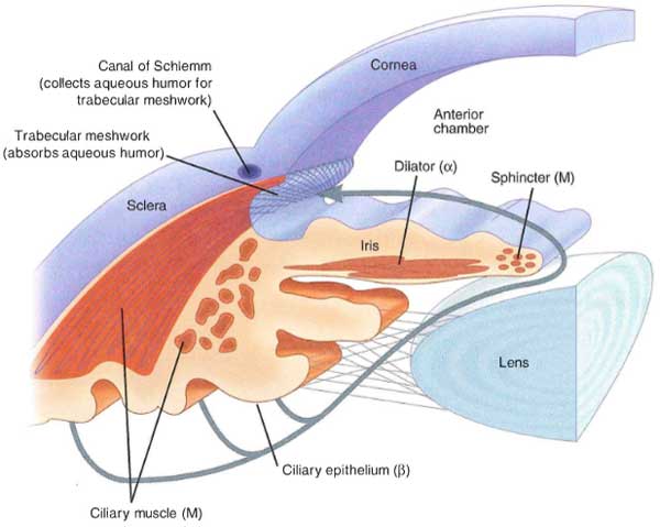
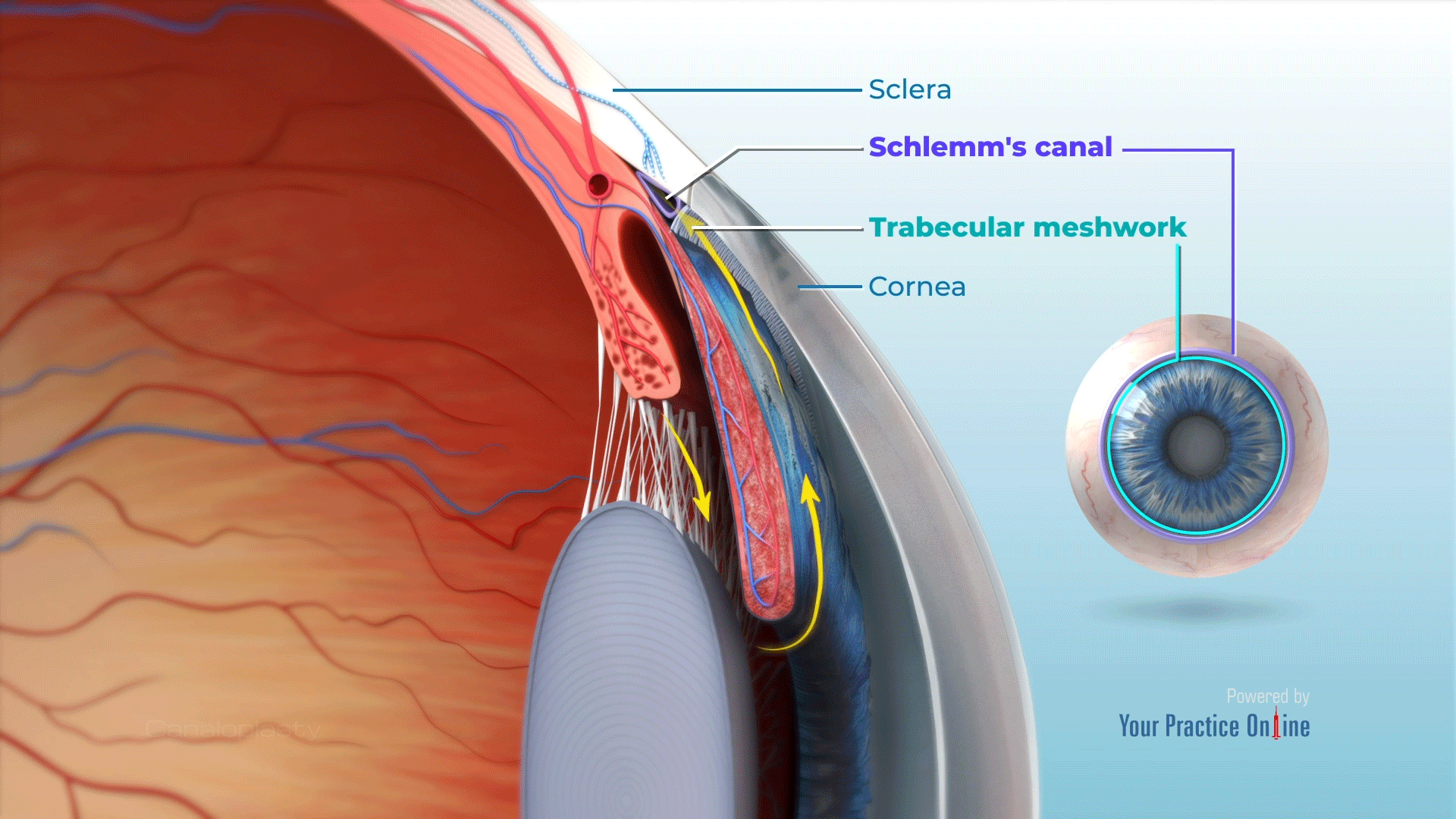
Schlemm's canal is a circular lymphatic-like vessel in the eye that collects aqueous humor from the anterior chamber and delivers it into the episcleral blood vessels via aqueous veins.
The aqueous humor flows in and out of the front of your eye. Its job is to keep proper ocular pressure (eye pressure) in your eye. The pressure is maintained by having the same amount of fluid come in as the amount of fluid leaving the front of your eye. This fluid fills the anterior and posterior chambers of your eye.The vitreous humor makes up the vitreous body located in the vitreous cavity between your lens and retina of your eye. The vitreous humor is contained in a protective layer called the vitreous membrane. Vitreous humor is more viscous than water but still lets light through. Vitreous humor is responsible for about 80% of the volume of your eye.[16]


It is named after Friedrich Schlemm (1795–1858), a German anatomist.
Nearly 200 years ago, a gifted German anatomist stumbled upon a rather macabre discovery. While examining a man who died at his own hand, by hanging, the anatomist unearthed a thin-walled canal, forming a delicate ring around the whites of the eyes. Ordinarily, such a structure might have escaped notice, but in this corpse, it was engorged with blood.
Although no drawings were made to document this seminal observation, the canal bears the name of its discoverer, Friedrich Schlemm, and is now known to play important roles in normal eye physiology and disease.
Schlemm was known for his pathological studies on cadavers. He was the first to discover the corneal nerves of the eye, which he describes in his 1830 treatise named Arteriarum capitis superficialum icon nova. He is known today for the eponymous. Schlemm's canal, which is a channel in the eye that collects aqueous humor from the anterior chamber and delivers it into the bloodstream.
کانال اشلم (Schlemm's canal) یا سینوس سیاهرگی صلبیه (Venous sinus of sclera) مجرایی حلقوی است در محل اتصال صلبیه و قرنیه که ماده زلالیه از اتاق پیشین چشم به درون آن وارد میشود. در داخل کرهی چشم دو ناحیهی دارای مایع وجود دارند که توسط عدسی چشم از یکدیگر جدا شدهاند. بخش بزرگتر که در پشت عدسی قرار دارد زجاجیه نام دارد که حاوی یک مادهی شفاف ژله مانند است. بخش جلویی که کوچکتر از زجاجیه است و حاوی یک مایع آبکی شفاف است زلالیه نام دارد.
زلالیه از دو قسمت تشکیل شده است:
یک بخش که در جلوی عنبیه قرار دارد اتاق قدامی نام دارد و بخش دیگر که در پشت عنبیه است اتاق خلفی نام دارد.
- زلالیهی چشم در جسم مژگانی تولید میشود و از طریق کانال اشلم تخلیه میشود.
- محل اتصال محيط قرنيه و ريشه عنبيه را زاويه اتاق قدامى مىنامند.
اجزاء اصلى سازنده زاويه عبارتند از:
- خط شوالب (Schwalbe's Line) انتهاى اندوتليوم قرنيه
- شبکه ترابکولار (trabecular meshwork) شبکهاى از صفحات سوراخدار که از طریق منافذ آن زلالیه به کانال اشلم مىرسد
- خار صلبيهاى (Scleral Spur)
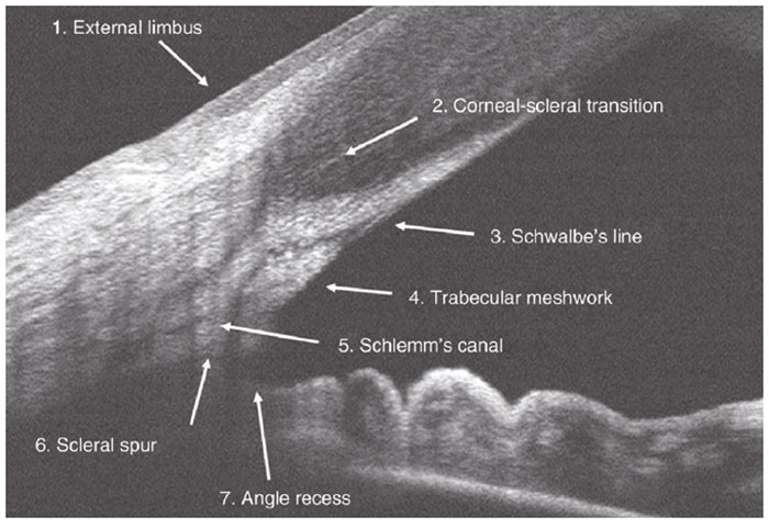
An AS-OCT scan of a wide open A/C angle showing key anatomical landmarks - the cornea, Scleral spur, trabecular meshwork, and iris.
زلاليه وارد شده به کانال اشلم توسط ۳۰ کانال جمعکننده و نهايتاً ۱۲ وريد زلالى به سيستم وريدى اپىاسکلرا وارد مىشود.[7]
وقتی این کانال مسدود شود یک بیماری به نام گلوکاما(glaucoma) یا آبسیاه بروز میکند
Schlemm's canal (SC) functions to maintain proper intraocular pressure (IOP) by draining aqueous humor and has emerged as a promising therapeutic target for glaucoma, the second-leading cause of irreversible blindness worldwide.[13]
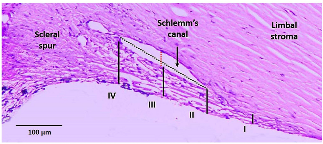
Schlemm's canal is defined as a vascular sinus with a flattened elliptical lumen that encircles the globe of the eye, measuring approximately 190–370 μm in length and about 36 mm in circumference, which plays a role in regulating intraocular pressure.[15]
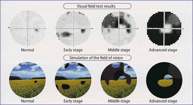
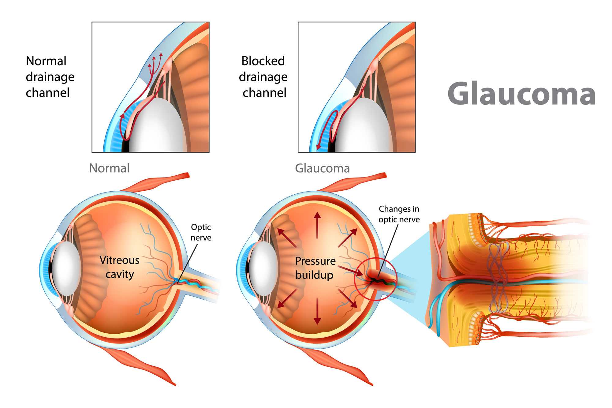
Glaucoma is a group of eye conditions that damage the optic nerve, the health of which is vital for good vision. This damage is often caused by an abnormally high pressure in your eye.
Glaucoma is one of the leading causes of blindness for people over the age of 60. It can occur at any age but is more common in older adults. Many forms of glaucoma have no warning signs. The effect is so gradual that you may not notice a change in vision until the condition is at an advanced stage.
The canal is essentially an endothelium-lined tube, resembling that of a lymphatic vessel. On the inside of the canal, nearest to the aqueous humor, it is covered by the trabecular meshwork; this region makes the greatest contribution to outflow resistance of the aqueous humor.
Schlemm’s canal (SC) is comprised of endothelial cells surrounded by connective tissue like a vein. SC possesses internal collector channels and is connected to episcleral and conjunctival veins through the external collector channels, the intrascleral venous plexus, the deep scleral plexus and the aqueous veins.
Conventionally, the canal has been considered a blood vessel, but recent studies have revealed that the molecular identity of Schlemm's canal is strikingly reminiscent to the one of lymphatic vasculature.[1]
Schlemm's Canal Is a Unique Vessel with a Combination of Blood Vascular and Lymphatic Phenotypes that Forms by a Novel Developmental Process -- journals.plos.org
Schlemm's canal serves as a drainage tube for fluid from the anterior chamber of the eye and is directly relevant to glaucoma, a disease that causes vision loss in over 70 million people. Aqueous humor enters the canal and then drains into connected veins. Molecular understanding of the development of Schlemm's canal and its drainage functions has remained limited.Schlemm's canal is thus a unique vessel with a combination of blood vascular and lymphatic characteristics.
Both Schlemm's canal and the aqueous humor are vital to the health and function of the eye. It is critical for the posterior and anterior portions of the eye to maintain a careful balance of aqueous humor production and drainage to ensure proper pressure of two chambers. The balance of production and drainage also serves to promote the correct spatial distances between the various organelles of the eye. If this important balance is not maintained, it is common for eye disorders relating to distortion of the size and shape of the eyeball to present, and there is an increased potential for seriously impaired vision to occur.

There are two ways in which the canal of Schlemm can be adversely affected, causing dysfunction and possible damage. In addition to eye disease, physical injury to the area can also create an imbalance of the aqueous humor and damage the scleral venous sinus. If too much aqueous humor is produced, the intraocular pressure is likely to rise, causing the potential for serious eye diseases, such as glaucoma. This disease is characterized by optic nerve atrophy, impaired, blurry vision, and eye retina detachment. Left untreated, glaucoma can also result in permanent vision loss.[11]

Schwalbe's line is the anatomical line found on the interior surface of the eye's cornea, and delineates the outer limit of the corneal endothelium layer. Specifically, it represents the termination of Descemet's membrane. In many cases it can be seen via gonioscopy.
Some evidence suggests that the corneal endothelium actually possesses stem cells that can produce endothelial cells, especially after injury, albeit on a limited scale.
Although Schlemm's canal (SC) has central roles in ocular physiology and homeostasis, its development, mature phenotype, and molecular processes are poorly understood. SC has a critical role in aqueous humor drainage (AQH) from the eye, a process that regulates the intraocular pressure (IOP). Abnormal resistance to AQH drainage results in IOP elevation, a key factor contributing to glaucoma. Glaucoma is one of the most common neurodegenerative diseases and will affect an estimated 80 million people by the end of this decade. SC is also important for anterior chamber associated immune deviation (ACAID), a form of immune tolerance. During ACAID, immune cells are exposed to an antigen in the eye and then exit the eye via SC. From SC they return to the systemic circulation via blood vessels to which SC is connected. After exiting SC, these cells induce a systemic suppression of immune responses to that antigen. Thus, SC is a unique and important vessel that needs to be better understood.

Aqueous outflow - A continuum from trabecular meshwork to episcleral veins
Schlemm's canal. (A) SC is an endothelium-lined channel that encircles the cornea and provides an exit route for aqueous humor. (B) Aqueous humor is produced from the ciliary body and drained into aqueous and episcleral veins through the trabecular meshwork and SC. (C) Aqueous humor is drained transcellularly and transported from the basal to luminal side through SC ECs, causing formation of giant vacuoles. SC ECs have an intermediate blood-lymphatic EC phenotype and express Prox1, VEG FR3, Tie2, and integrin α9, but not LYVE1 or podoplanin. Angpt1 + stromal cells adjacent to the SC LECs may produce proteins of trabecular meshwork. When SC function is impaired, aqueous humor drainage is impeded and intraocular pressure is increased, ultimately leading to glaucoma. Angpt-Tie2 signaling maintains SC integrity, and loss of such signaling induces primary congenital and open-angle glaucoma. AHO, aqueous humor outflow; E & A vein, episcleral & aqueous vein; VEC, venous endothelial cell.[3] eResearch by Navid Ajamin -- spring 2013
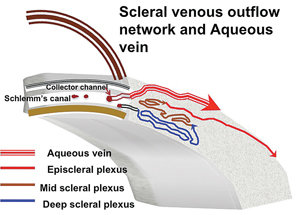
This diagram depicts the route of aqueous humor once it leaves Schlemm’s canal. Aqueous must pass through the collector channels to the deep scleral plexus (blue), then to the mid plexus (brown), finally arriving at the episcleral plexus (red). We are currently unable to visualize this pathway preoperatively. However, we can see episcleral flow during surgery (assuming the intrascleral pathway is intact) by visualizing the episcleral venous wave.
Note: The large laminated red vein—an aqueous vein of Ascher—originates from the canal and bypasses the entire intrascleral network (deep and mid). This large vein can often be seen postoperatively at the slit lamp and usually correlates with low IOP.[6]

from the anterior chamber through a sievelike layer of tissue in the lining of the eyeball at the outer periphery of the iris into a circular channel, the canal of Schlemm, from which the aqueous humour flows (by way of vessels called aqueous veins) into blood vessels. Blockage of the aqueous humour flow causes increased pressure in the posterior chamber, and this pressure is transmitted by way of the vitreous to the optic nerve head and the retina. Abnormally high intraocular pressure that is unrelieved causes vision impairment.
The thin coats of the eye are not sufficiently rigid in themselves to withstand distortion following the pull of the extraocular muscles…
There are two types of blockage that result in glaucoma.
- The blockage may occur in the porous tissue between the anterior chamber and the canal of Schlemm, in the canal itself, or in the aqueous veins. This blockage is continuous, and the effect is chronic glaucoma.
- In persons in whom the angle at the periphery of the anterior chamber is acute—i.e., in whom the outer rim of the iris is close to the wall of the eyeball—the pressure of aqueous humour upon the back of the iris may force the outer part of the iris against the wall, so as to cover the outlet into the canal of Schlemm. Glaucoma caused by this type of obstruction is called acute or narrow-angle glaucoma. When the pupil contracts, as during sleep, it tends to pull the iris away from the entrance into the canal of Schlemm and allow passage of aqueous humour, so that the high intraocular pressure may be intermittent in this type of blockage.
Chronic glaucoma does not cause symptoms in its early stages, and it is diagnosed by observation of the abnormally high intraocular pressure or the physical effects of abnormal pressure upon the optic disk (the point where the optic nerve leaves the eyeball). Treatment is primarily medical—the reduction of intraocular pressure by means of drugs that contract the pupil (miotic drugs) and allow greater outflow of the aqueous humour.

Narrow-angle glaucoma causes pain in the eye, headaches, and sometimes nausea and vomiting. The affected person may see halos around lights. Treatment of an acute attack is similar to that of chronic glaucoma, but permanent elimination of the high pressure requires surgery; i.e., an opening is cut through the iris at its outer periphery to allow passage of the aqueous humour.[4]
The picture shows a conjunction between the Schlemm canal and Singh canal. Singh canal is the lucid interval, what we can see in cases of arcus senilis. There is present an anatomical basis of physiology and pathology. The fluid is moving from Schlemm canal to Singh canal and hydrates the cornea through the network of channels in the cornea.[5]
Questions:
- Which part of the eye is responsible for producing aqueous humour?
- Is trabecular meshwork the same as canal of Schlemm?
- What happens if the canal of Schlemm is obstructed?
- What happens if the canal of Schlemm is blocked?
- How does glaucoma affect the canal of Schlemm?
- What happens during a Schlemm's canaloplasty?
- How does aging affect the chamber angle?
- What is the function of Schlemm's Canal?
- What is the size of the Schlemm Canal?
- Where does Schlemm's Canal drain?
- How to identify Schlemm's canal?
- What does schlemm mean?
Reference:
- en.wikipedia.org/wiki/Schlemm%27s_canal
- journals.plos.org/plosbiology/
- researchgate.net/figure/Schlemms-canal-A-SC-is-an-endothelium-lined-channel-that-encircles-the-cornea-and_fig3_321809038
- britannica.com/science/glaucoma
- medtube.net/ophthalmology/medical-pictures/9825-schlemm-canal-and-singh-canal-connection
- reviewofophthalmology.com/article/the-fluid-wave-evaluating-canal-surgery
- vista.ir
- openophthalmologyjournal.com/VOLUME/4/PAGE/52/FULLTEXT
- jax.org/news-and-insights/2014/december/eyeing-the-eye
- en.wikipedia.org/wiki/Friedrich_Schlemm
- wisegeek.com/what-is-the-canal-of-schlemm.htm
- mayoclinic.org/diseases-conditions/glaucoma/symptoms-causes/syc-20372839
- pubmed.ncbi.nlm.nih.gov/38367239
- ypo.education/ophthalmology/canaloplasty-t719/video
- sciencedirect.com/topics/medicine-and-dentistry/schlemms-canal
- my.clevelandclinic.org/health/body/24611-aqueous-humor-vitreous-humor
See also:
 وبلاگ تخصصی عینک شامل مجموعه مطالب پزشکی است که اطلاعات مفیدی در رابطه با عینک , چشم، لنز، سلامتی چشم و راه های پیشگیری از بیماریهای چشمی، کنترل و درمان آن را در اختیار شما کاربر محترم می گزارد.