The iris (plural: irides or irises) is a thin, circular structure in the eye, responsible for controlling the diameter and size of the pupils and thus the amount of light reaching the retina. "Eye color" is the color of the iris, which can be green, blue, or brown. In some cases it can be hazel (a combination of light brown, green and gold), grey, violet, or even pink. In response to the amount of light entering the eye, muscles attached to the iris expand or contract the aperture at the center of the iris, known as the pupil. The larger the pupil, the more light can enter.
Etymology
The word iris is derived from the Greek goddess of the rainbow, due to the many colours of the iris.
Embryology
The iris develops from the anterior two layers of an embryonic neuroectoderm structure called the optic cup. The optic cup also produces the iris sphincter and dilator muscles.
General structure
The iris consists of two layers: the front pigmented fibrovascular tissue known as a stroma and, beneath the stroma, pigmented epithelial cells.
The stroma connects to a sphincter muscle (sphincter pupillae), which contracts the pupil in a circular motion, and a set of dilator muscles (dilator pupillae) which pull the iris radially to enlarge the pupil, pulling it in folds. The back surface is covered by a heavily pigmented epithelial layer that is two cells thick (the iris pigment epithelium), but the front surface has no epithelium. This anterior surface projects as the dilator muscles. The high pigment content blocks light from passing through the iris to the retina, restricting it to the pupil. The outer edge of the iris, known as the root, is attached to the sclera and the anterior ciliary body. The iris and ciliary body together are known as the anterior uvea. Just in front of the root of the iris is the region referred to as the trabecular meshwork, through which the aqueous humour constantly drains out of the eye, with the result that diseases of the iris often have important effects on intraocular pressure, and body provide a lesser secondary pathway for the aqueous humour to drain from the eye.
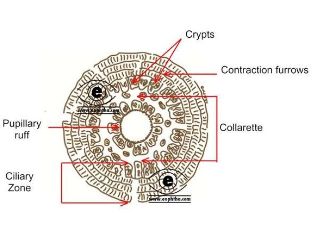
The iris is divided into two major regions:
The pupillary zone is the inner region whose edge forms the boundary of the pupil.
The ciliary zone is the rest of the iris that extends to its origin at the ciliary body.[1]
The layers of the iris eResearch by Navid Ajamin -- spring 2012
From anterior to posterior the iris is organised in the following layers:
1. Endothelial
2. Anterior marginal layer
3. Vascular layer = Stroma
4. Posterior marginal layer = Dilatator layer
5. Epithelial pigment = Stratum pigmenti iridis
6. Retinal layer = Pars iridica retinae
1. The question of the existence of the Endothelial layer is not completely settled. Many researchers assume an anterior membrane of the human iris, others dispute it.
2. The anterior marginal layer is composed predominantly of cells, between which lie numerous nerve endings but few blood vessels. Cells bearing colour material—Chromatophoren—may be present, which together with the stroma gives rise to certain colour changes in the iris. Where the marginal layer is missing, smaller or larger dark-shining openings—so-called crypts—tissue spaces, allow a view of the interior of the spongy iris—stroma. These crypts will be considered later as lacunae.
3. The Vascular layer, or iris-stroma, constitutes the principal bulk of the iris. It consists mainly of numerous blood vessels which radiate in spokes, and therefore run radially from the outer margin of the iris towards the pupil. The blood vessels are enveloped in a thick adventitia of connective tissue fibre, and are surrounded by a loose ramifying network and pigment cells, which fill out the spaces between the blood vessels.
These blood vessels appear as spiral formations below the anterior marginal layer. In these formations they can adapt to the conditions of expansion and contraction of the iris.
Besides the radiating blood vessels of the iris stroma, there is in the iris an arterial ring arising from the annular anastomosis of the ciliary blood vessels—the Circulus arteriosus iridis minor. It is situate at the border between the pupillary zone and the ciliary zone, and is called in Iridology the Iris-wreath.
In a very light iris one can also see a grey band at the pupillary margin. This is composed of smooth muscle fibres which surround the pupil in a ring-formation. They form the sphincter of the iris—Sphincter pupillae —which lies in the iris-stroma.
4. The posterior marginal layer—Dilatator layer—joins on to the posterior surface of the vascular layer. It consists of a continuous layer of spindle-shaped smooth muscle fibres, extending from the outer margin of the iris to the ciliary border near to the pupillary margin. Here it unites with the connective tissue of the sphincter.
5-6. The epithelial pigment forms the posterior surface of the iris and extends to the pupillary margin, around which it runs to the anterior surface of the iris, thereby giving rise to the frequently visible dark-yellow to black-brown pupillary margin.
This margin, where the fibres reflect back, is the only structure in the human body, which as the embryological representation of the central nervous system, provides a surface accessible to view.

This posterior pigment layer consists of two layers of epithelial cells which pass over into one another to the pupillary margin. (Stratum pigmenti iridis with Pars iridica.) Both together form the continuation of the retina as far as the pupillary margin. Thus, this layer of the iritis denoted Retinal, in contrast to the anterior layer which is called the Uveal. (Pars retinalis iridis, and Pars uvealis iridis.)
Apart from the structure referred to above, examination frequently reveals a number of light or dark concentric arc lines. These are seen particularly frequently in a brown iris where they stand out because of their light colour on a dark background. These are the 'contraction rings' of the iris, which in Iridoscopy have a special meaning.
Quite remarkable are the groups of white flakes seen at the periphery of the ciliary zone, and sometimes scattered regularly around the whole iris like a rosary. These will be discussed later under the heading 'Acute or chronic inflammation of the mucous membranes'.
At the periphery, there appears a partial, or frequently entire, dark almost black circle (Scurf rim). In old age it becomes obscured by a silver-grey rim projectingfromthesclera(Sclerotic rim). The black circle is formed by the crypts of the ciliary margin, and the silver-grey rim results from fatty infiltration—it is a sign of senile change (Arcus senilis).[2]

Reference:
- en.wikipedia.org/wiki/Iris_(anatomy)
- ipeerx.com/articles/category/10/message/204/
 وبلاگ تخصصی عینک شامل مجموعه مطالب پزشکی است که اطلاعات مفیدی در رابطه با عینک , چشم، لنز، سلامتی چشم و راه های پیشگیری از بیماریهای چشمی، کنترل و درمان آن را در اختیار شما کاربر محترم می گزارد.