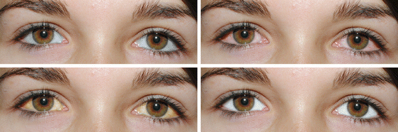
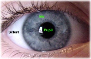
The sclera (/ˈsklɪərə/) (from the Greek skleros, meaning hard), also known as the white of the eye, is the opaque, fibrous, protective, outer layer of the eye containing collagen and elastic fiber. In humans the whole sclera is white, contrasting with the coloured iris, but in other mammals the visible part of the sclera matches the colour of the iris, so the white part does not normally show. In the development of the embryo, the sclera is derived from the neural crest.
In children, it is thinner and shows some of the underlying pigment, appearing slightly blue. In the elderly, fatty deposits on the sclera can make it appear slightly yellow.
The sclera forms the posterior five-sixths of the connective tissue coat of the globe. It is continuous with the dura mater and the cornea, and maintains the shape of the globe, offering resistance to internal and external forces, and provides an attachment for the extraocular muscle insertions. The sclera is perforated by many nerves and vessels passing through the posterior scleral foramen, the hole that is formed by the optic nerve. At the optic disc the outer two-thirds of the sclera continues with the dura mater (outer coat of the brain) via the dural sheath of the optic nerve. The inner third joins with some choroidal tissue to form a plate (lamina cribrosa) across the optic nerve with perforations through which the optic fibers (fasciculi) pass.
The thickness of the sclera varies from 1mm at the posterior pole to 0.3 mm just behind the rectus muscle insertions. The sclera's blood vessels are mainly on the surface. Along with the vessels of the conjunctiva (which is a thin layer covering the sclera), those in the episclera render the inflamed eye bright red.
 In many vertebrates, the sclera is reinforced with plates of cartilage or bone, together forming a circular structure called the scleral ring. The cornea and sclera make up the outer tunic of the eye. Each is a connective tissue containing collagen fibrils embedded in a proteoglycan-rich extrafibrillar matrix, but whereas the cornea is uniquely transparent, the sclera is totally opaque.

The eyes of all non-human primates are dark with small, barely visible sclera.
The sclera is made up of three divisions: the episclera, loose connective tissue, immediately beneath the conjunctiva; sclera proper, the dense white tissue that gives the area its color; and the lamina fusca, the innermost zone made up of elastic fibers.

The sclera is covered by the conjunctiva, a clear mucus membrane that helps lubricate the eye. It is thickest in the area surrounding the optic nerve.

The sclera is made up of three divisions:
- the episclera, loose connective tissue, immediately beneath the conjunctiva
- sclera proper, the dense white tissue that gives the area its color
- the lamina fusca, the innermost zone made up of elastic fibers.

There are a number of abnormalities associated with the sclera. eResearch by Navid Ajamin -- spring 2012
Some are genetic and include:
- Melanosis: excess deposits of melanin (pigment) on the surface of the sclera, which can become inflamed and uncomfortable
- Scleral Coloboma: missing tissue that results in notching and bulging of the sclera (lesions)
- Ectasia: a thinning and bulging of the sclera
What does a healthy sclera look like?
A healthy sclera is white. But what does it mean when the sclera takes on a different hue? If your whites become yellow or otherwise discolored, consult with your ophthalmologist. In some cases, this could signal an underlying health condition.
 Reference:
- eaglet-eye.com
- en.wikipedia.org/wiki/Sclera
- healthline.com/human-body-maps/sclera#1
- link.springer.com/chapter/10.1007/978-0-387-73906-9_13
- morancore.utah.edu/section-04-ophthalmic-pathology/sclera
- aao.org/eye-health/tips-prevention/discolored-sclera-whites-of-my-eyes-turn-yellow
 وبلاگ تخصصی عینک شامل مجموعه مطالب پزشکی است که اطلاعات مفیدی در رابطه با عینک , چشم، لنز، سلامتی چشم و راه های پیشگیری از بیماریهای چشمی، کنترل و درمان آن را در اختیار شما کاربر محترم می گزارد.