About half of all adults in the USA aged 20 and older have refraction errors in their eyes, a study carried out by researchers at the National Eye Institute revealed.[1]
Astigmatism is a defect in the curvature of the cornea (the dome-like transparent structure which covers the iris and the pupil) or in the shape of the eye lens.
Normally, the cornea and the lens are regular and are curved in the same shape throughout. This helps to focus light clearly onto the retina at the back of the eye. Nevertheless, if the cornea or the lens are not smooth or do not have a regular curve, the rays of light do not refract correctly, which causes a refraction problem.
Types of astigmatism
Based on asymmetry of structure
- Corneal astigmatism - astigmatism due to an irregularly shaped cornea (like an American football or rugby ball instead of a soccer ball)
- Lenticular astigmatism - astigmatism due to an irregularly shaped lens
Based on Axis of the Principal Meridians
- Regular astigmatism
- Against-the-rule astigmatism
- With-the-rule astigmatism
- Oblique astigmatism
- Irregular astigmatism

Based on focus of the principal meridians
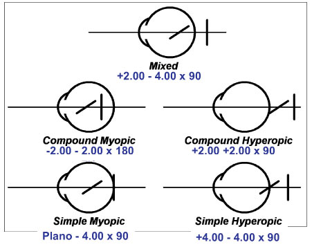
Simple astigmatism
Simple hyperopic astigmatism
Simple myopic astigmatism
Compound astigmatism
Compound hyperopic astigmatism
Compound myopic astigmatism
Mixed astigmatism [2]
Regular and irregular astigmatism [4]
- Regular astigmatism. The principal meridians are perpendicular to each other and form a 90º angle. Most astigmatisms are regular and are of the cornea.
- Irregular astigmatism. The principal meridians are not perpendicular. It may be the consequence of an injury or surgery that has caused the scarring of the cornea. In addition, it may be caused by a keratoconus, an eye problem which causes the thinning and deformity of the cornea.

Does Astigmatism Get Worse With Age ?
Simple and compound astigmatisms
Simple astigmatism
- Simple myopic astigmatism. One of the two principal meridians of the eye focuses light rays in front of the retina. The other focuses correctly onto the retina.
- Simple hypermetropic astigmatism. One of the two principal meridians focuses rays of light behind the retina. The other focuses correctly onto the retina.
Compound astigmatism
- Compound myopic astigmatism. The two main meridians of the eye focus light rays in front of the retina.
- Compound hypermetropic astigmatism. The two principle meridians focus light rays behind the retina.
- Mixed astigmatism. One principle meridian focuses the light in front of the retina and the other behind.

Take a look at the above image. Move back from your screen, until you get some blur on the lines. Now move closer again, until the lines are just barely clear. Now, assess: Do all the lines look the same? Any more bold than others?
If all lines look the same or nearly the same, then you may not need an astigmatism correction. [3]
The lines address the “axis” part of your current astigmatism correct. The degree is where you would get additional correction, and the cylinder expresses how much correction is added. There is a bit more to it, though for our purposes here this is all we really need. [3]

What are the common signs and symptoms of astigmatism? [5]
In eyes without astigmatism, light enters the eye and hits the retina—which is a sensitive layer located at the back of the eye that is responsible for sending information to the optic nerve in your brain. As light enters your eye, your retina triggers nerve impulses to your brain. When your optic nerve becomes triggered, your brain is able to process a visual image for your eyes to see.
However, when you have astigmatism, your retina isn't able to function properly, which can affect your vision. The reason this happens is because astigmatism causes your eyes to change shape. As a result, you aren't able to properly focus on the objects in front of you, which can make your vision seem blurry or distorted. eResearch by navid ajamin -- spring 2011
Why exactly astigmatism happens is currently unknown. Some researchers theorize that normal changes in vision as you age or having an underlying eye condition can all increase your risk of developing astigmatism.[7]
- Blurry and/or distorted vision when looking at near and far objects
- Squinting when trying to focus at near and far objects
- Experiencing eye strain
- Double vision when looking at near and far objects
- Headaches
- Trouble seeing clearly at night, especially when driving
- Trouble seeing in the dark
What are the causes of stigma? [9]

The eyeball has two curved surfaces through which light passes to reach the retina. The first surface is the cornea, the tissue that covers the eye. The second is crystallinity, the lens of the eye, a clear structure inside the eye that is responsible for focusing near objects.
A perfectly shaped eye has both surfaces curved round. Then the light is reflected evenly resulting in a clear image directly on the retina.
The error occurs when either the cornea or the lens of the eye has an oval curvature. When the curves are not the same, the light rays do not reflect the same, which means that two different images will be formed. These images overlap, resulting in blurred vision.
Depending on the surface that has the curvature defect, astigmatism can be corneal or lenticular.
Some people are born with astigmatism. For others, it occurs as a result of eye trauma, disease, or surgery.
Contrary to popular belief, it does not occur and is not worsened by reading in low light, viewing screens from a short distance.
Diagnosis
If you notice changes to your vision, it's good practice to see your eye care provider (such as an optometrist or ophthalmologist) for testing. During your eye exam, your provider can learn more about your symptoms, check your vision, and rule out other conditions that may be causing blurriness or eye strain.
Your provider can perform a number of diagnostic tests to learn more about your condition and test for other eye conditions (such as nearsightedness or farsightedness).
These tests include:
- Visual acuity: Tests how well you see objects close by and far away by reading letters of different sizes on a chart
- Refraction: Assesses how much refractive error (lack of focus in the eyes) you have
- Retinoscopy: Uses a handheld device called a retinoscope to test the level of refractive error in your eye
- Keratometry: Involves using a device known as a keratometer to measure the curvature and shape of the cornea
- Pachymetry: Measures the thickness of the cornea while using a device called a pachymeter to determine if you need eye surgery
Factors that may increase your chance of Astigmatism [8]
- Heredity – a family history of astigmatism
- A disease history of corneal scaring or thinning
- Low birth weight
- Advancing age
- Corneal scarring due to injury
- Corneal thinning
- Pre-existing refractive errors of the eye such as Myopia or Hypermetropia
- Severe allergies resulting in constant rubbing of the eyes and Diabetes
- History of excessive nearsightedness or farsightedness

Reference:
- medicalnewstoday.com
- psychology.wikia.com
- endmyopia.org/test-need-astigmatism-correction-normalized-prescriptions
- icrcat.com/en/eye-conditions/astigmatism
- plano.co/eye-conditions/other-eye-conditions/astigmatism
- optometryzone.com/2017/02/18/how-to-find-type-of-astigmatism-through-prescription
- health.com/astigmatism-7547383
- optography.org/etiology-of-astigmatism
- vitreum.ro/en/conditions/astigmatism
- reviewofoptometry.com/news/article/rethink-glasses-in-infants-with-high-astigmatism-hyperopia
 وبلاگ تخصصی عینک شامل مجموعه مطالب پزشکی است که اطلاعات مفیدی در رابطه با عینک , چشم، لنز، سلامتی چشم و راه های پیشگیری از بیماریهای چشمی، کنترل و درمان آن را در اختیار شما کاربر محترم می گزارد.