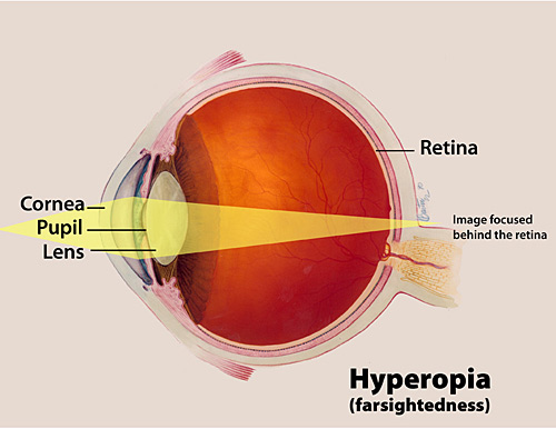
Hyperopia, also known as farsightedness, longsightedness or hypermetropia, is a defect of vision caused by an imperfection in the eye (often when the eyeball is too short or the lens cannot become round enough), causing difficulty focusing on near objects, and in extreme cases causing a sufferer to be unable to focus on objects at any distance. As an object moves toward the eye, the eye must increase its optical power to keep the image in focus on the retina. If the power of the cornea and lens is insufficient, as in hyperopia, the image will appear blurred.[1]
What does it mean to show farsightedness?
People with hyperopia can experience
blurred vision,
asthenopia,
accommodative dysfunction,
binocular dysfunction,
amblyopia, and strabismus.
Classification of hyperopia
Simple hyperopia
Pathological hyperopia
Functional hyperopia
Ornithological hyperopia
Causes
Hyperopia can be caused bysinus infections, injuries, migraines, aging or genetics.
How is farsighted vision corrected? eResearch by Navid Ajamin -- summer 2011
Farsightedness
Farsighted (also called hyperopia) is a term to describe an eye condition that lets you clearly see objects “far” or distant in your field of vision, while objects that are near appear blurry or hazy. Due to the nature of this type of vision problem, farsightedness can affect vision in different ways.
Farsightedness happens in eyes that are incorrectly focusing images behind the retina rather than directly on it. The retina is the light-sensitive tissue at the back of the human eye responsible for processing images.
Farsighted vision is treated with corrective lenses like eyeglasses or contact lenses, and can also be treated surgically with types of surgery. Farsighted vision can develop in children or adults, and between 5 and 10 percent of all Americans are considered to be farsighted.
Persons who are extremely nearsighted, have diabetes, or have had cataract surgery are also more likely to report eye floaters.
Farsightedness Symptoms
Symptoms of farsightedness include eyes that feel tired or strained, headaches, squinting and blurred vision, especially when viewing objects that are near. But symptoms can vary person to person based on the degree of farsighted vision; some may notice little visual impairment, while others may have blurred or hazy vision for objects at distance and nearby.
Farsighted vision can develop at any time, and happens in both children and adults.
Farsightedness develops when the eyeball becomes “shorter” than it should be, moving the “focal point” of the images we see from on top of the retina, to behind the retina. Abnormalities in the eye’s lens or cornea can also cause farsighted vision.
Your child always seems to have tears streaming down their face. And they can’t stop rubbing their eyes. But they aren’t sad — so what’s going on?
Babies usually don’t make tears for at least a few weeks after birth.
After that time your child may have a blocked tear duct if they have:
Eye mucus or discharge with a yellowish color.
Redness or crusting around their eye.
Tears that build up near the inner corner of their eye but don’t drain.
Tears that run down your their cheek when they’re not crying (epiphora).
Crusting and discharge on their eyelashes when they wake up in the morning.
اصولا علل اشک ریزش را می توان این طور دستهبندی کرد :
1) تولید اشک زیادتر از حد طبیعی باشد (وجود ایرادی در غدد اصلی سازنده اشک)
2) اشکال در مسیر خروج اشک باشد که اصطلاحا انسداد مجرا نامیده میشود (دلیل آن اختلالی در کار خروج اشک از چشم و هدایت آن به داخل بینی است.)
یکی از علل مهمی که شیرخواران را به مطب چشم پزشک میآورند، همین اشکریزش است. حدود 10 درصد نوزادانی که متولد میشوند، مبتلا به انسداد مجرای اشکی هستند، یعنی موقع تولد، هنوز مجرای اشکی آنها به داخل بینی باز نشده و این شیرخواران با اشکریزش و گاهی با ترشحات چرکی مواجه میشوند.
تجمع مواد کنار مژهها و گاه چسبیدن مژهها به هم خصوصا هنگام خواب و لزوم دستکاری پلکها در موقع بیدار شدن از مشکلات عمدهای است که موجب نگرانی والدین میشود و معمولا مجبورند پلکها را تمیز کنند تا چشمها از یکدیگر بازگردد.
Blocked Tear Duct in Children
The most common cause is tear duct (the tube that drains tears away from the eyes) blockage. This condition occurs in up to 20% of children in the first year of life, as the tear duct is slow to develop or the lower end of the tract is not fully opened.
Watery eyes can be due to many factors and conditions.
In infants, persistent watery eyes, often with some matter, are commonly the result of blocked tear ducts. The tear ducts don't produce tears, but rather carry away tears, similar to how a storm drain carries away rainwater. Tears normally drain into your nose through tiny openings (puncta) in the inner part of the lids near the nose. In babies, the tear duct may not be fully open and functioning for the first several months of life.
In older adults, persistent watery eyes may occur as the aging skin of the eyelids sags away from the eyeball, allowing tears to accumulate and flow out.
Sometimes, excess tear production may cause watery eyes as well.
Allergies or viral infections (conjunctivitis), as well as any kind of inflammation, may cause watery eyes for a few days or so.
Medication causes
Chemotherapy drugs
Epinephrine
Eyedrops, especially echothiophate iodide and pilocarpine
Common causes
Allergies
Blepharitis (which is eyelid inflammation)
Blocked tear duct
Common cold
Corneal abrasion (scratch): First aid
Corneal ulcer
Dry eyes (caused by decreased production of tears)
Ectropion (a condition in which the eyelid turns outward)
Entropion (a condition in which the eyelid turns inward)
Foreign object in the eye: First aid
Hay fever (allergic rhinitis)
Ingrown eyelash (trichiasis)
Keratitis (which is inflammation of the cornea)
Pink eye (conjunctivitis)
Stye (sty) (a red, painful lump near the edge of your eyelid)
Tear duct infection
Trachoma
Other causes
Bell's palsy
Blow to the eye or other eye injury
Burns
Chemical splash in the eye: First aid
Chronic sinusitis
Granulomatosis with polyangiitis
Inflammatory diseases
Radiation therapy
Rheumatoid arthritis
Sarcoidosis
Sjogren's syndrome
Stevens-Johnson syndrome
Surgery of the eye or nose
Tumors affecting the tear drainage system
به طور کلی انسداد مادرزادی مجرای اشکی در بچهها به سه صورت دیده میشود:
- اولین حالت، اشک ریزش مداوم بدون همراه بودن ترشحات چرکی است.
- دوم، اشکریزش مداوم همراه با ترشحات چرکی به خصوص هنگام ماساژ کیسه اشکی است که این بیانگر انسداد کامل مجرای اشکی میباشد (این فرم شایعی است).
- سوم، اشکریزش متناوب به خصوص هنگام سرماخوردگی است که بیانگر مشکلات بینی می باشد که هنگام سرماخوردگی شدت یافته و به طور موقت مجرای اشکی را میبندد.
هریک از سه حالت فوق باید توسط والدین مورد پیگیری قرار گیرد و بچه توسط چشمپزشک معاینه شود.
معمولا این بچهها نسبت به نور حساس نیستند، ولی قرنیه، شفاف و کره چشم سالم است و تنها انسداد مجرا مطرح است.
حدود 90 درصد این بچهها تا سن یک سالگی انسدادشان برطرف میشود و والدین نباید نگران باشند. تنها لازم است معاینه شوند و دستور ماساژ صحیح و درمان طبی توسط پزشک معالج به والدین داده شود.
هیچ اقدام خاصی برای باز شدن مجرا لازم نیست و نباید در درمان جراحی سونداژ بیمورد عجله شود و باید منتظر بمانیم تا مجرا باز شود. تنها در موارد عفونت حاد کیسه اشکی است که سونداژ را زودتر از موعد توصیه میکنیم.
سونداژ یک عمل کاملا ساده و موفقیتآمیز است که در بیمارستان تحت بیهوشی عمومی انجام میشود.
بهترین زمان انجام سونداژ حوالی یک سالگی است و تاخیر میتواند شانس موفقیت را کم کند، ولی تا پنج سالگی هم ، بهتر است اولین اقدام سونداژ (به تنهایی) و یا سونداژ همراه با لولهگذاری باشد. eResearch by Navid Ajamin
Teary eyes: other causes
Another common cause of tearing in children is epiblepharon, where an extra horizontal fold of muscle causes the lashes to turn inwards.
This is usually more common in the lower lids. The constant rubbing of the lashes against the cornea causes irritation, tearing, redness and glare, and increases the risk of scarring and infection of the cornea. Mild cases can resolve with age as the facial structures elongate and mature, while severe cases need to be corrected with surgery. Surgery is usually performed to direct the lashes outwards, away from the cornea. Viral or bacterial infections of the eye can also cause tearing. These are usually transient and resolve once the acute infection resolves. Viral conjunctivitis can be preceded by a cold or there may be a positive contact history with someone who has a similar infection. However, if a newborn has red and sticky eyes from birth, bacterial infections such as chlamydia have to suspected. This is usually passed from the mother to the baby through the birth canal. Hence patients with severe conjunctivitis should be evaluated by an eye specialist, who will advise on the appropriate treatment. A rare but serious cause of tearing in infants that needs to be excluded is congenital glaucoma. It is commonly diagnosed between the ages of three and six months and typically presents with – apart from tearing – over-sensitivity to light, involuntary closure of the eyes, redness, cloudiness or enlargement of the eyes. A thorough examination of the eye pressure and other parts of the eye is needed to diagnose the condition and its severity. Delayed diagnosis or treatment of this condition would result in potentially irreversible blindness. Generally, once the more serious causes have been excluded, tearing in children is a relatively harmless condition that can be managed conservatively. Unless there is a definite bacterial infection, usually just keeping the lids clean and wiping away any discharge without antibiotic eyedrops is sufficient.
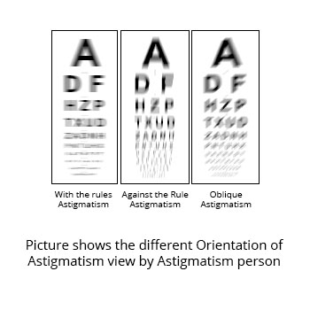
آستیگماتیسم یکی از شایعترین مشکلات اپتیکی چشم است، و معمولاً علت آن نامنظمی شکل و انحنای قرنیهاست. گاهی نیز علت آن نامنظمی شکل و انحنای لنز که در پشت عنبیه قرار دارد است. آستیگماتیسم حالتی است که چند تا از دیوپترهای چشم کرویت خود را از دست دادهاست.
اگر چشم را به عنوان یک عدسی کروی در نظر بگیریم. هرگاه این عدسی از حالت کروی خارج شود و به سمت حالت بیضوی برود (شبیه خربزه). در این صورت دارای دو کانون خطی به جای یک کانون نقطهایی خواهد بود. در نتیجه تصاویر بدلیل انکسار نامساوی در قسمتهای مختلف قرنیه کاملا بر روی شبکیه متمرکز نمیشوند و تصاویر چه دور و چه نزدیک تار میشوند. بنابراین افرادیکه دچار درجات بالایی از آستیگماتیسم هستند نه تنها همانند افراد نزدیکبین اشیای دور را تار میبینند، بلکه اشیای نزدیک را هم تار میبینند.
انواع آستیگماتیسم
در عمل چشمهای آستیگمات به سه شکل خود را بروز میدهند:
آستیگماتیسم ساده
آستیگماتیسم مرکب
آستیگماتیسم مخلوط
در تقسیمبندی که بر مبنای محور دو خط کانونی انجام میشود:
آستیگماتیسم منظم
آستیگماتیسم غیر منظم[1]
آستیگماتیسم (astigmatism) یك نقص خفیف و به راحتی قابل درمان انحنای چشم شماست که باعث تاری دید میشود. آستیگماتیسم هنگامی به وجود می آید كه لایه خارجی و شفاف جلوی چشم یعنی قرنیه و یا عدسی چشم كه درون چشم قرار دارد، انحنایش در یك جهت كمی متفاوت از انحنایش در جهت دیگر است. به این ترتیب سطح قرنیه یا عدسی در بعضی نواحی مسطحتر یا منحنیتر از نواحی دیگر است.
The American Academy of Pediatrics has issued standards for visual acuity at different ages, including:
20/40 for children 3-4 years old
20/30 for older children
20/20 for school-age children
Many children usually suffer from astigmatism right from birth.Astigmatism may be present from birth, or it may develop after an eye injury, disease or surgery. Astigmatism isn't caused or made worse by reading in poor light, sitting too close to the television or squinting.[7]
In addition to their visual acuity, how a child's two eyes compare to each other is also important.
At any age, if there is a two-line difference between the eyes, then that might indicate a serious loss of vision, like for example, if one eye is 20/20, but the other eye is 20/40. Or if one eye is 20/30 and the other eye is 20/50.[3]
The doctor may use tests to diagnose astigmatism and figure out how severe it is: Vision test. You'll read the letters on a standard eye chart from 20 feet away. If your vision is 20/20, you can see from 20 feet what a normal eye can see from 20 feet.
If you have less than 0.6 diopters of astigmatism, your eyes are considered normal. Between this level and 2 diopters, you have a small degree of astigmatism. Between 2 and 4 is moderate astigmatism, and above 4 is considered significant astigmatism.[4]
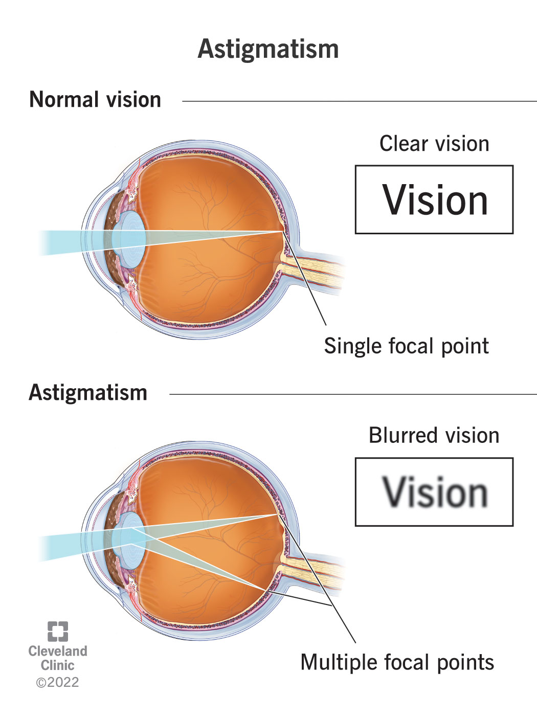
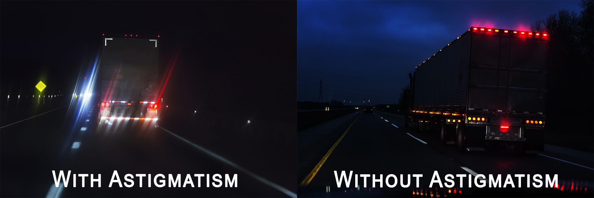
Astigmatism is a common vision problem caused by an error in the shape of the cornea. With astigmatism, the lens of the eye or the cornea, which is the front surface of the eye, has an irregular curve. This can change the way light passes, or refracts, to your retina. This causes blurry, fuzzy, or distorted vision.[6]
Children with astigmatism might experience:[5]
Difficulty focusing on the printed words or/and lines
Eyestrain, headaches and tired eyes
Discomfort or irritation in eyes
Distorted or blurred vision
Squinting eyes so as to see objects
Inability to clearly see both near objects, and far objects without squinting
Sensitivity to light
Striped Circle Visual Acuity Chart; A Novel Visual Acuity Chart
Can astigmatism be corrected in children?
Yes. Astigmatism can usually be corrected with properly prescribed eyeglasses or contact lenses, although these may not be necessary before the child starts grade school. Some children who have only a slight degree of astigmatism and no nearsightedness or farsightedness may not need corrective lenses at all.[11]
Astigmatism can occur in children and adults. Your risk of developing astigmatism may be higher if you have any of the following:
a family history of astigmatism or other eye disorders, such as keratoconus (degeneration of the cornea)
scarring or thinning of your cornea
excessive nearsightedness, which creates blurry vision at a distance
excessive farsightedness, which creates blurry close-up vision
a history of certain types of eye surgery, such as cataract surgery (surgical removal of a clouded lens)[6]
Probably the most important thing to note about astigmatism is that it can worsen due to eye rubbing. Admittedly, some unknowingly wake up in the morning, rub their eyes and think nothing of it, however this seemingly benign habit can prove quite harmful over time.
By rubbing your eyes, you are damaging your corneas, increasing eye pressures, and altering the shape of the eye resulting in unwanted astigmatism. Eye rubbing can also lead to Keratoconus.[8]
Rubbing your eyes may seem like a relatively harmless thing to do. Most of us do it regularly, whether we are suffering from hay fever or a common cold, or are just feeling tired and groggy. Rubbing stimulates tears to flow, lubricating dry eyes and removing dust and other irritants.
The best ways to prevent yourself from touching your eye area is to use eye drops to keep your eyes hydrated and prevent itching. Artificial tears are a non-medicated yet highly sophisticated imitation of natural tears.[9]
Your eye has two structures with curved surfaces that bend (refract) light onto the retina, which makes the images:
The cornea, the clear front surface of your eye along with the tear filmastigmatism can affect your vision at night
The lens, a clear structure inside your eye that changes shape to help focus on near objects
In a perfectly shaped eye, each of these elements has a round curvature, like the surface of a smooth ball. A cornea and lens with such curvature bend (refract) all incoming light equally to make a sharply focused image directly on the retina, at the back of your eye.Astigmatism may be present from birth, or it may develop after an eye injury, disease or surgery.[10]
Astigmatism isn't caused or made worse by reading in poor light, sitting too close to the television or squinting.
آستیگماتیسم با مطالعه در نور كم یا تماشای تلویزیون از فاصله نزدیك بهتر یا بدتر نمیشود.
هنگامی كه قرنیه دارای اعوجاج باشد، شما مبتلا به "آستیگماتیسم قرنیهای" هستید. هنگامی كه عدسی دارای اعوجاج باشد" آستیگماتیسم عدسی" دارید.
هر دو نوع آستیگماتیسم، تاری دید ایجاد میكند، اما اغلب موارد آستیگماتیسم ناشی از نایكنواختی انحنای قرنیه است.
فرد مبتلا به آستیگماتیسم هم در فاصله نزدیك و هم در فاصله دور تاری دید دارد.
آستیگماتیسم معمولاً از هنگام تولد وجود دارد و ممكن است با دوربینی یا نزدیك بینی تركیب شود. معمولاً این عارضه ثابت میماند و در طول زمان بهتر یا بدتر نمی شود. بسیاری از افرادی كه دارای مقدار اندكی آستیگماتیسم هستند که مقدار آن قدر زیاد نیست كه نیاز به عمل تصحیحی داشته باشد.
Astigmatism is most easily seen above and below the plane of best focus. Column “A” shows a bead in focus as well as above and below that plane of best focus. Notice the symmetry of the bead above and below the plane of best focus. The images in columns “B” and “C” are from objective lenses with moderate astigmatism. Notice the apparent vertical elongation above and horizontal elongation below the plane of best focus. While the elongation due to astigmatism is not necessarily horizontal or vertical, the elongations are typically orthogonal above and below the plane of best focus. These intensities in these images were squared to enhance the images for visualization. [1]
علائم و نشانه های آستیگماتیسمشامل موارد زیر است:
common astigmatism questions
اعوجاج در بخش هایی از میدان بینایی.
تاری خطوط عمودی، افقی یا مایل.
در چشم شما دو بخش وجود دارد كه مسئول متمركز كردن تصاویر هستند:قرنیه و عدسی.
▪ در چشم طبیعی این عناصر كانونی كننده انحنایی یكدست مانند سطح یك توپ لاستیكی دارند. ▪ قرنیه و عدسی با داشتن چنین سطح منحنی همه شعاع های نور وارد شده به چشم را به یك میزان خم میكنند (میشكنند) و یك تصویر متمركز واضح بر روی پرده حساس پشت چشم یعنی شبكیه ایجاد میكنند. اما اگر انحنای قرنیه یا عدسی یكدست نباشد، شعاع های نور به طور یكسان نمیشكنند، در این حالت شما دچار خطای انكسار نور هستید.
آستیگماتیسم یكی از اشكال مختلف خطاهای انكسار نور در چشم است. در آستیگماتیسم، قرنیه یا عدسی در یك جهت انحنای بیشتری از جهت دیگر دارد.
آستیگماتیسم تصحیح نشده باعث تاری دید می شود. در این حالت تاری دید در یك جهت _ افقی، عمودی یا مایل _ بیش از جهت دیگر وجود دارد.
آستیگماتیسم ممكن است در تركیب با سایر خطاهای انكساری مثل نزدیك بینی یا دوربینی رخ دهد:
What is Astigmatism In Children
▪ در نزدیك بینی (میوپی) انحنای قرنیه بیشتر از حد عادی است یا كره چشم درازای بیش از حد طبیعی دارد. در نتیجه شعاع های نور به جای آنكه دقیقاً روی شبكیه متمركز شوند، در جلوی شبكیه به هم می رسند و اشیای دور تصویری مبهم خواهند داشت. ▪ در دوربینی (هیپروپی) انحنای قرنیه كمتر از حد عادی است یا كره چشم طول كمتر از حد طبیعی دارد، در نتیجه حالت عكس نزدیك بینی رخ میدهد. نور در پشت چشم متمركز میشود و تصویر اشیای نزدیك تار میشود اما دید دور عادی باقی میماند. ▪ در اغلب موارد آستیگماتیسم از هنگام تولد وجود دارد. ممكن است آستیگماتیسم در نتیجه وارد شدن آسیب به چشم بیمار یا جراحی رخ دهد.
چه هنگامی باید به چشم پزشك مراجعه كرد
اگر درجه آستیگماتیسم چشم شما آن قدر باشد كه در كاری كه می خواهید انجام دهید اختلال ایجاد كند یا اگركیفیت بینایی تان مانع رضایت شما از نحوه فعالیت هایتان است، به چشم پزشك مراجعه كنید.
چشم پزشك درجه آستیگماتیسم شما را تعیین می كند و در مورد اینكه چه روشی را برای تصحیح بینایی تان انتخاب كنید به شما مشاوره خواهد داد. تغییر درجه آستیگماتیسم چشم در طول زندگی اگر اصولاً رخ دهد، بسیار تدریجی و كند است. انجام معاینات منظم چشم، راه مناسبی برای شناسایی تغییرات حدت بینایی است تا در صورت لزوم عینك یا لنز تماسی برای شما تجویز شود یا شماره آنها تصحیح شود.یك فرد بزرگسال سالم باید تا ۵۰ سالگی هر سه تا پنج سال یك بار معاینه چشم انجام دهد. پس از ۵۰ سالگی فواصل معاینات كمتر کنید. اگر دچار مشكلات انكساری مانند آستیگماتیسم هستید، هر دو سال یك بار به هر تعدادی كه چشم پزشكتان توصیه می كند، به او مراجعه كنید.
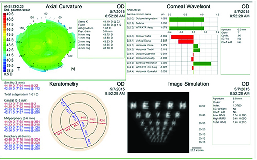
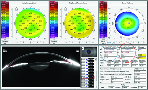
● تشخیص بیماری چشم پزشك شما ممكن است از ابزارهایی كه در زیر می آید برای معاینه چشم شما استفاده كند: ▪ قرنیهسنج (Keratometer) : چشم پزشك در قرنیه سنجی با استفاده از دستگاهی به نام قرنیه سنج یا كراتومتر میزان و جهت گیری آستیگماتیسم قرنیهای را با اندازه گیری میزان نور منعكس شده از سطح قرنیه مشخص میكند. ▪ كراتوسكوپ و ویدئوكراتوسكوپ: این ابزارها برای تشخیص و تعیین مقدار انحنای سطح قرنیه در صورت وجود آستیگماتیسم مورد استفاده قرار می گیرند. كراتوسكوپ حلقههای نورانی را روی قرنیه می افكند. سپس انعكاس این حلقه های نورانی روی قرنیه از طریق كراتوسكوپ مورد مشاهده قرار میگیرد و برحسب شكل و فواصل این حلقهها می توان میزان آستیگماتیسم قرنیه را محاسبه كرد. با اتصال كراتوسكوپ به یك دوربین ویدئویی، ویدئوكراتوسكوپ ساخته شده است، كه با آن می توان تصویر قرنیه را روی یك صفحه تلویزیونی دید. ویدئوكراتوسكوپ رایج ترین وسیله مورد استفاده برای تعیین مقدار انحنای سطح قرنیه در آزمونی است كه مكاننگاری (توپوگرافی) قرنیه نامیده می شود. [2]
وبلاگ تخصصی عینک شامل مجموعه مطالب پزشکی است که اطلاعات مفیدی در رابطه با عینک , چشم، لنز، سلامتی چشم و راه های پیشگیری از بیماریهای چشمی، کنترل و درمان آن را در اختیار شما کاربر محترم می گزارد.


 وبلاگ تخصصی عینک شامل مجموعه مطالب پزشکی است که اطلاعات مفیدی در رابطه با عینک , چشم، لنز، سلامتی چشم و راه های پیشگیری از بیماریهای چشمی، کنترل و درمان آن را در اختیار شما کاربر محترم می گزارد.