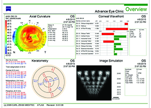
Residual astigmatism can be caused by incorrect placement of the IOL, incorrect marking of the cornea, inaccurate preoperative measurements, unanticipated surgically induced astigmatism, posterior corneal curvature, or rotation of the IOL itself.
incorrect placement of the IOL:
Most times, a primary malposition occurs because of surgeon error. In these instances, part of the lens may be sitting in the capsular bag and part in the sulcus. Alternately, the lens haptic may break during insertion, and rather than centering properly within the bag, the IOL sits lopsided.
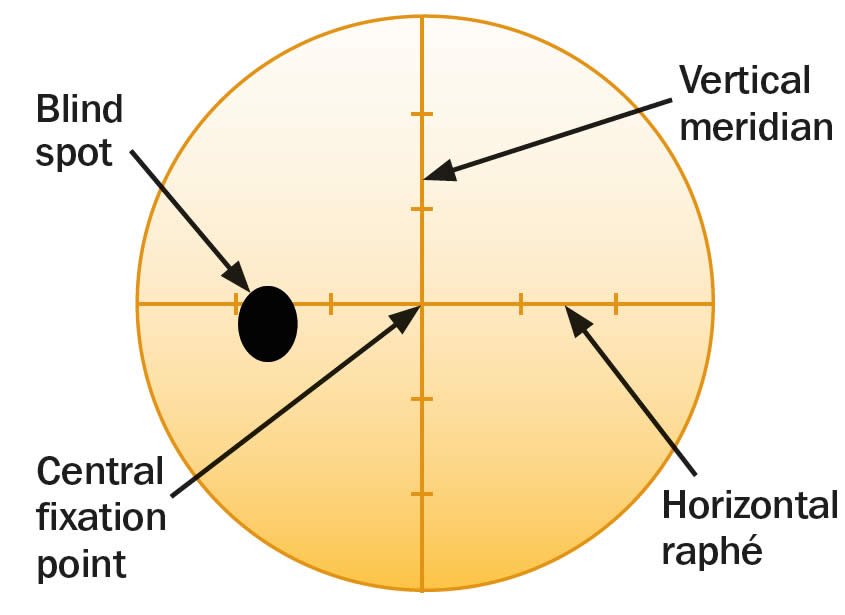
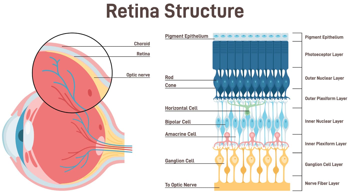
The eye’s retina receives and reacts to incoming light and sends signals to the brain, allowing you to see. One part of the retina, however, doesn't give you visual information—this is your eye’s “blind spot.”
At the back of your eye is the retina. Your retina is made up of light-sensitive cells which send messages to your brain about what you see. Everyone has a spot in their retina where the optic nerve connects. In this area there are no light-sensitive cells so this part of your retina can’t see. We call this the blind spot.
Most of the time you don’t notice your blind spot because the spot in one eye doesn’t match the spot in the other eye. Each eye supplies information to the brain, filling in what’s missing. Also, sometimes the brain will fill in the missing information with what it thinks should be there. That causes one kind of optical illusion. eResearch by Navid Ajamin -- Autumn 2025
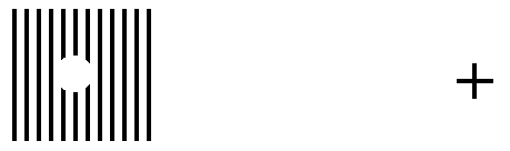
Instructions:
R ... L
Place your eye a distance from the screen approximately equal to three times the distance between the R and the L. Move your eye towards or away from the screen until you notice the other letter disappear. For example, close your right eye, look at the "L" with your left eye, and the "R" will disappear.
Blind Spot meaning and anatomy
(Upper row) A cross section of the eye showing the blind spot and retinal veins, as well as the fact that light goes through all the retinal layers before hitting the photoreceptors. (Lower row) Top-down view of the retina, showing how big the blind spot and retinal veins are relative to the fovea, which is the high-resolution region of the retina. (Images adapted with permission from Webvision-University of Utah).
Night blind spot
It is estimated that once fully adapted to darkness, the rods are 10,000 times more sensitive to light than the cones, making them the primary receptors for night vision. Since the cones are concentrated near the fovea, the rods are also responsible for much of the peripheral vision. The concentration of cones in the fovea can make a night blind spot in the center of the field of vision.
Description: Students will make a simple prop and use it to find their blind spot
Purpose: To locate and identify the blind spot
Length of Activity: 20 minutes
Materials:
One 3 x 5 inch card (or other stiff paper) per student.
Black markers.
1 ruler per student.
Steps:
1. Students should be instructed to make a dot and an X on the white side of the index card as pictured.
2. They should then hold the card so the X is on the right side and raise it to eye level about an arm's length away.
Night blind spot
3. Have students close their right eye.
4. Student should look directly at the X with their left eye only. They should note that they can also see the dot, but should not focus on it.
5. While looking at the X, and keeping an awareness of the dot, have students bring the cards slowly towards their faces. At some point they should be aware that the dot has disappeared and then reappeared.
6. Now have students repeat but this time close their left eyes. They should use their right eyes to look at the dot while keeping aware of (but not looking directly at) the X. This time the X will disappear and then reappear as the card is slowly brought towards their faces.
7. Now have students take their markers and ruler to draw a straight line through the center of both the dot and the X.
8. Repeat the activity. Note that this time the line seems to be continuous, with no gap, even as the X or dot disappears.
What’s Going On?
At the back of your eye is the retina. Your retina is made up of light-sensitive cells which send messages to your brain about what you see. Everyone has a spot in their retina where the optic nerve connects. In this area there are no light-sensitive cells so this part of your retina can’t see. We call this the blind spot. The point at which the mark on the card disappears is where your blind spot is.
When you draw a line through the dot and X you set up an optical illusion. The brain knows that a line is there and fills in the gap, even as it loses sight of the dot or X.
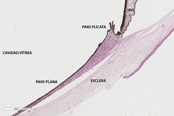
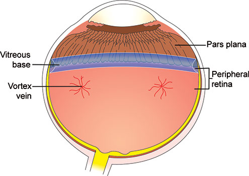
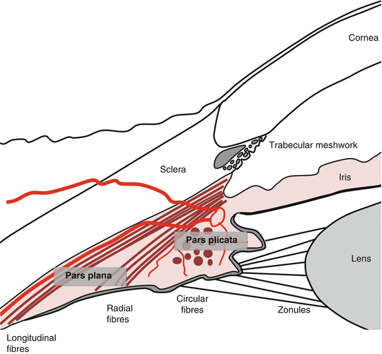
Pars plana refers to a region of the eye that is characterized by clear, smooth, cystoid cavities known as pars plana cysts, which exist between the pigmented and nonpigmented epithelial layers.
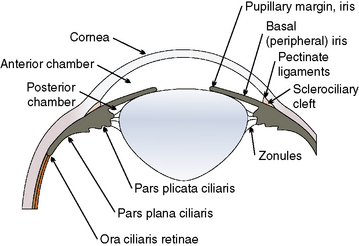
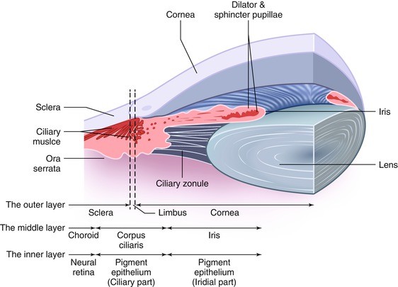
The pars plana (also known as orbicularis ciliaris) (Latin: flat portion) is part of the ciliary body in the uvea (or vascular tunic, the middle layer of the three layers that comprise the eye). It is about 4 mm long, located near the junction of the iris and sclera, and is scalloped in appearance.
The pars plana constitutes the two-thirds of the ciliary body as the posterior portion. It is a 4 mm wide, smooth surface structure. The pars plana is positioned between the retina and pars plicata and is avascular. Avascular pertains to having little or no blood vessels.
The pars plicata is 2 mm wide and consists of 70 ciliary processes, each approximately 0.5–0.8 mm high and 0.5 mm wide.
The pars plicata (also known as corona ciliaris) (Latin: folded portion) is the folded and most anterior portion of the ciliary body of an eye. The ciliary body is a part of the uvea, one of the three layers that comprise the eye. eResearch by Navid Ajamin -- autumn 2025
What is the difference between pars plana and plicata?
The pars plicata gives rise to the ciliary processes to which the zonules of the lens attach and it surrounds the periphery of the iris. The pars plana has a scalloped posterior border that fits into the scalloped edge of the retina at the ora serrata.
What is the difference between vitrectomy and pars plana?
A vitrectomy performed for diseases of the posterior segment is called a posterior or pars plana vitrectomy. This kind of vitrectomy is performed by a retina specialist. Anterior Vitrectomy: In rare cases, the vitreous gel comes through the pupil into the anterior (front) chamber of the eye.
What are the symptoms of Pars Planitis?
Patients with Pars Planitis usually do not have frank eye pain. What they do notice though is floaters or “stuff” in their vision. In some cases patients with Pars Planitis may go on to develop cataracts and resultant blurry vision.
At pars plana vitrectomy (PPV), changes in ciliary body dimensions with age may affect how sclerotomies are placed so as to avoid iatrogenic damage to the crystalline lens and peripheral retina.
Pars plana vitrectomy is defined as a surgical procedure used to address complicated proliferative diabetic retinopathy and other retinal conditions such as non-clearing vitreous hemorrhage and retinal detachment.
Patients with pars planitis present with minimal symptoms, for example, floaters or blurry vision. In most cases, there is the absence of photophobia and pain. Occasionally patients may present with sudden loss of vision due to retinal detachment or acute vitreous hemorrhage.
Pars plicata refers to the area of the ciliary body that is located between the pars plana and the iris, measuring 2 mm wide. It gives rise to the ciliary processes, which are responsible for attaching the zonules of the lens, and surrounds the periphery of the iris. The non-pigmented epithelium of pars plicata plays a crucial role in the production of aqueous humor, and damage to this area can lead to hypotony.
Researchers have shown that the green color has the ability to soothe our nervous system. Indeed, since it is the easiest color to perceive for our eyes, our whole body can then relax when this color surrounds us. That's why green is so prevalent in hospitals, schools or offices.
THE COLOUR GREEN
Soothing, and restful on the eye.
Relaxing mentally as well as physically.
Helps alleviate depression, nervousness and anxiety.
Offers a deep sense of renewal, self-control and harmony.
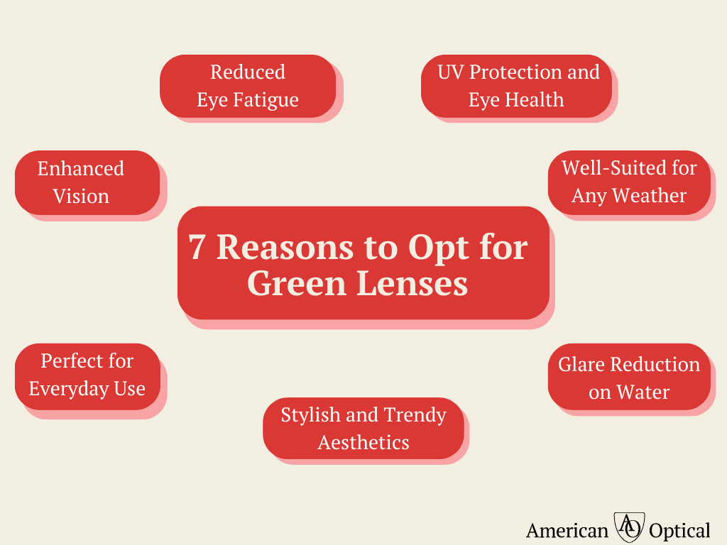
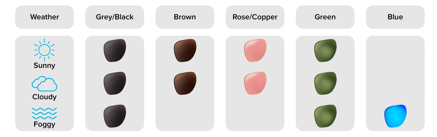
What are green lenses good for?
Sunglasses with green lenses provide better contrast than gray lenses.
transmit color accuracy better than brown lenses, and are ideal for both sunny and low-light environments.
Perfect for water or field sports such as cycling or skiing, green lenses protect and comfort your eyes on foggy, cloudy, or bright days.
What are green lenses used for?
Another reason why green tinted glasses can be a good choice is that they help reduce the temperature as well as the glare that is eliminated by excessive lights. This reduction in the glare can create a soothing ambience and help you focus and work for longer hours without any strain or headaches.
What are the benefits of green cut lenses?
Rayban Aviator Sunglasses
Green lens sunglasses can reduce the high and low-energy light that reaches the eyes while emphasizing the middle of the visible light spectrum. They result in a more comfortable experience, enhance contrast, and make colors look more natural compared to other tinted lenses.
Are Green Lens Sunglasses Right For You?
Green lens sunglasses can reduce the high and low-energy light that reaches the eyes while emphasizing the middle of the visible light spectrum. They result in a more comfortable experience, enhance contrast, and make colors look more natural compared to other tinted lenses.
Green sunglasses are suitable for a wide range of activities, including driving, hiking, fishing, golf, and more. They’re also ideal for protecting sensitive eyes from glare and eye fatigue.
There are a few factors to consider when shopping around for green sunglasses, including anti-glare and UV protection coatings, and a comfortable fit.
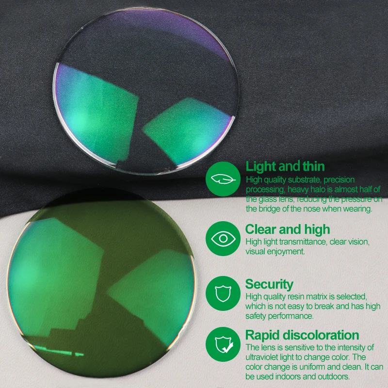
Green Coating
To set them apart from uncoated lenses, the original coated lenses were produced in green colour. The reflection gives a 2 percent enhancement at 515 nm, which makes the contrast visible, and the reflection appears as a green film.
The common reflection reduction film, which is currently excellent and old, makes up this layer of film. In terms of UV protection and alleviation of eye fatigue, the green film layer performs somewhat better, and the light green colour, which is currently the common colour of the lens coating layer, is harder to find on the lens.
Advantages
When viewed from specific angles, green coatings exhibit a greenish-teal hue.
In the green and yellow regions of the visible light spectrum, they are effective at reducing reflections. In bright outdoor environments, green coatings can improve visual clarity and contrast.
For sunglasses and outdoor activities, they are a popular choice.
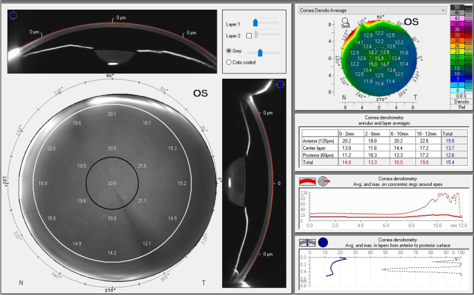
Corneal densitometry (CD) uses the biological properties of the corneato visualize the morphology of the cornea and determine the degree of corneal transparency. At present, it is an emerging metric that has shown promise in various clinical diagnosis and evaluation of eye diseases and surgeries. We introduce the different methodologies used to measure CD.
Furthermore, we systematically categorize the diagnostic value of CD into high, medium, and low levels based on its clinical significance.
By analyzing a wide range of conditions, including keratoconus, postrefractive surgery changes, and other corneal pathologies, we assess the utility of CD in each context. We also discuss the potential implications of these classifications for disease monitoring and prognosis evaluation. Our review underscores the importance of integrating CD assessments into routine clinical practice to enhance the accuracy and effectiveness of diagnostic processes for corneal disorders.
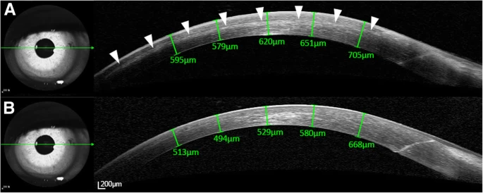
Spectral-domain optical coherence tomography scanning of the cornea before (a) and after (b) performing suture lysis using an argon laser. Note that the diffuse and thin fluid pocket in the corneal interface region (arrowheads) resolved when the intraocular pressure was lowered from 14 to 9 mmHg
We suggest a theory to frozen light, which was first registered in 2000 by Lene Hau.
Lene Vestergaard Hau is a Danish physicist and educator. She is the Mallinckrodt Professor of Physics and of Applied Physics at Harvard University.She was also awarded tenure in 1999 and is now Mallinckrodt Professor of Physics and Applied Physics at Harvard. In 2001 she became the first person to stop light completely, using a Bose–Einstein condensate to achieve this. For her doctoral studies in quantum theory, Hau worked on ideas similar to those involved in fibre optic cables carrying light, but her work involved strings of atoms in a silicon crystal carrying electrons. While working towards her doctorate, Hau spent seven months at CERN, the European Laboratory for Particle Physics near Geneva. She received her doctorate from the University of Aarhus in 1991 at the age of 32, but by this time her research interests had changed direction.
Frozen light is explained here as a new state of matter. The explanation is given through space-time terms of the General Theory of Relativity. We consider a fully degenerate region of space (space-time), which is the ultimate case of the isotropic region (home of photons), where the metric is particularly degenerate. Both the space-time interval, the observable time interval, and the observable three-dimensional interval are zero in a fully degenerate region.-- Harvard University
فوتون(به انگلیسی: Photon) که معمولاً با نماد γ نمایش داده میشود، یک ذره بنیادی است. فوتون یک کوانتوم یا بهعبارتی کمترین مقدار قابل اندازهگیری در یک میدان الکترومغناطیسی مانند تابش الکترومغناطیسی (نور و امواج رادیویی) محسوب میشود و همچنین در نقش حامل نیرو برای نیروی الکترومغناطیس نیز عمل میکند. فوتون جرم ندارند. (جرم ذاتی یا سکون ندارند) اگرچه سرعت فوتون به محیط بستگی دارد اما در محیط خلأ، همواره با سرعتی برابر با سرعت نور، معادل ۲۹۹٬۷۹۲٬۴۵۸ متر بر ثانیه حرکت میکنند.
فوتون تفاوتهایی اساسی نسبت به ذراتی همچون «کوارک» (quarkquark) یا الکترون دارد. جرم ساکن این ذره برابر با صفر بوده، از این رو سرعت این ذره در خلاء دقیقا برابر با سرعت نور است. شاید مهمترین تاثیری که فوتون در زندگی یک فرد عادی دارد، تاثیر آن در دیدن محیط اطراف است. در حقیقت بدون وجود فوتون قادر نخواهیم بود محیط اطرافمان را مشاهده کنیم.
فوتون هم خواص موج و هم خواص ذره را دارد. برای نمونه یک فوتون میتواند منعکس شده یا تداخل ویرانگر ایجاد کند. تداخل ویرانگر به حالتی گفته میشود که دو موج برخوردی یکدیگر را خنثی میکنند. بنابراین این ویژگیها نشانگر موجی بودن فوتون است. از طرفی به عنوان یک ذره، فوتون تنها میتواند با انتقال مقدار مشخصی از انرژی، با دیگر مواد کنش داشته باشد.
What is the different nature of light?
Answer: Light has a dual nature, implying that it is made up of both waves and particles. Although Einstein believed that light is a particle (photon), quantum physics has revealed that light may operate as both a particle and a wave at the same time.
Light behaves in many different ways when it comes in contact with something.
What are the 7 natural sources of light?
Natural sources of light include the sun, stars, fire, and electricity in storms. There are even some animals and plants that can create their own light, such as fireflies, jellyfish, and mushrooms. This is called bioluminescence.
Light is a transverse, electromagnetic wave that can be seen by the typical human. The wave nature of light was first illustrated through experiments on diffraction and interference. Like all electromagnetic waves, light can travel through a vacuum. The transverse nature of light can be demonstrated through polarization. eResearch by Navid Ajamin -- autumn 2025
ماهیتهای متفاوت نور
ماهیت ذرهای: ایزاک نیوتن در کتاب خود در رسالهای دربارهٔ نور نوشت: پرتوهای نور ذرات کوچکی هستند که
? How Does A Photon Experience The Universe
از یک جسم نورانی نشر میشوند. احتمالاً نیوتن نور را به این دلیل به صورت ذره در نظر گرفت که در محیطهای همگن به نظر میرسد در امتداد خط مستقیم منتشر میشوند، این امر را قانون مینامند و یکی از مانندهای خوب برای توضیح آن، به وجود آمدن سایه است. برخی دیگر از دانشمندان نیز اظهار داشتهاند که نور از ذرات در ارتعاش شدید تشکیل یافته است. نیوتن معتقد بود نور از درون واسطهای به نام اتر گذر میکند که غیر مادّی است و دیده نمیشود. بر اساس نظریه اتر، فضا آکنده از این واسطه است. هماکنون این نظریه باطل شده است و معتبر نیست.
ماهیت موجی:همزمان با نیوتن، کریستیان هویگنس(۱۶۹۵–۱۶۲۹ میلادی) طرفدار توضیح دیگری بود که در آن حرکت نور به صورت موجی است و از چشمههای نوری به تمام جهات پخش میشود. هویگنس با به کار بردن امواج اصلی و موجکهای ثانوی، قوانین بازتاب و شکست را تشریح کرد. حقایق دیگری که با تصور موجی بودن نور توجیه میشوند پدیدههای تداخلیاند، مانند به وجود آمدن فریزهای روشن و تاریک در اثر بازتاب نور از لایههای نازک یا پراش نور در اطراف مانع، مانند آزمایش دوشکاف.
ماهیت الکترومغناطیس: بیشتر به خاطر نبوغ جیمز کلارک ماکسول (۱۸۷۹–۱۸۳۱) است که ما امروزه میدانیم نور نوعی انرژی الکترومغناطیسی است که معمولاً به عنوان امواج الکترومغناطیسی توصیف میشود. گستره کامل امواج الکترومغناطیسی شامل: موج رادیویی، تابش فروسرخ، نور مرئی از قرمز تا بنفش، تابش فرابنفش، پرتو ایکس و پرتو گاما میباشد.
ماهیت کوانتومی نور: طبق نظریه مکانیک کوانتومیِ نور، که در دو دهه اول سده بیستم به وسیلهماکس پلانک، آلبرت انیشتین و نیلز بور برای اولین بار پیشنهاد شد. انرژی الکترو مغناطیسی کوانتیده است، یعنی جذب یا نشر انرژی میدان الکترو مغناطیسی به مقدارهای گسستهای به نام فوتون انجام میگیرد. انرژی است.
نظریه مکملی
نظریه جدید نور شامل اصولی از تعاریف نیوتن و کریستیان هویگنس است. بنابرین گفته میشود که نور رفتار دوگانهای داردبرخی از پدیدهها مثل تداخل و پراش رفتار موجی آن را نشان میدهد و برخی دیگر مانند پدیده فتوالکتریک و پدیده کامپتون با رفتار ذرهای نور قابل توضیح هستند.
Therefore, we refer to such a region and particles which inhabit it as zero-space and zero-particles.
Moving to the coordinate quantities inside zero-space shows that real speed therein is that of light, depending on the gravitational potential and the rotation of space. It is shown that the eikonal equation for zero-particles is a standing wave equation: zero-particles are standing light waves, while zero-space is filled with a system of standing light waves (light-like hologram). With these, zero-particles appear to a regular (external) observer as mere stopped light. This paper has been submitted to The Abraham Zelmanov Journal. The Abraham Zelmanov Journal
This journal is named after Abraham Zelmanov (1913-1987), a prominent scientist working in the General Theory of Relativity and cosmology, whose main goal was the mathematical apparatus for calculation of the physical observable quantities in the General Theory of Relativity (it is also known as the theory of chronometric invariants).
Frozen Light, aurora
Which country recently freezes light?
Generally, light exists only as a particle or wave. But recently, a team of researchers from Italy's University of Pavia and CNR Nanotec reported successfully 'freezing' light by manipulating photons in a meticulously arranged ultra-cold environment.
How long was light frozen?
A newly designed trap freezes a beam of light for 1 second. Researchers have frozen a pulse of light in place for a full second, a thousand times longer than the previous record.
Did scientists freeze light fact check?
“Freezing light” means slowing it down or stopping it for a short time. Scientists do this by making light interact with super-cold atoms or special materials, causing it to pause and then continue moving. It's not actually frozen like ice. As for “light being the source of all matter,” that's not true.
Is it possible to solidify light?
It has been theorized that solid light could exist. Some experiments claim to have created solid photonic matter or molecules by inducing strong interaction between photons. Potential applications of solid light could include logic gates for quantum computers and room-temperature superconductor development.
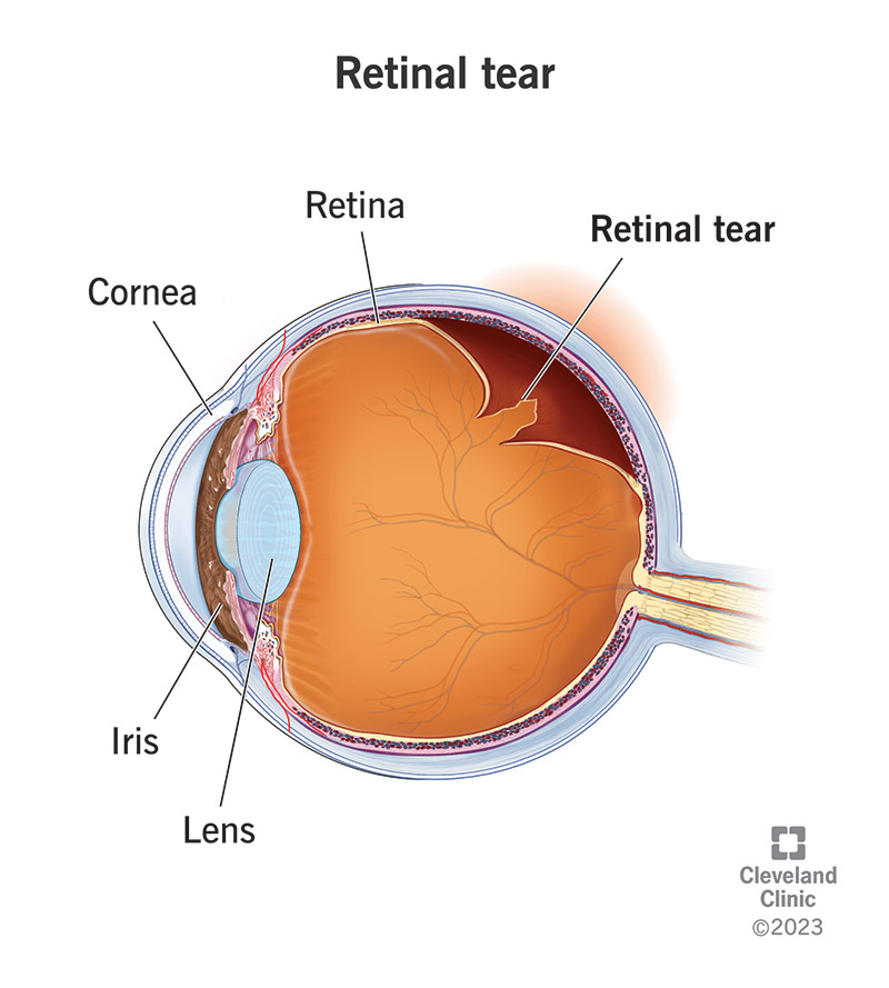
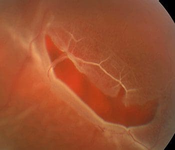
A retinal tear or break happens when the gel-like vitreous in your eye pulls on your retina and causes a split. Your retina is a thin layer of tissue that’s sensitive to light found at the back of your eye.
A retinal tear isn’t the same as a retinal detachment. A retinal tear could lead to a retinal detachment if the tear isn’t treated. A retinal detachment happens when the retina pulls away from the tissues that support it.
You can also develop a hole in your retina when your retina gets thinner. Retinal holes are less likely to lead to retinal detachment.
Retinal tears and any injury that damages your retina threatens your eyesight and is a medical emergency. Contact your eye care provider as soon as you have retinal tear symptoms or any type of eye injury.
How common is a retinal tear?
Retinal tears are common, with nearly one in ten people developing one at some point during their lifetime. Retinal detachments, on the other hand, are less common, occurring in approximately one in 300 people.
Can stress cause a retina tear?
Even though stress can't cause retinal detachment, it can be harmful to your eye health in other ways. In addition to cortisol during times of stress, the body also produces epinephrine or adrenaline. This causes the pupils to dilate so you are able to see the world more clearly and be protected from danger.
A retinal tear is less severe than a retinal detachment, but you still may need treatment. You probably won’t feel pain, but you may have blurry vision and a lot of eye floaters and light flashes. Your provider can repair a tear before it leads to a detached retina.
What are the symptoms of a retinal tear?
Symptoms of a retinal tear may include:
Flashes of light (photopsia).
Suddenly seeing more black spots or floaters than usual.
Darkening vision.
Blurred vision.
You might have a retinal tear and have no symptoms.
A concussion can affect vision by damaging the eyes themselves, muscles that surround the eyes or parts of the brain involved in vision. Even small vision changes can affect your daily life by making it difficult to read, drive, exercise, and carry out work or school activities.
As an athlete, experiencing a concussion can be a scary and unsettling experience. The road to recovery can sometimes feel daunting, especially if you’re worried about returning to your pre-injury level. One aspect that many athletes may be concerned about is the effect of a concussion on their vision, particularly their vision stamina.
The good news is that with proper treatment and monitoring, vision stamina can be restored as part of your concussion recovery plan. So, if you’re grappling with a concussion and worry about your vision, know that there are steps you can take to regain your visual strength. With patience, support, and the right professional guidance, like the expert care you’ll receive at Parker Performance Institute, you’ll be back in the game in no time.
The Impact of Concussions on Vision Stamina
When determining the best concussion recovery plan, each athlete’s vision stamina, or their ability to sustain visual focus and clarity over time, should be thoroughly examined. Understanding post-concussion visual stamina is crucial for a successful recovery.
Signs Your Vision Stamina May Be Affected by a Concussion
After you’ve received a concussion, you may find that your eyes tire more quickly, and this is negatively affecting your overall quality of life. Other signs that your vision stamina has been compromised include:
Frequent headaches
Blurred or double vision
Difficulty focusing on objects (close up or far away)
Eye strain and fatigue
Sensitivity to light
If you notice any of these signs, it’s essential to address them proactively.
Phantom Eye Syndrome is NOT a multi-million dollar summer blockbuster movie. It is a real medical condition that many patients report after one eye has been surgically removed.
What is a phantom vision?
Phantom vision was manifest by the transient belief that visual sensations were present in the absent eye. This phenomenon was never spontaneously divulged; in all instances the visual sensations had to be specifically elicited.
Can watching TV cause glaucoma?
Impact on Eye Pressure
While screen time alone isn't likely to cause glaucoma, it can contribute to increased IOP, which can worsen the condition in glaucoma patients. It's a good idea to be mindful of your screen habits and ensure you're giving your eyes enough rest to avoid unnecessary pressure.
What is phantom glasses syndrome?
This eerie phenomenon is called Phantom Glasses Syndrome, and it's a spooky trick our brains play—especially when we go without glasses after years of wearing them. 👓🎃 Phantom Glasses Syndrome can make you feel like your frames are still resting on your nose or even that there's a “ghostly” lens smudge to clear away!
What causes phantom glasses syndrome?
The longer you've worn glasses, the more likely your brain has gotten used to them as part of you, creating this spooky sensation even when they're not there!
Much like Phantom Limb Syndrome, in which a person might still feel like they have their arm or leg following an amputation, a person whose eye has been removed might still get sensations of pain or visual perceptions. A recent study at the University of Liverpool involved 239 patients who had undergone eye removal due to cancer. Sixty percent of respondents reporting symptoms such as pain, visual sensations like colors or shapes, or the impression of actually seeing with the missing eye. Some even reported seeing objects or people that were not actually present. People who suffer severe vision loss but without eye removal can experience similar visual hallucinations. This is called Charles Bonnet Syndrome.
How do you treat phantom eye syndrome?
Treatment. Treatment on painful phantom eye syndrome is limited and does not point out a standard treatment protocol but possible treatment pathways include resting techniques, pharmacologic, non-pharmacologic, surgery, drug therapy, and psychological.
Can glaucoma cause you to hallucinate?
About one in five people with retinal conditions such as macular degeneration experiences hallucinations, which can also occur in people with other macular diseases and ocular conditions such as glaucoma, as well as stroke. CBS is more common in people aged 80 years and above, but can occur at any age.
Glaucoma” is Greek in origin and describes a “blue-grey” or a “blue-green” colour. How does a word describing colours come to be used in naming of an eye condition?
Well, one of the prominent features of glaucoma is a build up of pressure in the eye. As the pressure increases, the clear window at the front of the eye, the cornea, starts to have trouble in keeping itself transparent.
The inner surface of the cornea then “fractures” and fluid starts to accumulate in its substance. So that when light reflects off this damaged surface, the onlooker starts to see a bluish-green or bluish-grey colour instead of being able to look straight into the eye.
When looking at a vibrant field of flowers, if all is well with your eyes the colors may pop. But what if you begin to notice that in some parts of the visual field the colors are somewhat muted or things are starting to look gray? You know you haven't experienced any kind of eye injury but feel as if something must be amiss.
Dimness of vision can be a symptom connected to a variety of eye conditions.
Who is more likely to suffer from eye strain?
Those with more probabilities of suffering from eye strain are:
People working long hours, especially in front of screens.
People exposed to environments with poor lighting.
People who misuse glasses or don’t use them at all but they need them.
What is eye strain?
Also known as asthenopia or visual fatigue, it is a vision disorder that has increased considerably due to changes in lifestyle habits in our society. The main causes are anxiety and the prolonged use of electronic screens, although it is also related to a sedentary life and long working hours.
How can I avoid eye strain?
There are a few recommendations that can help prevent eye strain:
People who work with computers or screens are advised to take short regular breaks to rest their eyes.
When looking at a screen, it is important to maintain an adequate distance so as not to strain the eyes. It is recommended that the computer screen be placed 10 cm below the visual axis.
The use of indirect light, when using screens, will prevent us from straining our eyes.
Visual exercises can be performed to relax the eyesight, such as looking into the distance to relax accommodation, and blinking exercises to properly hydrate the eyes.
It is advisable to moisturize the eyes and maintain good eye and eyelid hygiene.
After using screens, it is important to perform outdoor activities to exercise distant vision.
Good health habits, such as getting a good night’s rest, eating a balanced diet and, above all, regular physical exercise, will help to avoid visual stress.
Your depth perception is an important part of your vision. It helps you see objects in three dimensions and understand how far away they are from you. Because your depth perception depends on information from both your eyes and your brain, anything affecting your overall vision can impact your depth perception.
Have you ever been on the sports field and not been able to fully judge how far away your teammate is? Or have you ever felt nervous while driving your car home because it seems like that car is really far away, but it’s actually right in front of you? If either of these situations seem familiar to you then it may not be as simple as broken glasses, you might be experiencing common problems with what is called depth perception.
Depth perception is your ability to see objects in three dimensions, including their size and how far away they are from you. It’s made possible by lots of parts in your eyes and your brain working together to process information, estimate their location and create the images you see.
If something’s affecting your ability to see out of one or both eyes, there’s a good chance your depth perception will be affected, too.
What is the purpose of depth perception?
Depth perception is an important part of your overall vision.
It helps you understand an object’s size and distance from you. Depth perception is what lets you see in three dimensions. That’s where it gets its name — you’re perceiving an object’s depth.
For example, if you’re reading this article on your phone or a computer, depth perception is what you’re using to tell the difference between this flat text on your screen and the device you’re reading it on. Without your depth perception, the words you’re reading and the device you’re reading on would both look totally flat.
In addition to letting you see objects fully, your depth perception keeps you safe. Everything from walking around your home to knowing when to swing the bat to hit a baseball relies on your depth perception. Correctly judging how far away something is lets you drive safely or know where other people around you are on the sidewalk.
How does depth perception work?
Many parts of your eyes and brain work together to maintain your depth perception. It’s part of your visual pathway. Your visual pathway works in a few steps:
Light and information enter your eye.
Your retinas at the back of your eye interpret it and send that information to your optic nerves.
Your optic nerves meet in an X-shaped area at the front of your brain called the optic chiasm.
Your brain’s visual cortex (the area responsible for all your sight) takes the information from your optic nerves and combines it into the images you see.
Depth perception usually comes from having binocular vision (seeing with two eyes). It’s possible to see with only eye (monocular vision), but your depth perception might be less accurate than it usually is. However, some people who have good vision in one eye but not the other still have good depth perception because their brains adjust to overcome their limited ability to see.
Because your depth perception depends on information from your eyes and your brain, anything affecting your overall vision can impact your depth perception. The most common issues include:
Low vision.
Strabismus (crossed eyes).
Amblyopia (lazy eye).
Injuries to your eye or trauma can also affect your depth perception, especially if your optic nerves are damaged.
Pain behind the eye can stem from common issues like migraines, eye strain, or sinus infections. However, it can also signal more serious conditions such as optic neuritis, which is an inflamed optic nerve, or glaucoma, an eye condition related to increased internal eye pressure. If you experience consistent or severe pain, it's crucial to see a healthcare professional to get a proper diagnosis and appropriate treatment.
درد پشت چشم میتواند علل گوناگونی داشته باشد، از جمله میگرن، سردرد تنشی، سینوزیت، خشکی چشم، و مشکلات بینایی مانند نزدیکبینی یا آستیگماتیسم. اگر درد شدید، همراه با علائم دیگری مانند تب، تاری دید، یا مشکلات تنفسی باشد، یا اگر به طور ناگهانی رخ دهد، لازم است به پزشک مراجعه کنید.
Common Causes
Headaches:
Migraines (a type of severe headache), cluster headaches (excruciating pain, usually on one side), and tension headaches (a feeling of pressure) can all cause pain behind the eyes.
Eye Strain:
Spending long hours looking at screens, reading, or driving can tire your eye muscles, leading to pain behind the eyes.
Sinusitis:
Infections or inflammation in the sinus cavities, located behind the nose and eyes, can increase pressure and cause pain.
Dry Eye Syndrome:
Insufficient tear production can make the cornea dry and irritated, leading to a gritty or painful sensation.
More Serious Conditions
Optic Neuritis:
Inflammation of the optic nerve can cause pain behind the eye and is a significant reason to see an eye doctor.
Glaucoma: This condition involves increased pressure inside the eye (intraocular pressure), which can cause discomfort and pain.
Graves' Disease:
Also known as thyroid eye disease, this condition can cause pressure and pain behind the eyes.
When to Seek Medical Help
Persistent or severe pain:
If the pain is regular, intense, or doesn't improve, you should have it evaluated.
Accompanying symptoms:
Look out for other signs like changes in vision, redness, sensitivity to light, or a stiff neck, which may point to a more serious problem.
Early diagnosis: Getting your eyes checked by an optometrist or other healthcare provider is essential for peace of mind and to ensure proper treatment for any underlying condition.
Conjunctivitis: Also called pink eye, conjunctivitis
is inflammation of the thin membrane lining the eye and eyelid (conjunctiva). This condition is caused by an infection (viral or bacterial) or allergies. Pink eye symptoms include burning or soreness with discharge. Bacterial conjunctivitis leads to eye pain, while allergic conjunctivitis also causes itchy eyes and puffy eyelids.
Blepharitis
Blepharitis is inflammation of the eyelash follicles, typically arising from excess bacteria at the base of the lashes. Symptoms include swollen, itchy eyelids, eyelash problems, light sensitivity, dandruff-like flakes, and a sensation of something in the eye. Symptoms are often worse in the morning.
Tear duct infection: Also called dacryocystitis
this is an infection in the tear drainage system, usually caused by bacteria. Symptoms include pain, redness, and swelling near the inner corner of the eye, along with excess tears or pus draining from the eye. In severe cases, a fever may also occur.
Leber congenital amaurosis (LCA) is a rare, inherited eye disorder that causes severe vision loss from birth or early childhood. It's the most common cause of inherited childhood blindness. LCA affects the retina, specifically the photoreceptor cells (rods and cones) responsible for detecting light and color, leading to severe vision impairment.
Leber’s congenital amaurosis is a rare eye disease that affects babies’ retinas. Some babies have blindness at birth. Others have very poor vision. Changes in genes that develop and form retinas cause Leber’s congenital amaurosis. Treatment for children with very poor vision includes eyeglasses or magnifying glasses.
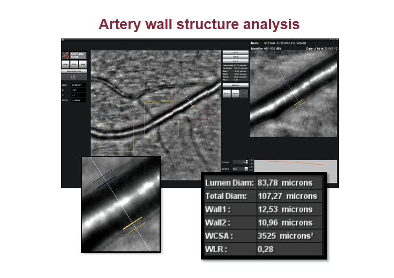
Pulsatile blood flow in the central retinal artery
Some babies with LCA are born with blindness. Other babies with the condition develop symptoms when they’re about 6 months old. You may not notice any changes in your baby’s vision right away. For example, babies often rub their eyes when they’re tired. But your baby frequently rubbing or poking at their eyes may be an early symptom of LCA. Other symptoms include:
Misaligned eyes (strabismus)
Sensitivity to light (photophobia)
Shaking eyes (nystagmus)
Your child’s pupils don’t adjust to changes in light (slow or missing pupillary response)
People with LCA typically experience very poor vision from birth or early infancy, often with vision worse than 20/400 (meaning they can only see at 20 feet what someone with normal vision can see at 400 feet). Some individuals may have only light perception or no vision at all.
One of the factors that affects your child’s eye development and visual function is the genes they inherit from each parent. Normal genes will work together to create a functioning eye. Unfortunately, abnormal genes can be passed on and can disrupt your child’s eye development. In Leber’s Congenital Amaurosis, mutated genes cause the retina to develop improperly and can lead to severe vision loss or blindness.
Leber’s Congenital Amaurosis is a disease in which the retina of affected infants develops incorrectly, leading to severe visual impairment.
LCA is one of multiple inherited retinal diseases and must be passed down by both parents for the child to have it.
The genes responsible for LCA can be detected in parents and their children with specialized testing and are part of how children are officially diagnosed with LCA.
Visual aids can help children adapt to their level of vision loss, and support groups help parents and children deal with the realities of LCA.
Several clinical trials using gene therapy have shown promising results for helping stop LCA progression.
LCA causes
Leber’s congenital amaurosis happens when your baby inherits certain genetic variations that affect how their retinas develop.
The genetic variations affect the process that creates images that your baby sees. Normally, light-detecting cells (photoreceptors) in your baby’s retinas turn light into electrical signals. Your baby’s brain turns the signals into images that they see. Leber’s congenital amaurosis affects that process so there’s less electrical activity (signals). The less electrical activity there is, the less sight your baby has.
Variations in almost 30 different genes can cause Leber’s congenital amaurosis. The variations affect the following genes:
CEP290
CRB1
GUCY2D
RPE65
LCA is usually an autosomal recessive condition. That means both biological parents have one or more of the changed genes that can cause Leber’s congenital amaurosis.
Diagnosis:
Early detection: Parents may notice a lack of visual response in the first few months of life.
Electroretinogram (ERG): This test measures the electrical activity of the retina and can help confirm LCA.
Genetic testing: Can help identify specific gene mutations causing LCA.
Yes, heat can affect eye vision. Exposure to excessive heat can lead to various vision-related issues, including blurred vision, dry eyes, and eye strain. Heat can also exacerbate existing eye conditions like dry eye and conjunctivitis.
Here's a more detailed explanation:
Blurred Vision:
Heat exhaustion and heatstroke can cause blurred vision, double vision, or difficulty focusing due to dehydration and the body's response to overheating, according to Specsavers.
Dry Eyes:
High temperatures can cause the tear film on the eye to evaporate faster, leading to dry eyes, irritation, and blurred vision, says The Eye Foundation.
Eye Strain:
Sustained exposure to extreme heat, especially with poor eye protection, can cause eye strain, leading to headaches, blurry vision, and difficulty concentrating.
Exacerbation of Existing Conditions:
Heat can worsen pre-existing conditions like dry eye, conjunctivitis, and sensitivity to light, notes Dr Agarwals Eye Hospital.
Corneal Sensitivity:
Heat can increase the sensitivity of the cornea (the clear outer layer of the eye), making it more prone to irritation and discomfort.
Heat Distortion:
In extreme heat, layers of air with varying temperatures can distort light and affect how we perceive distance and shapes, explains Great Big Photography World.
Uhtoff's Phenomenon:
For people with Multiple Sclerosis, increased body temperature due to heat can temporarily worsen vision, but this effect is usually temporary reports Specsavers.
Conjunctivitis:
Heat and humidity can increase the prevalence of conjunctivitis (pink eye), which is a contagious eye infection.
To protect your eyes from heat:
Stay hydrated: Drink plenty of fluids to prevent dehydration.
Wear sunglasses: Protect your eyes from harmful UV rays and bright light with UV-protective sunglasses, recommends eye-q india.
Use artificial tears: If you experience dry eyes, use lubricating eye drops to keep your eyes moist.
Take breaks: If working or reading in hot conditions, take regular breaks to rest your eyes.
Consider a hat: Wear a hat with a wide brim to provide extra shade and reduce sun exposure.
Seek shade: Limit your time in direct sunlight, especially during peak hours.
Consult a doctor: If you experience persistent or severe vision problems, consult an eye doctor
وبلاگ تخصصی عینک شامل مجموعه مطالب پزشکی است که اطلاعات مفیدی در رابطه با عینک , چشم، لنز، سلامتی چشم و راه های پیشگیری از بیماریهای چشمی، کنترل و درمان آن را در اختیار شما کاربر محترم می گزارد.


 نمایش داده میشود، یک


:max_bytes(150000):strip_icc()/GettyImages-1339352439-ebb5bda15d354e88828a21b693b44659.jpg)


:max_bytes(150000):strip_icc()/VWH-EVERGREEN-Left-Eye-Pain-Why-It-Hurts-and-How-to-Relieve-It-1-FINAL-TEXT-1-1-f39feb0d4c644f6db182c332c7e922ca.png)


 وبلاگ تخصصی عینک شامل مجموعه مطالب پزشکی است که اطلاعات مفیدی در رابطه با عینک , چشم، لنز، سلامتی چشم و راه های پیشگیری از بیماریهای چشمی، کنترل و درمان آن را در اختیار شما کاربر محترم می گزارد.