ابزار وبمستر

Different Types of Glaucoma
There are several different categories of glaucoma which include: Open Angle Glaucoma, Narrow Angle Glaucoma, Congenital Glaucoma and Secondary Glaucoma.
[1]

Types of glaucoma [2]

There are four main types of glaucoma:

  • primary open angle glaucoma
  • primary angle closure glaucoma
  • secondary glaucoma
  • developmental glaucoma
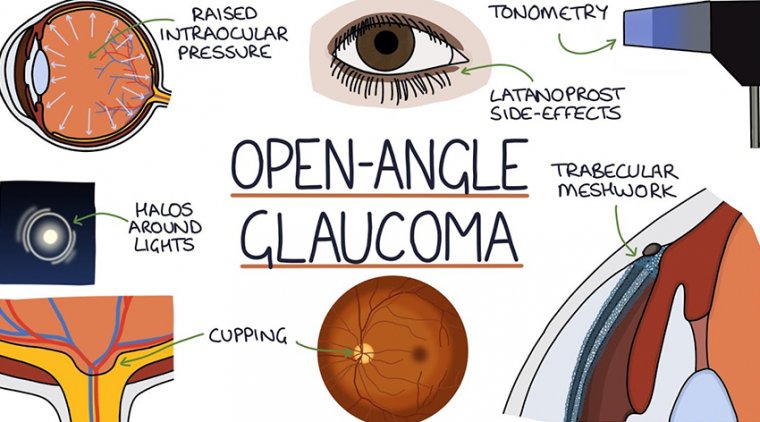
Open Angle Glaucoma
Open Angle Glaucoma is the most common form of glaucoma and is a result of the trabecular meshwork becoming less efficient over time. In this case, the eye continues to produce the same amount of aqueous humor, but the trabecular meshwork is not able to keep up. This imbalance of fluid production and drainage results in an excess amount of aqueous fluid. As the fluid builds up, the intraocular pressure (IOP) elevates. As a consequence to this elevation in eye pressure, the optic nerve becomes damaged.

For most people with open angle glaucoma, there are no perceivable symptoms in the early stages of the disease. However, as the optic nerve is continually subjected to an environment of increased eye pressure, it becomes damaged. This damage is eventually perceived as peripheral visual field defects. If the eye pressure is not monitored or controlled, open angle glaucoma can eventually lead to blindness. eResearch by Navid Ajamin -- autumn 2011

An ideal intraocular pressure is different for each individual. Some patients are able to tolerate a higher eye pressure while others cannot. It is important to be followed regularly by your eye doctor to determine an appropriate target IOP and ensure that this pressure is maintained over time.

Symptoms of glaucoma often do not show up until considerable visual loss has occurred. This is not the case for diabetics, however. Someone with diabetes is at much higher risk for vision loss.

Narrow Angle Glaucoma
Narrow Angle Glaucoma is less common than Open Angle Glaucoma. People who are farsighted (hyperopic) are more susceptible to this form of glaucoma because they typically have smaller eyes, although Narrow Angle Glaucoma can occur in nearsighted eyes. A smaller eye correlates to more crowding of the structures within the eye. In a normal eye there is sufficient space between the iris (the colored part of the eye) and the trabecular meshwork. However, in a crowded eye, the space between the iris and the trabecular meshwork is narrowed.

The iris controls the amount of light that enters the eye. Under bright light conditions the iris contracts to cover more space and make the pupil smaller (minimizing the amount of light that enters the eye). In dark lighting conditions, the iris dilates to allow more light to enter the eye. In a person with narrow angles, as the pupil dilates the iris can completely block the trabecular meshwork drainage angle. This can result in a rapid rise in eye pressure and can lead to an angle closure glaucoma attack.

An individual suffering from an angle closure attack typically experiences a severe headache above the eye brow, eye pain, significant eye redness, the pupil becomes fixed and dilated, the vision is significantly blurred, halos are seen around lights and nausea and vomiting can occur. An angle closure glaucoma attack is an emergency and must be treated immediately. Unfortunately, the damage done to the optic nerve during an angle closure glaucoma attack can be significant and is permanent. In some cases, Angle Closure Glaucoma can present insidiously however, without sudden symptoms.

Preventing Angle Closure
Fortunately, there is a means of preventing angle closure attacks. Narrow angles can be diagnosed during a routine eye exam. At that time, the doctor will look at the trabecular meshwork drainage angle through a special contact lens. This testing, referred to as gonioscopy, allows the doctor to determine the degree of narrowing. If the angles are very narrow, the doctor may recommend a laser procedure known as Laser Peripheral Iridotomy. During this in-office (out-patient) procedure, a small hole is created in the iris. This microscopic hole allows for another route for the aqueous fluid to enter the anterior chamber, and allows the iris to fall away from the drainage angle. The doctor makes the hole on the top part of the iris that is usually covered by the eyelid.

As with any surgical procedure, there are some risks to Laser Peripheral Iridotomy. However, the risks of the procedure are minimal and if the procedure is recommended, the benefit of the procedure in reducing the risk of angle closure glaucoma outweighs the risk of the procedure. Some of the risks include blurred vision, slight bleeding or inflammation within the eye. Some people notice a twinkle or flicker of light or shadow image after the procedure. These symptoms can arise from the affect of light entering the small hole in the iris. This side effect of the procedure is very unusual; in most cases, the upper eyelid covers the location of the iridotomy and the optical side effect is not perceived. Occasionally, the procedure needs to be repeated if the iridotomy heals over.

Imaging the angle. A slice through the front of two eyes taken with a special camera shows an open angle above and a closed angle below (images made with anterior segment optical coherence tomography). The arrow points to where the iris is separate from the cornea in the open angle (upper picture). In the lower picture, the iris is so close to the cornea that it nearly touches the trabecular meshwork (in front of arrowhead).[3]

Congenital Glaucoma
Congenital Glaucoma is a result of the malformation of the trabecular meshwork drainage angle during fetal development. The defective trabecular meshwork impedes the proper drainage of the aqueous fluid within the eye causing an increase in intraocular pressure. The elevated eye pressure eventually leads to damage of the optic nerve, which may result in blindness. For this reason it is imperative that children afflicted by the disease be seen by an ophthalmologist regularly in hopes of preserving as much of the child’s vision as possible.

Congenital glaucoma is generally diagnosed within the first few months of a child’s life. Infants with congenital glaucoma often have symptoms of tearing, red eye, light sensitivity and a cloudy appearance of the eye. In the far majority of congenital glaucoma cases, both eyes are affected by the disease. Males are more commonly afflicted with this disease than females; however, congenital glaucoma is rare, affecting approximately 0.05% of children.

glaucoma.jpeg

Secondary Glaucoma
Secondary glaucoma is an acquired form of glaucoma and may present itself as open angle or angle closure glaucoma. This type of glaucoma can be a result of trauma, inflammatory diseases, tumors or use of corticosteroids. Secondary glaucoma may be temporary.

Traumatic Secondary Glaucoma
In the circumstance of either blunt or penetrating ocular trauma, the trabecular meshwork can be damaged. This damage leads to the trabecular meshwork inefficiently draining the aqueous fluid out of the eye. As a result, the amount of aqueous fluid in the eye builds and increases the intraocular pressure. This increase in eye pressure leads to damage of the optic nerve. Eventually, the optic nerve damage results in visual field defects, and in some cases blindness.

Inflammatory Glaucoma and Steroid Responders
Inflammatory diseases such as uveitis can also lead to secondary glaucoma. Additionally, most cases of uveitis are treated with corticosteroids. Unfortunately, one of the side effects of corticosteroid use can be a significant increase in intraocular pressure. This cyclic condition is often a tight-rope-walk for the ophthalmologist to effectively treat the ocular inflammation while trying to maintain an adequate intraocular pressure.

Pseudoexfoliative Glaucoma
One of the more common forms of secondary glaucoma is referred to as pseudoexfoliation glaucoma. This type of secondary glaucoma occurs when the eye forms material that can affect the drainage angle. Like a clogged sink, the fluid is unable to properly drain and fluid accumulates within the eye. This accumulation of fluid can lead to an increase in eye pressure and eventually damage the optic nerve.

Pigmentary Glaucoma
Similarly, pigmentary glaucoma is another form of secondary glaucoma. Pigmentary glaucoma is a result of the iris pigment breaking off. These pigment granules can then clog the drainage angle and induce glaucoma.

Neovascular Glaucoma
Neovascular glaucoma is another form of secondary glaucoma. This form of glaucoma typically affects individuals with diabetes, or eyes that have circulation problems in the retina or in the arteries that supply the eye. Neovascularization refers to the new growth of abnormal blood vessels. The new blood vessels can block the trabecular meshwork and lead to high eye pressure. This form of glaucoma can be difficult to treat.

ICE Syndrome
ICE is an acronym for, “IridoCorneal Endothelial” syndrome. This is a very rare form of glaucoma that typically affects one eye. The condition occurs when the endothelial layer (the most inner layer) of the cornea grows over the trabecular meshwork and across the surface of the iris. This causes an increase in intraocular pressure and leads to damage of the optic nerve. These abnormal endothelial cells form adhesions that attach the iris to the cornea, which in turn blocks the drainage angle further.[1]

Reference:

  1. ocvermont.com
  2. glaucoma-association.com/about-glaucoma/types-of-glaucoma
  3. hopkinsmedicine.org/wilmer/services/glaucoma/book

اورژانس چشم

تعریف: اورژانس های چشم و بینائی به مواردی گفته میشود که بایستی در اسرع وقت و فوری اقدام به درمان نمود و عدم درمان به موقع باعث وارد شدن صدمات جبران ناپذیر به چشم و سیستم بینائی می شود.

What to Do in an Eye Emergency

An eye emergency occurs any time you have a foreign object or chemicals in your eye, or when an injury or burn affects your eye area.

Remember, you should seek medical attention if you ever experience swelling, redness, or pain in your eyes. Without proper treatment, eye damage can lead to partial loss of
vision or even permanent blindness.

Preventing eye injury
Eye injuries can happen anywhere, including at home, work, athletic
events, or on the playground. Accidents can happen during high-risk activities, but also in places where you least expect them.

What are 5 ophthalmic emergencies?

All ocular emergencies, including a penetrating globe injury, retinal detachment, central retinal artery occlusion, acute angle-closure glaucoma, and chemical burns, should be referred immediately to the emergency department or an ophthalmologist.

Careful eye examination and simple tests can help primary care physicians make decisions about appropriate treatment and referral. All patients with eye problems should be tested for visual acuity and ocular movements. Confrontation visual field examination, pupillary examination, and direct ophthalmoscopy of both eyes also should be performed. Ocular injury from high-velocity trauma or from chemicals may be easily misdiagnosed. After a chemical burn, thorough eye washing for at least 30 minutes or until the pH of the eye is within physiologic range is critical to prevent further damage. Use of an eye shield is required in patients with a ruptured globe to protect the injured eye and preserve the patient's vision.

There are things you can do to decrease your risk of eye injuries, including:

  • Wear protective eyewear when you use power tools orengage in high-risk sporting events. You’re at an increased risk any time you’re around flying objects, even if you’re not participating.
  • Follow the directions carefully when working with chemicals or cleaning supplies.
  • Keep scissors, knives, and other sharp instruments away from young children.
  • Teach older children how to use them safely and supervise them when they do.
  • Don’t let your children play with projectile toys, such as darts or pellet guns.
  • Childproof your home by either removing or cushioning items with sharp edges.
  • Use caution when cooking with grease and oil.
  • Keep heated hair appliances, like curling irons and straightening tools, away from your eyes.
  • Keep your distance from amateur fireworks.
  • To decrease your chances of developing permanent eye damage,you should always see an eye doctor after you experience an eye injury.

Symptoms of eye injury

Eye emergencies cover a range of incidents and conditions, each with their own distinct symptoms.

You should contact your doctor if it feels like you have something in your eye, or if you experience any of the following symptoms:

  • loss of vision
  • burning or stinging
  • pupils that are not the same size
  • one eye is not moving like the other
  • one eye is sticking out or bulging
  • eye pain
  • decreased vision
  • double vision
  • redness and irritation
  • light sensitivity
  • bruising around the eye
  • bleeding from the eye
  • blood in the white part of the eye
  • discharge from the eye
  • severe itching
  • new or severe headaches

If there’s an injury to your eye, or if you have sudden vision loss, swelling, bleeding, or pain in your eye, visit an emergency room or urgent care center.

Serious complications can occur from an eye injury. You shouldn’t attempt to treat yourself.

Although you may be tempted, be sure not to:

  • rub or apply pressure to your eye
  • try to remove foreign
  • objects that are stuck in any part of your eye
  • use tweezers or any other tools in your eye (cotton swabs can be used, but only on the eyelid)
  • put medications or ointments in your eye

If you wear contact lenses, don’t take them out if you think you’ve suffered an eye injury. Attempting to remove your contacts can make your injury worse.

The only exceptions to this rule are in situations where you have a chemical injury and your lenses didn’t flush out with water, or where you cannot receive immediate medical help.

The best thing you can do in an eye emergency is to get to your doctor as soon as possible.

Image result for eye injuries

Chemical injuries to the eye

Chemical burns result when cleaning products, garden chemicals, or industrial chemicals get into your eyes. You can also suffer burns in your eye from aerosols and fumes.

If you get acid in your eye, early treatment generally results in a good prognosis. However,
alkaline products like drain cleaners, sodium hydroxide, lye, or lime can permanently damage your cornea.

If you get chemicals in your eye, you should take the following steps:

  • Wash hands with soap and water to remove any chemicals that may have gotten on your hands.
  • Turn your head so the injured eye is down and to the side.
  • Hold your eyelid open and flush with clean cool tap water for 15 minutes. This an also be done in the shower.
  • If you’re wearing contact lenses and they’re still in your eye after flushing, try to remove them.

Get to an emergency room or urgent care center as quickly as possible. If possible, continue to flush your eye with clean water while you’re waiting for an ambulance or traveling to the medical center.

Small foreign objects in the eye

If something gets in your eye, it can cause eye damage or a loss of vision. Even something as small as sand or dust can cause irritation.

Take the following steps if you have something small in your eye or eyelid:

  • Try blinking to see if it clears your eye. Don’t rub your eye.
  • Wash your hands before touching your eye. Look into your eye to try to locate the object. You may need someone to help you with this.
  • If necessary, look behind your lower lid by pulling it down gently. You can look under your upper lid by placing a cotton swab on the lid and flipping the lid over it.
  • Use artificial tear eye drops to help rinse out the foreign body.
  • If the foreign object is stuck on one of your eyelids, flush it with water.
  • If the object is in your eye, flush your eye with cool water.
  • If you cannot remove the object or if the irritation continues, contact your doctor.

Large foreign objects stuck in your eye

Glass, metal, or objects that enter your eye at high speed can cause serious damage. If something is stuck in your eye, leave it where it is.

Do not touch it, do not apply pressure, and do not attempt to remove it.

This is a medical emergency and you should seek help immediately. Try to move your eye as little as possible while you wait for medical care. If the object is small and you’re with another person, it may help to cover both eyes with a clean piece of cloth. This will reduce your eye movement until your doctor examines you.

Sudden Blurry Vision or Vision Loss

Blurry vision can happen if you have a refractive error like myopia or hyperopia, but sudden blurred vision is concerning. Not all instances of sudden blurry vision are emergencies but seek treatment regardless. Only your eye doctor can determine the cause of your blurry vision.

Sudden blurry vision can be a symptom of:

  • Detached retina
  • Concussion
  • Stroke
  • Hyphema
  • Age-related macular degeneration
  • Macular hole
  • Eye infection
  • Migraine
  • Eyestrain

If you ever experience sudden vision loss, seek immediate medical attention. Don’t assume this issue will resolve itself—protect your vision by seeing your optometrist.

Corneal Abrasion

A corneal abrasion is a scratch on your cornea. This injury can happen because of contact with dirt, dust, sand, wood, metal, contact lenses, and other materials. You may have a corneal abrasion if you’re experiencing:

  • Pain
  • A gritty feeling in your eye
  • Watery eyes
  • Redness
  • Light sensitivity
  • Headache

You require an assessment after scratching your cornea. In the meantime, you can rinse your eye with clean water to remove as much material as possible and blink several times to get rid of smaller particles.

اورژانس های چشم عبارتند از:

  • گلوکوم با زاویه بسته AACG
  • انسداد ورید و شریان مرکزی چشم
  • سوختگی های شیمیائی
  • سوختگی های تشعشعی
  • سوختگی های حرارتی
  • اجسام خارجی در چشم
  • پارگی های قرنیه و صلبیه
  • صدمات اوربیت
  • نارسائی پیوند چشم

1) گلوکوم

اصطلاحی است برای گروهی از بیماری های چشم به دلیل بالا رفتن فشار داخلی چشم (IOP) آسیب میبیند و در نتیجه بینائی شخص کاهش میلبد و اگر درمان نشود شخص بینائی خود را از دست میدهد.این افزایش IOP به دلیل گیر افتادن مایع زلالیه که بین عدسی و عنبیه جریان دارد می باشد(مایع زلالیه عدسی و قرنیه را تغذیه میکند).

چندین نوع گلوکوم وجود دارد اما یکی از موارد خطرناک گلوکوم که یک اورژانس چشمی است گلوکوم با زاویه بسته می باشد. که عنبیه راه خروج مایع زلالیه را میبندد در نتیجه مایع زلالیه که دائما در حال تولید می باشد.در چشم تجمع پیدا میکند و فشار داخل چشم را به سرعت بالا میبرد.

Image result for site:arenaeyesurgeons.com emergency

علائم:

کاهش سریع و پیشرونده بینائی و تاری دید،درد پیرامون چشم،پر خونی ملتحمه(چشم شخص قرمز به نظر میرسد)و احتقان درد ممکن است با تهوع و برادیکاردی(کاهش ضربان قلب) توام باشد،کاهش دید مرکزی،مردمک ها به شکل بیضی عمودی در می آیند و در نیمه متسع تثبیت می شوند و به نور واکنش نمی دهند و قدرت تطابق خود را از دست میدهند.در صورت عدم درمان شخص ظرف ۱-۲ روز بینائی خود را از دست میدهد.

درمان:

جراحی لیزری فوری برای یاز کردن راه خروجی مایع زلالیه و همچنین از داروهای کاهنده IOP مثل پیلوکاربین استفاده کرد. چشم دیگر هم باید بوسیله پیلوکاربین یاجراحی درمان شود تا از حملات بعدی جلوگیری شود.

۲ )نارسائی پیوند چشم:

علائم اولیه:تیرگی دید ،ناراحتی،اشک ریزش،و قرمزی چشم

اگر شخص پیوند چشم زده باشد و دچار نشانه های بالا شد باید هر چه سریعتر به چشم پزشک مراجعه کند.

درمان: تجویز فوری کورتیکوستروئید های موضعی به صورت ساعتی و تزریق کورتیکوستروئیدها در پیرامون چشم می باشد.تمام تلاش ها متوجه جلوگیری از حفظ پیوند است که ممکن است در موارد شدید و مقاوم رد پیوند از عوامل تضعیف کننده سیستم ایمنی بدن استفاده شود.

۳)انسداد ورید و شریان مرکزی چشم:

علائم:دید چشم بطور ناگهانی بدون احساس هیچ دردی از بین میرود.از علت های آن می توان بیماری های دریچه ای قلب،رکود خون وریدی،افزایش غلظت خون و….را نام برد.این بیماری معمولا در افراد با فشار بالا و یا افرادی که دچار ارترواسکلروزیس هستند اتفاق بیافتد.اگر از بین رفتن بیماری به دلیل انسداد ورید و شریان مرکزی چشم باشد و وقت آن از ۱ ساعت بگذرد دیگر کاری نمیتوان انجام داد و بینائی برای همیشه از بین میرود،پس در این گونه موارد فوت وقت اصلا جایز نیست و باید شخص را هر چه سریعتر به چشم پزشک رسانید.

Image result for eye emergencies

۴ )سوختگی های شیمیائی:

شامل سوختگی های اسید و باز می باشد که هر دو خطرناک است ولی باز از اسید خطرناک تر است و در میان بازها آمونیاک از همه خطرناکتر. بازها چون میتوانند بیشتر در چشم نفوذ کنند و تا مدت بیشتری به آسیبهای خود ادامه دهند.و iop را افزایش دهند.اسیدها به نسبت بازها خطر کمتری دارند زیرا پروتئین های مربوط به بافت های آسیب دیده رسوب کرده و به مانعی در برابر نفوذ بیشتر اسید تبدیل می شوند.

اساس درمان در این مواقع شتشوی چشم تا حدی است که ph چشم نرمال و ph قرنیه به حدود۷/۳-۷/۶ برسد.بهترین مایع برای شتشو محلول نرمال سالین و یا رینگر و اگر نبود آب استریل و یا آب جوشیده سرد شده و یا آب معمولی.باید پشت پلک ها را هم شست و سریع مصدوم را به مرکز تخصصی چشم انتقال داد.

۵)سوختگی های تشعشعی:

در این نوع سوختگی ها معمولا در اثر قرار گرفتن طولانی مدت قرنیه چشم در معرض اشعه های مضر
مانند اشعه ماورای بنفش اتفاق میافتد و باعث سوختگی و زخم قرنیه میشود.اهم آن سوختگی در جوشکاران بدون عینک محافظ،اسکی بازان و سوختگی ناشی از لامپ های خورشیدی است.

علائم:درد شدید در چشم،حساسیت شدید به نور و ریزش اشک زیاد می باشد.

اساس درمان در این نوع سوختگی بستن چشم همراه با استراحت و استفاده از مسکن جهت کنترل درد و استفاده از قطره های آنتی بیوتیک و سیکلوپلژیک ها(برای اتساع مردمک چشم)که البته با تجویز پزشک انجام میشود.

اگر لایه های عمقی قرنیه در گیر نشده باشد معمولا ۲۴-۴۸ ساعت بهبودی حاصل میشود.از قطره های تتراکائین منحصرا در بیمارستان برای بیحسی موقت چشم برای عمل جراحی،معاینه استفاده میشود.و در حد یکی دو بار هم مصرف نمیگردد(شخص در هنگام استفاده از این قطره ها نباید چشم خود را بمالد چون ممکن است به قرنیه آسیب بزند.شروع اثر دارو ۲۰ ثانیه تا ۱ دقیقه و اثر آن ۱۰-۲۰ دقیقه باقی میماند.استفاده ی بیش از یکی دو بار به شدت به سلولهای قرنیه آسیب میرساند و زمینه را برای زخم وعفونتهای قرنیه فراهم میسازد،که نتیجه ی نهائی(ایجاد یک بافت جوشگاه)در قرنیه و افت شدید بینائی است.معمولا جوشکارها و تراشکارها ی بدون محافظ که دچار سوختگی قرنیه شده اند از این دارو سر خود و به دفعات زیاد مصرف میکنند و در نتیجه دچار مشکل میشوند.این افراد حتی بدون درمان ۶-۸ ساعته خوب میشوند و درد آنها تسکین می یابد.این افراد باید در طول ۶-۸ ساعت چشمها را بسته و از مسکنهایی مثل استامینوفن با یا بدون کدئین استفاده نمایند و استراحت کنند.

۶)سوختگی های حرارتی:

در سوختگی های حرارتی به علت بسته شدن غیر ارادی پلک ها اغلب تنها پلک ها آسیب میبینند و قرنیه آسیبی نمیبیند.تورم و قرمزی پلکها از علائم مهم این عارضه است.

کمک های اولیه شامل شستن چشم ها با آب فراوان است تا حرارت چشم گرفته شود و بعد باید چشم را پانسمان کرد و شخص را به مرکز درمانی مربوطه رساند.

اساس درمان در سوختگی های چشم مانند درمان سوختگی های سایر نقاط بدن است.یعنی استفاده از مسکنهای سیستمیک مانند مرفین و استفاده از پمادهای آنتی بیوتیک مانند تتراسیکلین،و یکی از مهمترین اقدامات دوختن پلک های بالا به پلک های تحتانی با نخ بخیه ۴ صفر سیلک است.تا از بروز چروکیدگی پلک و برگشتن پلک ها به سمت خارج جلوگیری شود.

۷)اجسام خارجی در چشم:

چنانچه اجسام خارجی در سطح قرنیه یا ملتحمه باشد و فرو نرفته باشد میتوان آن را از سطح قرنیه برداشت ولی اگر احتمال فرو رفتن آن باشد نباید دستکاری کرد و باید یک لیوان کوچک یا کاپ چشمی روی چشم گذاشت و بعد چشم را با باند بست.این عمل به این خاطر است که پانسمان روی چشم فشار وارد نکند و باعث تخلیه محتویات چشم نشود.

اجسامی مثل آهن،استیل،مس،و سبزیجات میتوانند موجب واکنش التهابی شدید شوند.اگر اجسامی مثل میخ یا چاقو در چشم فرو رفته باشد باید ابتدا در دو طرف آن ۲ باند گذاشت و یا با باند امامه ای آن را فیکس کرد و بعد ۱ لیوان روی آن گذاشت تا به چاقو یا جسمی که در چشم فرو رفته فشار نیاید و بیشتر در چشم فرو نرودبعد لیوان را با باند فیکس کرد.

در تمام موارد بالاباید هر دو چشم بست زیرا حرکت چشم سالم چشم آسیب دیده را هم تکان میدهد و باعث آسیب بیشتر آن میشود.در صدمات نافذ چشم آسپیرین منع مصرف دارد زیرا باعث تداوم خونریزی میشود.در صدمات نافذ چشم در صورت عدم حس نور تخلیه ی اولیه کره ی چشم انجام میشود که طی آن محتویات چشم با مقداری عصب بینائی خارج میشود. این عمل باید ظرف ۲ هفته از صدمه
ی اولیه انجا م شود و گرنه باعث بروز افتالمی سمپاتیکی (ophthalmia sympathetic) میشود که التهاب ناشی از صدمه چشم مبتلا به چشم سالمتر نیز سرایت میکند و آن را نابینا میگرداند.

۸ )پارگی های قرنیه و صلبیه:

بسیار خطرناک هستند زیراقسمت های درونی چشم در معرض عفونت قرار میگیرند و در نتیجه دچار
آسیب میشوند.در هنگام وارد شدن ضربه های سنگین و احتمالیه پارگی قرنیه و صلبیه نباید به چشم فشار آورد یا آن را پانسمان کرد زیرا ممکن است محتویات چشم در اثر فشار بیرون بریزد پس باید به شیوه ای که در بخش اجسام خارجی گفته شد با یک لیوان کوچک یا کاپ چشمی چشم را بست.

درمان شامل دوختن پارگی و پیشگیری از عفونت است که توسط پزشک انجام میشود.

۹)صدمات اوربیت:

در صدمات اوربیت چون صدمه به جمجمه خورده است اول باید علائم مغزی راچک کرد و وضعیت او را پایدار کرد بعد بینائی را چک نمود.بعد از پایداری وضعیت مصدوم برای وسعت صدمات چشم در اولین فرصت بینائی شخص را کنترل کرد که در چه حدی است.کره چشم باید از نظر صدمات بافت نرم مورد نظر قرار گیرد.

در ضربات کند مثل ضربه مشت ،زانو،آرنج،توپ گلف، و تنیس چون یک مرتبه اثر این ضربات فشار داخل اوربیت زیاد میشود استخوان کف اوربیت که کمترین مقاومت را دارد میشکند. در صدمات اوربیت نیز اگر خونریزی دارد باید هر چه سریعتر مصدوم را به مرکز تخصصی چشم انتقال داد.

پیشگیری از صدمات چشم:

  1. هنگام سرخ کردن غذا در ماهی تابه کاملا آن را به روغن آغشته کنید تا از پاشیده شدن روغن به بیرون جلوگیری شود.
  2. هنگام استفاده از مواد شیمیائی قوی از عینک ایمنی استفاده کنید و بعد از استفاده دست های خود را خوب بشورید.
  3. برای حفاظت چشم در برابر اشعه های نور خورشید از عینک استفاده نمائید.
  4. ۴-در باغ یا جنگل مراقب برخورد شاخه درختها با چشم و صورت خود باشید.
  5. به کسی اجازه ندهید جلو یا کنار ماشین چمن زنی بایستد.سنگها را از جلوی ماشین چمن زنی بردارید زیرا ممکن است با برخورد و یا از زیر ماشین در برود و صدمات جدی و شدیدی را به چشم وارد کند
  6. برای مشاهده ی نور ماشین در شب از چراغ قوه استفاده نمائید و از کبریت یا شمع استفاده نکنید.
  7. در هنگام استفاده از کابل پرشی در استارت ماشین از عینک محافظ استفاده نمائید.ابتدا اطمینان پیدا کنید که شخص با ماشین در تماس نیست،مطمئن شوید که کابل های مورد استفاده با هم اتصال ندارندو هرگز کابل را به پایانه ی منفی باطری وصل نکنید.
  8. در ورزشهای تنیس،اسکواش و از این قبیل مراقب برخورد توپ با چشم باشید
  9. در هنگام آتش بازی به کودکان اجازه ندهید آتش را شعله ور کنند و مواد محترقه در آتش بریزنند.سعی نکنید خاکستر را دوباره شعله ور کنید و در پایان روی آن آب بریزید.

Reference:

  • هفته نامه نوین پزشکی
  • parastoonursing.com
  • queenswayoptometric.com
  • aafp.org/pubs/afp/issues/2007/0915/p829.html
  • healthline.com/health/eye-emergencies#chemical-injuries
  • sciencedirect.com/science/article/abs/pii/S1357303918302378
  • greatlakesvisioncare.com/blog/common-eye-emergencies-symptoms-and-treatment.html

See also:

  • Top 10 Eye Emergencies
  • Lasik Patients Should Be Warned of Complications
nicetoview.blogfa.com
عینک eyewear وبلاگ تخصصی عینک شامل مجموعه مطالب پزشکی است که اطلاعات مفیدی در رابطه با عینک , چشم، لنز، سلامتی چشم و راه های پیشگیری از بیماریهای چشمی، کنترل و درمان آن را در اختیار شما کاربر محترم می گزارد.

Always Be Healthy
همیشه سلامت باشید

Navid Ajamin نوید عجمین
eMail: navid.aj@outlook.com
موضوعات وب
پیوندها

unicef for every child

شمارنده