ابزار وبمستر

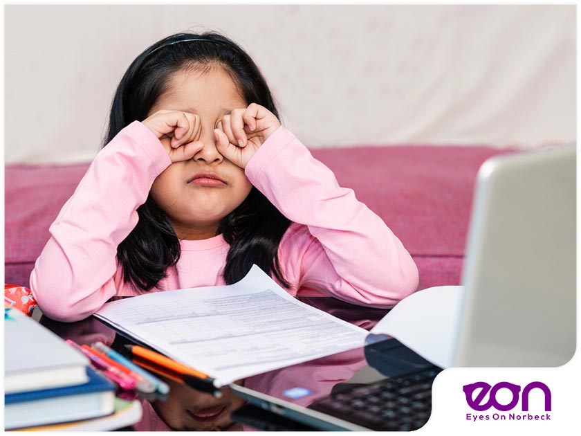
Visual Fatigue Syndrome (VFS) is caused by focusing on objects, such as computers, that are 1 to 3 feet away for extended periods of time.The symptoms of VFS are not only bothersome, they can also be painful, reduce the enjoyment of your day, and reduce the quality of your work.

Stress impacts us mentally and physically, but did you know it can affect our vision?

When we are severely stressed and anxious, high levels of adrenaline in the body can cause pressure on the eyes, resulting in blurred vision. People with long-term anxiety can suffer from eye strain during the day on a regular basis. If you become highly sensitised to any slight movement, over time the strain from other senses can cause muscular tensions and headaches.

SYMPTOMS OF STRESS-RELATED VISION IMPAIRMENT:

Parents blame smartphones, tablets for teens' sleep troubles ...

  • Tunnel vision. You may lose some of your peripheral vision and feel like you can only see straight in front of you.[4]
  • Sensitivity to light and movement; light may hurt your eyes or make it difficult for you concentrate, and focus.
  • Eye twitching; eyes can randomly spasm, with no pain but discomfort.
  • Very dry or very wet eyes; both can be a symptom, however, can also be caused by other issues.
  • Blurry vision; finding it hard to concentrate, or focus. If you have additional symptoms, visit your local GP.
  • Eye strain; discomfort and minor pain as your eyes feel tight and swollen.
  • Eye floaters; tiny spots that swim across your vision.

If you have any of these symptoms with no other medical issues, the best option for you is to get enough rest, eat healthily, use meditation, or any stress relief exercises that help you to relax. Taking at least a few minutes to consciously relax will help your body calm down.[3]

Risks and consequences of oxidative stress. The eye is an organ that is predisposed to great levels of oxidative stress. The eye is constantly exposed to factors such as radiation, chemicals, oxygen, drugs, which induce the formation of reactive oxygen species (ROS) that can ultimately damage cells. This figure is modified from Flammer J. Glaucoma, Glaucoma A Guide for Patients. An Introduction for Care-Providers. A Quick Reference. 3rd ed. Cambridge: Hogrefe & Huber; 2006. Figure S1.29; p 222.

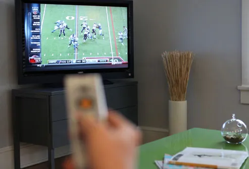
In recent years there has been a shift in the way we use our vision. Instead of using our eyes to see most things at distance, we spend most of the day viewing objects that are within arms reach. These items include the computer, television, cell phone, PDAs, even books are now available in digital format. Both the real world and written word have now been replaced by a constant barrage of illuminated, digital pixels. This new visual environment commonly induces visual fatigue. eResearch by Navid Ajamin -- spring 2011

Users of digital media may experience eyestrain, blurred vision, tired eyes, dry eyes, neck and back pain. Even those who can see 20/20 and those who do not normally wear glasses may experience visual fatigue. Studies have shown us that 83 percent of all individuals experience one or more symptoms of Visual Fatigue Syndrome.

Unfortunately most of those affected by visual fatigue are not getting the help they need.The reason is poor education. Most people do not understand why they are experiencing problems and, in many cases, eye doctors are not properly trained to recognize the symptoms of visual fatigue. Even when an eye doctor is well informed, he or she may lack the proper tools necessary to help patients combat symptoms.[2]

Driving is a complex task, requiring full concentration and a calm attitude. Heightened emotions such as stress, anger or upset are a form of cognitive distraction that can significantly impede drivers’ ability to spot and respond to hazards. Research has found that drivers who suffer from work-related stress are more likely to speed and take other risks while driving and more like to be involved in serious crashes [5]

Reference:

  1. eyewalk.net/innovation/antifatigue_en
  2. iowaeyeblog.com/2009/11/visual-fatigue-syndrome
  3. whitbyonline.com/about-us/news/stress-on-your-eyes
  4. allabouteyes.com/stressed-stress-affects-eyes
  5. brake.org.uk/facts-resources/15-facts/487-driver-stress

See Also:

  • Anti-Fatigue Lenses
  • What are anti-fatique lenses
  • Can Stress Cause Blurry Vision?
  • Overview Of Anti-Fatigue Lenses

Blinking, a normal involuntary reflex, occurs, on average, 12 times per minute or 17,000 times a day, Professor Alan Hedge of Cornell University states. Sometimes children begin blinking so frequently it seems that they're blinking 17,000 times a minute, rather than a day. Parents often get concerned over the causes of rapid eye blinking, but most blinking is benign and self-limiting, according to a 2001 study reported by lead author David Coats of Baylor College of Medicine in the journal "Ophthalmology."

Causes

Causes for frequent blinking vary greatly. Frequent blinking can occur because a child has visual problems such as convergence insufficiency, which is trouble focusing both eyes on an up-close object. Double or blurred vision caused by convergence insufficiency can lead to excessive blinking, Baylor College of Medicine explains. Problems with the eyelids or the anterior segment of the eye, which consists of the cornea, iris, anterior chamber and lens, accounted for 37 percent of excessive blinking, Coats reported in the 2001 "Ophthalmology" study. Uncorrected refractive errors caused 14 percent of cases, he added.

Habit tics accounted for 23 percent of excessive blinking in Coats' report. Tics may also occur in children with Tourette's syndrome, a type of tic disorder that usually begins between ages 3 and 10, Family doctor.org reports. Movement disorders, called dystonias, can also cause excessive blinking, the National Institute of Neurological Disorders and Stroke reports. Allergic reactions can also cause excessive blinking, pediatrician Jeffrey Hull states. Rapid eye blinking may also occur as part of epilepsy, notes the Milton S. Hershey Medical Center.

Excessive Blinking in Children

Characteristics

In 89 percent of children with excessive blinking, both eyes are affected. Boys with excessive blinking outnumber girls two to one, Coats reported.

Diagnosis

History and clinical examination are usually adequate to make a diagnosis, Coats says, with neurological examination and neuroimaging usually not being required.

Treatment

Treatment depends on the cause. Tics often require nothing more than ignoring the symptoms until they disappear, Hull advises. Medications may help relieve specific symptoms of Tourette's. Correcting refractive errors and vision problems helps stop blinking caused by trouble seeing. Anticonvulsants are used to treat epilepsy. Dystonias may also respond to medications such as anticonvulsants as well as drugs that correct neurotransmitter abnormalities.

Considerations

Excessive blinking rarely causes serious problems and often resolves without treatment. Blepharospasm, a type of dystonia that causes first one eye and then both, in most cases, to close forcefully and repeatedly, can cause functional blindness even though both eyes see normally, by causing the eyelids to eventually remain completely closed, the National Institute of Neurological Disorders and Stroke warns. Conditions such as epilepsy and dystonia require close medical evaluation and follow-up. eResearch by Navid Ajamin -- spring 2011

Excessive blinking can be caused by problems with the eyelids or anterior segment (front surface of the eye), habitual tics, refractive error (need for glasses), intermittent exotropia or turning out of the eye, and stress. It is very rare for excessive blinking to be a sign of an undiagnosed neurologic disorder.

A pediatric ophthalmologist will be able to diagnose the cause of the symptoms. A thorough exam will be performed. If there is a problem such as an ingrown eyelash, corneal abrasion (scratch on the front surface of the eye), conjunctivitis (pink eye), foreign body in the eye, allergies affecting the eye or eye dryness, this can easily be diagnosed by performing an examination with an instrument called a slit lamp. This is a special microscope used to magnify the eye. If glasses are needed, this can also be easily detected. Any strabismus (in turning or out turning of the eye) will be diagnosed when the ophthalmologist examines the eye movements.

A pediatric ophthalmologist will be able to diagnose the cause of the symptoms. A thorough exam will be performed. If there is a problem such as an ingrown eyelash, corneal abrasion (scratch on the front surface of the eye), conjunctivitis (pink eye), foreign body in the eye, allergies affecting the eye or eye dryness, this can easily be diagnosed by performing an examination with an instrument called a slit lamp. This is a special microscope used to magnify the eye. If glasses are needed, this can also be easily detected. Any strabismus (in turning or out turning of the eye) will be diagnosed when the ophthalmologist examines the eye movements.If an abrasion or conjunctivitis is diagnosed, eye drops, or ointment may be given.

Glasses may be prescribed if the excessive blinking is caused by blurry vision.

بلفارواسپاسم Blepharospasm به معنای پلک زدن غیر ارادی یا بسته شدن غیر ارادی پلک‌ها است.

بلفارواسپاسم یک عارضه پیش رونده عصبی است که بیشتر در زنان میانسال و سالمند اتفاق می‌افتد.

علت ایجاد بلفارواسپاسم چیست؟

به نظر می رسد كه بلفارواسپاسم ناشی از نوعی از اشكال در عملكرد “عقده‌های قاعده ای مغز” باشد. عقده های قاعده ای بخشی از مغز هستند كه در تنظیم حركات هماهنگ عضلات دخالت دارند. البته هنوز به طور دقیق نمی دانیم كه چه مشكلی باعث بروز بلفارواسپاسم می شود.

در بیشتر موارد بلفارواسپاسم به صورت خودبه‌خودی و بدون هیچ علت مشخصی ایجاد می شود.

البته در بسیاری از افراد، خشكی چشم عامل محركی باشد كه در افراد حساس باعث شروع بلفارواسپاسم شود.

گاهی بلفارواسپاسم یا انواع دیگر دیستونی در افراد یك خانواده بروز می كند، بنابراین شاید زمینه های ارثی و ژنتیكی در بروز آن مؤثر باشند.

توصیه‌های بیماری بلفارواسپاسم [3]

۱. آموزش بیمار و اطرافیان و حمایت موثر افراد خانواده از فرد مبتلا به بلفارواسپاسم می‌تواند نتایج مفیدی در کنترل علائم داشته باشد.

۲. استفاده از عینک‌های آفتابی تیره به دو علت به مبتلایان بلفارواسپاسم کمک می‌کند. اول آنکه عینک آفتابی با جلوگیری از تابش نور شدید به چشم، مانع از تحریک چشم شده و تا حدی از بروز حمله بلفارواسپاسم جلوگیری می‌کند.

ثانیاً عینک تیره با پنهان کردن چشم، مانع از آن می‌شود که سایرین متوجه پلک زدن غیر عادی فرد شوند، در نتیجه حضور در اجتماع را برای فرد آسان‌تر می‌کند.

۳. استفاده از قطره‌های اشک مصنوعی و مرطوب کننده‌های چشم برای درمان خشکی چشم و درمان التهاب پلک (بلفاریت) با کاهش تحریک سطح چشم ممکن است به کنترل حملات بلفارواسپاسم کمک کند.

لرزش پلک چشم چیست؟ [4]

لرزش چشم یا پرش پلک که با نام میوکیمیا نیز شناخته می شود، یکی از اسپاسم های ناخواسته عضلات پلک چشم است که معمولا در پلک بالا رخ می دهد، البته میوکیمیا ممکن است در پلک پایین هم رخ دهد. در این حالت پلک چشم می پرد و ممکن است این حرکت آنقدر خفیف باشد که شخص متوجه آن نشود و در حالتی دیگر نیز ممکن است آنقدر لرزش پلک چشم شدید باشد که باعث کلافه شدن فرد شود. دلیل عمده این نوع پرش ها، انقباضات غیر ارادی ماهیچه ها است که با روش هایی می توان از این انقباضات ماهیچه ای جلوگیری کرد.

دلایل

همانگونه که گفته شد، لرزش های پلک چشم معمولا به دلیل اسپاسم های عضلات چشم و پلک است که این اسپاسم ها می تواند به دلایل ذیل رخ دهند.

  • خستگی و سوزش چشم ها
  • کشیدگی عضلات اطراف چشم
  • کارهای سنگین فیزیکی
  • میزان استرس بالا
  • مصرف برخی از داروها
  • مصرف کافئین، تنباکو و الکل

در واقع موارد ذکر شده می توانند در بوجود آمدن و تشدید اسپاسم های عضلانی پلک بسیار موثر باشند. اما به جز موارد گفته شده،دلایل عصبی و مغزی دیگری نیز وجود دارند که باعث ایجاد عارضه پریدن پلک می شوند که این دلایل عبارتند از:

  • فلج صورت یکی از مشکلاتی است که ممکن است باعث لرزش غیر طبیعی پلک شود.
  • دیستونی که یکی از عواملی است که باعث ایجاد اسپاسم ناگهانی عضلات می شود.
  • بیماری پارکینسون می تواند منجر به لرزش پلک های چشم شود.
  • سندرم تورتس نیز می تواند باعث حرکت های ناخواسته و پرش پلک شود.
  • خراش های موجود بر روی قرنیه چشم نیز ممکن است باعث پرش پلک شود.
  • بیماری سیستم عصبی موسوم به مولتیپل اسکلروز نیز می تواند منجر به اختلالات حرکتی در پلک ها شود.

برای درمان لرزش پلک چشم، ابتدا باید عاملی که در به وجود آمدن لرزش پلک تاثیر داشته است را شناسایی کرده و آن را برطرف نمایید. اما راهکارهایی وجود دارند که به صورت کلی می توانند باعث درمان این عارضه شوند.

Reference:

  1. livestrong.com/article/199401-abnormal-eye-blinking-in-children
  2. aapos.org/glossary/excessive-blinking-in-children
  3. pezeshk.us
  4. binaii.com/ArticleDetail/10217

Smoking is prevalent among teenagers now. All that is written on the cigarette is that cigarette smoking is injurious to health. What is not mentioned is that it is very harmful for human sight.

Age-related macular degeneration (AMD) is one of the main socioeconomical health issues worldwide. AMD has a multifactorial etiology with a variety of risk factors. Smoking is the most important modifiable risk factor for AMD development and progression. The present review summarizes the epidemiological studies evaluating the association between smoking and AMD, the mechanisms through which smoking induces damage to the chorioretinal tissues, and the relevance of advising patients to quit smoking for their visual health.

Image result for smoking and macular degeneration

There is growing evidence of the effects of smoking on vision. And these effects can be very significant. Cigarette smoking enhances oxidants which are chemical byproducts in our body. Oxidants can damage cells, including those in the eyes. The links between smoking and eye diseases is strong, including cataracts and age related macular degeneration.

Believe it, smoking increases the chance of your getting blind by up to four times, due to age-related macular degeneration (AMD). AMD is irreversible and is a progressive eye condition in which the central part of the retina (macula) is damaged.

Causes of Macular Degeneration

Once someone has been diagnosed with age related macular degeneration (AMD) or any disease for that matter, the mind starts asking questions like, how did I get this condition, what caused it or perhaps, could I have done something to prevent it?

There are many different factors that contribute to a person developing macular degeneration – some that can be controlled and some that cannot. eResearch by Navid Ajamin -- spring 2011

Here are the most common causes and risk factors:

1. Age
Macular degeneration is the number one cause of vision loss in the senior population. The older a person gets, the higher their risk of developing AMD. According to the National Eye Institute, ”a large study found that people in middle-age have about a 2 percent risk of getting AMD, but this risk increased to nearly 30 percent in those over age 75.”

2. Gender
Women are more likely to develop age related macular degeneration than men.

3. Race
It occurs in all races but it is more common in Caucasians.

4. Iris Color
The research shows that there is less pigment in blue eyes, and green eyes for that matter, than there is in brown eyes, so more light is able to penetrate blue eyes. This makes lighter eyes more sensitive to light and is what makes people with blue eyes more likely to have age-related macular degeneration. Macular degeneration is caused when the light sensitive cells in the eyes start to die, which can eventually result in blindness.

5. Heredity
My husband’s father has wet macular degeneration as well as did many of my husband’s aunts and uncles. The lifetime risk of developing late-stage macular degeneration is 50% for people who have a relative with macular degeneration versus 12% for people who’s relatives do not have macular degeneration (4x the risk).

6. Smoking
Smoking is the single most controllable risk factor that contributes to the development of macular degeneration. Every cigarette that is smoked causes damage to one’s vision. Smoking causes vasoconstriction or narrowing of the blood vessels which reduces the blood supply to the eyes. Smoking also creates free radicals which causes cellular damage while decreasing ones levels of antioxidants.

7. Diet Low in Dark Leafy Greens
A Harvard study done by Dr. Johanna Seddon in 1994 concluded that those who ate at least 5 servings of dark leafy greens per week had a 43% lower risk of developing AMD than those who ate small amounts or none at all. These lutein rich greens include kale, collard greens, spinach, Swiss chard, parsley, mustard greens, romaine lettuce and beet greens.

8. Diet Low in Omega 3 Fatty Acids
Thanks again to Dr. Seddon and her researchers at Harvard University, they discovered that people with diets of a 3:1 ratio of omega-6 to omega-3 fatty acids have less macular degeneration. Almost all of our processed foods and vegetable oils contain omega-6 which means that the average American gets way to much omega-6. Because omega 6 competes with omega 3, if we have too much omega-6 then the omega-3 we do take isn’t utilized properly.

9. High Blood Pressure
Dr. Michael A. Samuel the author of Macular Degeneration: A Complete Guide for Patients and Their Families writes “If you have high blood pressure that is fairly well controlled, you have double the risk of wet AMD compared to someone who does not have hypertension at all. If you have uncontrolled high blood pressure that goes above 160/90 you are three times more likely to develop wet AMD.” (Ophthalmology 2003;110: 636-643).

10. Exercise
Those with an active lifestyle were 70% less likely to develop macular degeneration than those who did little or no exercise according to researchers from the University of Wisconsin. The Wisconsin study that began in 1988, tracked almost 4,000 men and women (between the ages of 43-86) over a 15 year period by conducting regular eye tests and recording levels of exercise.

After accounting for other risk factors such as age, sex, history of arthritis, systolic blood pressure, body mass index, smoking, and education, those with a baseline active lifestyle of walking three times or more a week, were 70% less likely to develop macular degeneration than those who did little exercise.

Reference:

  • blurtit.com
  • pmc.ncbi.nlm.nih.gov/articles/PMC3866712
  • enhancedvision.com/low-vision-info/eye-conditions/causes-of-macular-degeneration.html

There's no proven technique for preventing presbyopia. The gradual decline of the ability to focus on near objects affects everyone. However, you can help protect your vision with these steps: Get regular eye examinations.[4]

There are five types of presbyopia: eResearch by Navid Ajamin -- spring 2011

  • Incipient presbyopia. This is the very earliest stage, when it may be a bit more difficult to read small print.
  • Functional presbyopia. This occurs when you begin to notice more problems with near sight.
  • Absolute presbyopia. If you have this type, your eyes cannot focus on near objects at all.
  • Premature presbyopia. This term is used when presbyobia occurs before the age of 40 years.
  • Nocturnal presbyopia. When this occurs, focusing on near objects is particularly difficult in low light conditions.[6]

Nothing can be done to prevent presbyopia. It is an inevitable part of aging. However, people who do a lot of close visual work, such as working with a computer or intensive reading, may develop presbyopia earlier than others. If you do close work, take a 10-minute break every one to two hours to relieve strain on the eyes. Allow your eyes to focus on objects at a middle or long distance away to give your eyes a rest from close focusing. Be sure to use bright lighting when reading to help your eyes focus.[1]

As we age, the lens of the eye becomes increasingly inflexible, making it harder to focus clearly on near objects. This is called presbyopia. ... Beginning early in life — perhaps as early as age 10 — our lenses gradually stiffen and begin to lose the ability to change shape.

Presbyopia is a condition in which the lens of the eye loses its ability to focus, making it difficult to see objects up close.

Presbyopia cannot be cured. Instead, prescription glasses, contact lens, reading glasses, progressive addition lenses, or bifocals can help correct the effects of presbyopia. Bifocals are often prescribed for presbyopia. Bifocals are eyeglasses that have two different prescriptions in one spectacle lens.[3]

The cause of presbyopia is lens hardening by decreasing levels of α-crystallin, a process which may be sped up by higher temperatures.

In optics, the closest point at which an object can be brought into focus by the eye is called the eye's near point. A standard near point distance of 25 cm is typically assumed in the design of optical instruments, and in characterizing optical devices such as magnifying glasses.

There is some confusion over how the focusing mechanism of the eye works. In the 1977 book, Eye and Brain, for example, the lens is said to be suspended by a membrane, the 'zonula', which holds it under tension. The tension is released, by contraction of the ciliary muscle, to allow the lens to become more round, for close vision. This implies the ciliary muscle, which is outside the zonula, must be circumferential, contracting like a sphincter, to slacken the tension of the zonula pulling outwards on the lens. This is consistent with the fact that our eyes seem to be in the 'relaxed' state when focusing at infinity, and also explains why no amount of effort seems to enable a myopic person to see farther away.

Presbyopia

Mobile Phones Causing Presbyopia At An Early Age pc-tablet.com

The ability to focus on near objects declines throughout life, from an accommodation of about 20 dioptres (ability to focus at 50 mm away) in a child, to 10 dioptres at age 25 (100 mm), and levels off at 0.5 to 1 dioptre at age 60 (ability to focus down to 1–2 meters only).

The expected, maximum, and minimum amplitudes of accommodation in diopters (D) for a corrected patient of a given age can be estimated using Hofstetter's formulas:

expected amplitude (D) = 18.5 - 0.3 × (age in years),

maximum amplitude (D) = 25 - 0.4 × (age in years),

minimum amplitude (D) = 15 - 0.25 × (age in years).

Causes, incidence, and risk factors

The lens of the eye needs to change its length or shape to focus on smaller objects, or objects that get closer or farther away. This is called the elasticity of the lens. This elasticity is slowly lost as people age. The result is a slow decrease in the ability of the eye to focus on nearby objects.

People usually notice the condition at around age 45, when they realize that they need to hold reading materials further away in order to focus on them. Presbyopia is a natural part of the aging process and it affects everyone.

Presbyopia: Symptoms, Treatment, Definition

Presbyopia causes the following symptoms:

  • Words appear blurred at a reading distance that used to be comfortable.
  • Reading material or other objects must be held farther away from your eyes to gain clarity or see details.
  • Brighter light is needed to see clearly (bright light constricts the pupils, which changes the focus of the light on the retina).
  • You have difficulty reading late at night, or when you are tired or stressed.
  • Your eyes become uncomfortable, or you become tired or drowsy when doing close work because of the strain of eye muscles working to change the lens shape.
  • You may have headaches as a result of muscle tension.

Presbyopia eventually affects everyone, even people who are already farsighted (hyperopic) or nearsighted (myopic). Because people who are farsighted already need to work when focusing on near objects, they may experience presbyopia a little earlier in life. People who are nearsighted will still experience presbyopia when wearing their corrective glasses or contact lenses for distance. However, for looking at objects or letters closer to them, they will likely be able to see quite clearly when they take off their glasses. This is where the term "nearsighted" comes from.[5]

You can’t prevent or reverse presbyopia with natural treatments. However, quitting smoking (if you smoke) and upping your intake of vitamins A, C, and E may improve your eye health.

Presbyopia is a progressive loss of your ability to see clearly at a close distance. It’s a natural result of the aging process.

Presbyopia progresses as the lenses inside your eyes lose elasticity. The speed of progression, the severity of visual symptoms, and the exact age of onset for presbyopia vary. However, the degeneration of the lens that leads to presbyopia is a natural part of the aging process.

When you’re young, the lens in your eye is flexible and relatively elastic. It can change its length or shape with the help of a ring of tiny muscles that surround it.The muscles that surround your eye can easily reshape and adjust your lens to accommodate both close and distant images.

With age, your lens loses flexibility and begins to stiffen. As a result, your lens becomes unable to change shape and constricts to focus on close images.With this hardening of your lens, your eye gradually loses its ability to focus light directly onto your retina.

You’re at a higher risk of premature presbyopia if you have: [7]

  • anemia, which is a lack of enough normal blood cells
  • cardiovascular disease
  • diabetes, or difficulty metabolizing blood sugar
  • hyperopia, or farsightedness, which means you have a greater difficulty seeing objects nearby than objects that are far away
  • multiple sclerosis, which is a chronic illness that affects your spine and brain
  • myasthenia gravis, which is a neuromuscular disorder that affects your nerves and muscles
  • eye trauma or disease
  • vascular insufficiency, or poor blood flow

Some prescription and over-the-counter drugs can reduce your eye’s ability to focus on close images. Taking the following drugs can put you at a higher risk of premature presbyopia:

  • alcohol
  • antianxiety drugs
  • antidepressants
  • antihistamines
  • antipsychotics
  • antispasmodics
  • diuretics

Other factors that may put you at a higher risk of premature presbyopia are:

  • being female
  • having intraocular surgery, or surgery done on the inside of the eye
  • eating an unhealthy diet
  • having decompression sickness, or “the bends,” which results from rapid decompression and typically occurs in scuba divers that surface too quickly

The American Academy of Ophthalmology recommends that adults have a complete eye exam every:

  • Five to 10 years under age 40
  • Two to four years between ages 40 and 54
  • One to three years between ages 55 and 64
  • One to two years beginning at age 65

Reference:

  1. sparkpeople.com
  2. wikipedia.org/wiki/Presbyopia
  3. my.clevelandclinic.org
  4. healthline.com
  5. health.harvard.edu
  6. patient.info/eye-care/long-sight-hypermetropia/age-related-long-sight-presbyopia
  7. healthline.com/health/presbyopia#risk-factors

Who should not wear contact lenses?

You should not wear contacts if you:

  • Have an eye infection or inflammation (redness & swelling).
  • Have an eye disease, eye injury or dryness that interferes with contact lens wear.
  • Have a systemic disease that may be affected by or impact lens wear.
  • Have certain types of allergic conditions.
  • Are using certain medications, such as some eye medications.

Reference: bausch.com

nicetoview.blogfa.com
عینک eyewear 
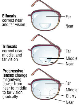
وبلاگ تخصصی عینک شامل مجموعه مطالب پزشکی است که اطلاعات مفیدی در رابطه با عینک , چشم، لنز، سلامتی چشم و راه های پیشگیری از بیماریهای چشمی، کنترل و درمان آن را در اختیار شما کاربر محترم می گزارد.

Always Be Healthy
همیشه سلامت باشید

Navid Ajamin نوید عجمین
eMail: navid.aj@outlook.com
موضوعات وب
پیوندها

unicef for every child

شمارنده