ابزار وبمستر

9 Ways to Reduce the Symptoms of Computer Eyestrain

Eyestrain is the number one complaint in office jobs, but there are many things workers and employers can do to reduce these symptoms. The best solution is number 1 below—see an eye doctor using the PRIO Vision Tester, and get a pair of eyeglasses specifically to wear when you use the computer.

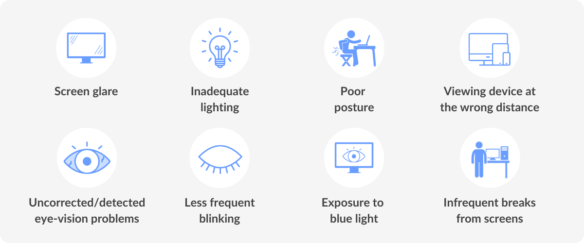
The potential impact of computer use on children’s vision involves the following factors:

  • Children often have a limited degree of self-awareness. Many children keep performing an enjoyable task with great concentration until near exhaustion (e.g., playing video games for hours with little, if any, breaks). Prolonged activity without a significant break can cause eye focusing (accommodative) problems and eye irritation.

Accommodative problems may occur as a result of the eyes’ focusing system “locking in” to a particular target and viewing distance. In some cases, this may cause the eyes to be unable to smoothly and easily focus on a particular object, even long after the original work is completed.

Eye irritation may occur because of poor tearflow over the eye due to reduced blinking. Blinking is often inhibited by concentration and staring at a computer or video screen. Compounding this, computers usually are located higher in the field of view than traditional paperwork. This results in the upper eyelids being retracted to a greater extent. Therefore, the eye tends to experience more than the normal amount of tear evaporation resulting in dryness and irritation.

  • Children are very adaptable. Although there are many positive aspects to their adaptability, children frequently ignore problems that would be addressed by adults. A child who is viewing a computer screen with a large amount of glare often will not think about changing the computer arrangement or the surroundings to achieve more comfortable viewing. This can result in excessive eye strain. Also, children often accept blurred vision caused by nearsightedness (myopia), farsightedness (hyperopia), or astigmatism because they think everyone sees the way they do. Uncorrected farsightedness can cause eye strain, even when clear vision can be maintained.
  • Children are not the same size as adults. Since children are smaller, computers don’t fit them well. Most computer workstations are arranged for adult use. Therefore, a child using a computer on a typical office desk often must look up further than an adult. Since the most efficient viewing angle is slightly downward about 15 degrees, problems using the eyes together can occur. In addition, children may have difficulty reaching the keyboard or placing their feet on the floor, causing arm, neck or back discomfort.
  • Children often use computers in a home or classroom with less than optimum lighting. The lighting level for the proper use of a computer is about half as bright as that normally found in a classroom. Increased light levels can contribute to excessive glare and problems associated with adjustments of the eye to different levels of light.

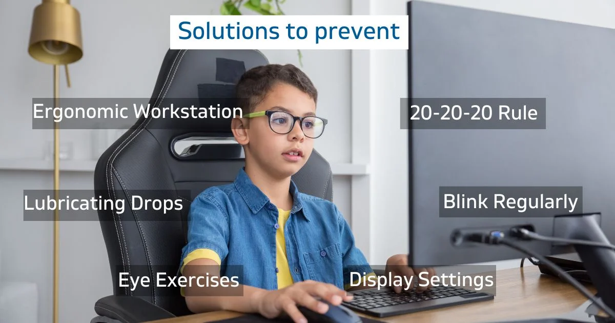
Here are nine additional tips for ways to reduce eyestrain.
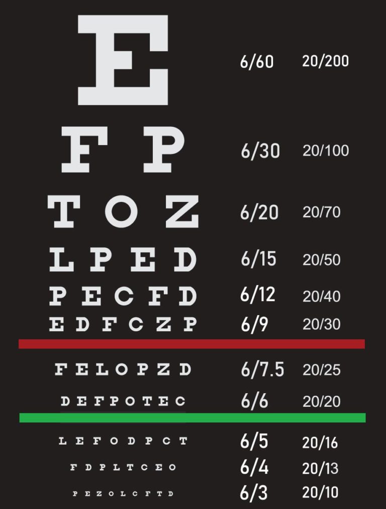
1. GET AN EYE EXAM!

This is the most important thing you can do to prevent or treat computer vision problems.

According to the National Institute of Occupational Safety and Health (NIOSH), computer users should have an eye exam before they start working on a computer and once per year thereafter.

2. USE PROPER LIGHTING

In your office you are likely to find several things that can cause eyestrain, including glare on walls and finished surfaces, reflections on the computer screen itself, excessively bright light coming in from outside, and excessively bright light inside

Eliminate exterior light and reflections by closing drapes or blinds.

When using computers, lighting should be about half that used in most offices. Reduce lighting by using fewer light bulbs or florescent tubes, or use lower intensity bulbs and tubes.

3. TAKE FREQUENT BREAKS

Full time computer users should take a 10-minute break every hour to reduce eyestrain problems according to experts. Part-time users should take frequent breaks, after sitting in front of their display for more than a hour.

4. REFOCUS YOUR EYES

Look away from your computer screen every 10-15 minutes and focus for 5-10 seconds on a distant object outside or down the hallway. This prevents the fixed gaze common among computer users. It also lets you blink, which wets your eyes.

5. BLINK MORE OFTEN

When staring at a computer, people blink less frequently—about 5 times less than normal, according to studies. Tears coating the eye evaporate more rapidly during long non-blinking phases and cause dry eyes. Office buildings may have excessively dry environments that also reduce tearing. For significant problems, ask your eye doctor about artificial tears or eye drops that you can use during the day.

6. MODIFY YOUR WORKSTATION

If you need to look back and forth between the printed or written page and the computer, this can cause eyestrain. Place written pages on a copy stand adjacent to the monitor. Properly light the copy stand. Adjust your workstation and chair to the correct height. Purchase ergonomic furniture to assure proper screen locations and posture.

Digital eye strain

7. MATCH THE COMPUTER SCREEN TO THE BRIGHTNESS OF THE ENVIRONMENT

Closely match the brightness of the environment with that of the computer screen. The contrast between the background and on-screen characters should be high.

8. MINIMIZE GLARE

Use window shades, blinds or drapes to block out excessive sunlight, or install an anti-glare screen, to minimize reflections on the screen itself. Reduce the internal ambient light if necessary. For conditions where outside light cannot be reduced, use a computer hood to cut glare and reflection. Have an Anti-Reflective coating applied to your glasses. This will prevent glare and reflections on the back side of your lenses form reaching your eyes.

9. EXERCISE EVEN WHEN SITTING

Anyone in a sedentary job, especially those using computers, should also stand up, move about, or exercise frequently. NIOSH recommends several sitting, stretching, and joint rotating exercises for computer users.

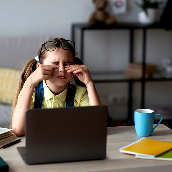
As parents or carers, it is essential to recognise the signs of Digital Eye Strain in children.

Some common symptoms include:

  • Eye discomfort: Complaints of tired, itchy, or burning eyes.
  • Squinting or blinking: Frequent squinting or blinking to refocus their vision.
  • Headaches: Recurring headaches, especially after screen time.
  • Dry eyes: Experiencing dryness or grittiness in the eyes.
  • Double vision: Temporary vision issues like double vision or blurred vision.

Reference:

  • yesite.co.za/2004/04/15/impact-of-computer-use-on-childrens-vision
  • visiondirect.com.au/optical-centre/eye-care/digital-eye-strain
  • gormleyopticians.com/protecting-childrens-eyes-digital-strain
  • prio.com/consumers/9ways.cfm

20/20 VISION

What does 20/20 vision mean?

20/20 vision is a term used to express normal visual acuity (the clarity or sharpness of vision) measured at a distance of 20 feet. If you have 20/20 vision, you can see clearly at 20 feet what should normally be seen at that distance. If you have 20/100 vision, it means that you must be as close as 20 feet to see what a person with normal vision can see at 100 feet.


Does 20/20 mean perfect vision?

No. 20/20 vision only indicates the sharpness or clarity of vision at a distance. There are other important vision skills, among them peripheral awareness or side vision, eye coordination, depth perception, focusing ability and color vision that contribute to your overall vision ability.

Is 15/15 vision better than 20/20?

No. 15/15 means normal sharpness of vision at 15 feet just as 20/20 indicates normal acuity at 20 feet. Most optometrists in Canada use 20 feet as the standard to express sharpness of vision.

Why do some people have less than 20/20 vision?

Visual acuity is affected by many factors. Less than optimum clarity may result from vision conditions like nearsightedness, farsightedness, or astigmatism, or from eye diseases.

Will clarity of vision vary with distance?

Some people can see well at a distance, but are unable to bring nearer objects into focus. This condition can be caused by farsightedness or presbyopia (a loss of focusing ability). Others can see items that are close, but cannot see those far away. This condition may be caused by nearsightedness.

If my vision is less than optimum, what can I do?

A comprehensive eye examination by a Doctor of Optometry should identify those causes, if any, that are affecting your ability to see well. In most cases, your optometrist can prescribe glasses, contact lenses or a vision therapy program that will help improve your vision. If the reduced vision is due to an eye disease, the use of ocular medication or other treatment may be needed. [1]

Eye Information : A-Z of Terms Explained [7]

Words your optometrist/optician might use...

The terms here are the most commonly used by your optician, most will only be of use in special cases or with people with 'complex' prescriptions. However, if you would like to know a little more about your eyes and the words used to describe their health please read on!

  • ABERRATION: Any defect or a distortion in any optical system.
  • ACCOMMODATION: The process by which the eye increases its power to focus on a specific object.
  • ACUITY (V.A. or visual acuity): Describes the clarity of vision.
  • AFOCAL: A lens or optical system with zero power.
  • AMBLYOPIA: (Also known as having a lazy eye) often occurs when the eye does not develop fully during early childhood and is not usually correctable via opticial means.
  • AMETROPIA: A defective refractive ability of the eye e.g short-sightedness or long-sightedness.
  • ANISOMETROPIA: Unequal refractice power in each eye, typically greater then 1.00D.
  • ANTIMETROPIA: A condition in which one eye is myopic and the other is hypermetropic.
  • APHAKIA: The ocular condition in which the lens of the eye is absent, or has been surgically removed.
  • AQUEOUS HUMOUR: Fluid that fills and separates the anterior (front) and posterior (back) of the human eye. It is a transparent gelatinous fluid.
  • ASPHERICAL: Not spherical, aspherical lenses have a curved surface.
  • ASTIGMATISM: When the cornea at the front of the eye is unevenly curved, often resulting in a rugby ball shaped eye, causing blurred vision.
  • BIFOCAL: A lens with two focal lengths, usually for distance/near vision.
  • BINOCULAR: Relating to both eyes.
  • BIOMICROSCOPE: An instrument designed for detailed examination of the eye, used particularly in contact lens practice. (Often referred to as a slit-lamp).
  • BLEPHARITIS: A chronic inflammation of the eyelid margins.
  • CANTHUS: Either corner of the eye where the eyelids meet.
  • CATARACT: Is when the clear lens inside your eye becomes cloudy or misty.
  • CONCAVE: A surface shaped like the inside of a sphere.
  • CONVERGENCE: Movement of the eyes turning inwards, i.e. towards each other.
  • CONVEX: Is when a lens is shaped outward.
  • CORNEA: The transparent anterior portion of the eye.
  • CRYSTALLINE LENS: The lens of the eye, which focusses light on to the retina.
  • DECENTRATION: The displacement, horizontal and/or vertical, of the centration point of a spectacle lens from the standard optical centre position.
  • DIOPTRE: The unit of measurement of refractive power of a lens.
  • DIPLOPIA: (Double vision) A condition where objects are seen 'double'.
  • DISC (OPTIC): The region of the retina, where the optic nerve joins the eyeball.
  • DISTORTION: Defect of an optical system resulting in an alteration of an objects original shape.
  • DIVERGENCE: Movement of the eyes, turning away from each other.
  • EMMETROPIA: An eye which gives perfect vision so that no glasses are required. Occurs when light from an object at infinity is sharply focussed on the retina.
  • FIXATION: Maintenance of visual gaze on a single location. The object is sharply formed on the retina.
  • FOCIMETER:An instrument that measures the power of a spectacle lens, or contact lens.
  • FUNDUS: The back of the eye. The fundus is viewed using an instrument called an ophthalmoscope.
  • GLAUCOMA: An eye disease characterised by increased intra-ocular fluid pressure which causes a restriction in field of vision.
  • GRAFT: Surgical procedure to replace damaged structures (e.g.cornea) with donor tissue.
  • HYDROGEL: A type of plastics material that contains water, commonly used in the manufacture of soft contact lenses.
  • HYPERMETROPIA (HYPEROPIA): Often referred to long-sightedness. When people are long sighted, they can see distance objects well but have difficulty focussing on objects that are close. This vision problem occurs when light rays entering the eye focuses behind the retina, rather than directly on it.
  • INJECTION (e.g. conjunctival ): Redness of the eye caused by the dilation (expanding) of superficial blood vessels in the sclera (white of the eye).
  • INTER-PUPILLARY DISTANCE: The horizontal distance measured between the pupil centres. Usually abbreviated to PD. Essential when making up spectacles.
  • IRIS: The coloured part of the eye surrounding the pupil.
  • IRITIS: A condition causing inflammation of the iris.
  • KERATITIS: An inflammation of the cornea.
  • KERATOMETER: An instrument used to measure the curvature of the front surface of the cornea. Often used in contact lens fitting.
  • LACRIMAL: Relating to the tears.
  • MEIBOMIAN GLANDS: Small glands in the eyelids known as meibomian glands are responsible for secreting oil that covers and protects the surface of the eye.
  • MIOSIS: Contraction of the pupil. (A drug that causes this is a miotic)
  • MONOCLE: A single lens, with or without a frame, worn by holding between the brow and the cheek.
  • MONOCULAR: Relating to one eye (or lens).
  • MULTIFOCAL: A lens with multiple powers eg a trifocal or varifocal lens.
  • MYDRIASIS: Dilation (enlarging) of the pupil. (A drug that causes this is a mydriatic)
  • MYOPIA: Often referred to short-sightedness. When people are short sighted, they can see near objects well but have difficulty focussing on objects that are far away. This vision problem occurs when light rays entering the eye focuses infront the retina, rather than directly on it.
  • NEAR VISION: The ability to read/carry out close work, usually at a distance of 33 - 40 cm.
  • NEUTRALIZATION: The process of determining the power of an unknown lens, using trial lenses of known power.
  • OCCLUDER: A device placed in front of an eye to effectively block vision.
  • OPACITY: Condition of a tissue or structure that is not transparent.
  • OPHTHALMOSCOPE: A hand held instrument for viewing the eye in particular the retina.
  • OPTICAL CENTRE: The point on a lens through which a ray of light will pass undeviated.
  • ORBIT: The bony socket of the skull where the eye is situated.
  • ORTHOPTICS: Is the diagnosis and non-medical management of abnormalities of binocular vision. This includes strabismus (squint), amblyopia (lazy eye).
  • PERMEABILITY: The ability of a material to allow the passage of a gas or fluid.
  • PHOTOCHROMIC: A lens made of a material that changes colour as a result of exposure to UV light and heat. For more information on photochromatic lenses please click here.
  • PRESBYOPIA: When the lens inside the eye loses some of its flexibility so that it becomes difficult to focus on close objects. Usually occurs with age.
  • PRISM: Is used in spectacles to correct a muscle imbalance.
  • PROSTHESIS: An artificial eye, or implant.
  • PUPIL: The opening within the centre of the iris. Thsi determines the amount of light which enters the eye.
  • PUPILLARY DISTANCE: This is the distance between the centre of the the pupils in each eye (measured in millimeters). Important in positioning the lenses of the glasses correctly in relation to the pupils so as to obtain the optimum vision.
  • REFLEX: Reaction of certain parts of the eye to a stimulus, e.g. the pupil reflex being the reaction of the pupil to a light stimulus.
  • RETINA: The light sensitive layer of cells lining the back of the eye. Often liked to the film of a camera.
  • RETINOSCOPE: Hand held instrument used to obtain an objective assessment of a patient's refractive condition (their prescription)
  • SALINE: A sterile solution of sodium chloride (salt) in water. Often used to rinse contact lenses
  • SCLERA: The white of the eye. A tough opaque fibrous tissue which serves as the eyes protective coat.
  • SCOTOMA: An area of partial or complete loss of vision surrounded by a normal field of vision.
  • SLIT - LAMP (SLIT-LAMP BIOMICROSCOPE): An instrument used to examine the eye under high magnification. Also used in contact lens practice.
  • STRABISMUS: A misalignment of the eyes so that the eyes are not directed towards the same point, when the patient is fixating. As images are formed at different points this often results in 'double vision'.
  • SURFACING: The process of generating, smoothing and polishing a spectacle lens surface to a given curvature.
  • TONOMETER: An instrument used for measuring the fluid pressure inside the eyeball (the intra ocular pressure (IOP)
  • TRIAL CASE: A case containing both spherical and cylindrical lenses which are either positive or negative in power. Used in testing eye sight.
  • TRIAL FRAME: An adjustable spectacle frame which can hold several lenses and is used in testing eye sight.
  • UNAIDED VISION: Vision without any form of correction i.e spectacles or contact lenses.
  • UNIOCULAR: Another expression for monocular, i.e. referring to one eye.
  • VERTEX DISTANCE:The distance (in mm) from the surface of the cornea, to the posterior surface of the spectacle lens or trial lens.
  • VISUAL ACUITY: The ability of the eye for seeing distinctly the details of an object at a specific distance.
  • VITREOUS HUMOUR: The clear gelly that fills the space between the lens and the retina of the eyeball.

Terms / Abbreviations

Meaning

Notes

dispensing optician

a person qualified to prescribe and dispense as well as to make glasses and contact lenses.

Dispensing opticians are trained to make up optical prescriptions written by Optometrists or Ophthalmologists. They also advise on, supply and fit glasses, and other optical aids to meet their patients' requirements.

A dispensing optician is a registered healthcare professional and plays various roles within an optical practice to help patients. The core role of a dispensing optician (DO) is to advise on, fit and supply the most appropriate spectacle frames and lenses for each person.[9]

optometrist

/ɒpˈtɒmɪtrɪst/

While opticians have received significant training, it is not to the same level as an optometrist who will have a degree in optometry. If an optometrist, having performed an eye test, recommends glasses or contact lenses, then an optician can help ensure that any visual correction provided works optimally.[8]

ophthalmologist

/ˌɒfθalˈmɒlədʒɪst/

DV: Distance VisionFar Vision
NV: Near VisionNear Vision
OD: Oculus DexterRight eye
OS: Oculus SinisteLeft eye
OU: Oculi UterqueBoth eyes
SPH: Spherical convergent or divergent refractive power+ / -
CYL: Cylindrical adding or subtracting power cylindrically+ / -
Axis: the angle in degrees 0..180
dioptre / diopter For example, a 3-dioptre lens brings parallel rays of light to focus at 1⁄3 metre.
Prism and BasePrism correction is measured in "prism diopters", and Base refers to the direction of displacement
Pupillary Distance (PD)is the distance between pupil centers, usually expressed in millimeters
Interpupillary Distance (IPD)It is written as two values if the prescription is for bifocals or progressive lenses

monocular pupillary distance ("monocular PD")

In countries such as the United Kingdom, PD measurement is not a legal requirement as part of the prescription and is often not included

Back vertex distance (BVD)

is the distance between the back of the spectacle lens and the front of the cornea

(the front surface of the eye)

This is essential in higher prescriptions (usually above ±4.00D) as slight changes in the distance between the spectacles and the eyes above this level can cause the patient to perceive a different power, leading to blur and/or other symptoms
SER: Spherical Equivalent RefractionEye care professionals use the term spherical equivalent refraction (SER) to refer to an eye's effective focusing power if only spherical aberration were present

SER = sph. + 1/2*cyl.

Distant vision

near vision.1

near vision.2

The DV portion of the prescription describes the corrections for distant vision. For most people under forty years of age, the NV or near-vision portion of the prescription is blank because a separate correction for near vision is not needed

for example :

Trifocals are mostly used by people with advanced presbyopia who have been prescribed 2 diopters or more of reading addition

The intermediate addition is normally half the reading addition. So, for someone with a distance prescription of -4 diopters and a reading addition of +3, the reading portion of their trifocals would have a net power of -1, and the intermediate segment would be -2.5 diopters. [3]

Corneal pachymetry

Corneal pachymetry is the process of measuring the thickness of the cornea. [5]A pachymeter is a medical device used to measure the thickness of the eye's cornea. It is used to perform corneal pachymetry prior to refractive surgery, for Keratoconus screening, LRI surgery and is useful in screening for patients suspected of developing glaucoma among other uses.[5]

eResearch by Navid Ajamin -- winter 2004

Description Term [6] Abbreviation

ACAnterior chamberfluid-filled space between the iris and the endothelium
AC 4/4Grade 4 anterior chamber angleopen angle between cornea and iris
AC 3/4Grade 3 anterior chamber angle
AC 2/4Grade 2 anterior chamber angle
AC 1/4Grade 1 anterior chamber angle
AC 0/4Grade 0 anterior chamber angleclosed angle between cornea and iris
AC/AAccommodative convergence / Accommodation ratiothe portion of the range of convergence that occurs in response to accommodation
AccAccommodationprocess of changing optical power to maintain focus as distance changes
AdAdvised
AddAddition
AITAfter-image transfer
ALTAlternating
ALT ETAlternating esotropia
ALT XTAlternating exotropia
ARCAnomalous retinal correspondence
A/VArteriole–venue ratio
BIOBinocular indirect ophthalmoscopy
BSVBinocular single vision
BVBinocular vision
BVDBack vertex distance
BVPBack vertex power
CDCentration distance
C/DCup–disc ratio
CFCount fingers vision – state distance
c/o or c.o.Complains of
CTCover test
c/uCheck up
CWClose work
ΔPrism dioptre
DDioptres
DCDioptres cylinder
DNADid not attend
DOBDate of birth
DSDioptres sphere
DVDistance vision
DVDDissociated vertical deviation
EFEccentric fixation
FBForeign body
FDFixation disparity
FFFoveal fixation
FHGFamily history of glaucoma
FMHFamily medical history
FOHFamily ocular history
F/UFollow up appointment
GHGeneral health
G(M)PGeneral (medical) practitioner
HAHeadaches
HARCHarmonious abnormal retinal correspondence
HMHand motion vision – state distance
HxHistory
IOLIntra-ocular lens
IOPIntra-ocular pressure
ISNTInferior, Superior, Nasal, Temporalrule used to assess optic disc appearance
KKeratometry

OS

Left eye
LHyperT or LHTLeft hypertropia
LHypoTLeft hypotropia
LOLenticular opacity
L/R FDL/R fixation disparity
L/RL hyperphoria
Left ETLeft esotropia
LVALow vision aid
MDUMallett distance unit
MNUMallett near unit
M.WingMaddox Wing
MRMaddox rod
NB: NADNo abnormality detected(is frequently used but is not recommended)
NCTNon-contact tonometer
NDNeutral density filter
NLPNo light perceptionNo light perception is considered total visual impairment, or total blindness; see Visual_impairment#Classification
NPCNear point of convergence or no previous correction
NRCNormal retinal correspondence
NVNear vision
NWTNormal wearing time
o symptomsZero symptoms
ϕHorizontal orthophoria
θVertical orthophoria
Horizontal and vertical orthophoria
OC'sOptical centres
Occ.Occupation
ODoculus dexter (right eye)
OHOcular history
OMBOculo motor balance
ONHOptic nerve head
OphOphthalmoscopy
OSoculus sinister (left eye)
OUoculus uterque (both eyes)
PDPupillary distance
PERRLAPupils equal, round, reactive to light and accommodation
PHPinholesee Pinhole occluder
PHNIPinhole No Improvementsee Pinhole occluder
PHVAPinhole Visual Acuity
PLPerception of light
POH / PrOHPrevious ocular history
PPAPeri-papillary atrophy
PtPatient
RAPDRelative afferent pupillary defect
ODRight eye
Ret.Retinoscopy
RHyperTRight hypertropia
RHypoT or RHTRight hypotropia
RNFLRetinal nerve fibre layer
RPERetinal pigment epithelium
RSOTRight esotropia
RxPrescription
SESpherical Equivalent
SLESlit lamp examination
SLMSlit lamp microscope
EPEsophoria
ETEsotropia
Supp.Suppression
VVision (unaided)
VAVisual acuity
VA OSLeft visual acuity
VA ODRight visual acuity
VDUVisual display unit
VFVisual field
VPSVariable prism stereoscope
WDWorking distance
X/12X months
X/52X weeks
X/7X days
XPExophoria
XTExotropia

Reference:

  1. foryoureyesonly.ca
  2. en.wikipedia.org/wiki/Eyeglass_prescription
  3. en.wikipedia.org/wiki/Trifocal_lenses
  4. careers.nhs.scot/careers/explore-our-careers/optometry/dispensing-optician
  5. wikipedia.org/wiki/Corneal_pachymetry
  6. en.wikipedia.org/wiki/List_of_optometric_abbreviations
  7. hdavisopticians.co.uk/eye-information-a-z-of-terms-explained
  8. specsavers.co.uk/eye-test/what-is-an-optometrist
  9. abdo.org.uk/for-the-public/what-is-a-dispensing-optician

Anti-Reflection Coatings

AR coatings are similar to the coatings found on microscopes and camera lenses. They consist of several layers of metal oxides applied to the front and back lens surfaces. Because of the layering effect, AR coatings sometimes have a hint of green or purple color, depending on the individual manufacturer's formula.

The most important benefits of anti-reflective (AR) coating may be the unseen advantage--relief from fatigue and eyestrain caused by glare. AR does have a big cosmetic advantage, but removing the distracting reflections and ghost images that contribute to eyestrain clearly is a bigger benefit. This increases comfort. Being more comfortable in anything we do is a benefit, especially wearing glasses. People always remark how uncomfortable glasses are. Well now you can offer a real solution to that problem. AR Coating!!

Image result for Anti-Reflection Coatings

Nighttime driving is also enhanced with AR coatings by eliminating the reflections from street lights, headlights from on-coming vehicles, and taillights. By applying an AR coating more light can pass through the lens providing greater image contrast and clearer vision.

Beyond these benefits and features, other ideal candidates are:

  1. High Index Wearers-AR helps reduce the concentric rings these strong prescriptions produce.

  2. Low Vision Patients-The increase of light transmission from AR coatings can benefit low vision patients.

  3. Light-sensitive patients

  4. Sunglass wearers

  5. First time wearers of glasses

  6. Professionals

  7. VDT users can benefit from AR's ability to reduce glare.

? How to see better at night driving

You get more out of life when you can enjoy it longer. Vision problems such as glare, reflections, ghost images and the fatigue they create reduces your enjoyment. AR coatings improve vision, reduce fatigue, and help you to enjoy every minute of life at work or at play.

AR coatings enhance appearance. It makes lenses look thinner by reducing distracting reflections. Also the wearers eyes are more visible and this improves eye contact for better communication.

Now, we have mention the cosmetic and comfort features but there are more than that. AR lenses when worn in daylight and indoors, most people find it provides noticeable better visual performance, making objects appear crisper and brighter. When reading a newspaper, magazine, or even a computer screen with AR coated lenses images and letters will appear sharper and crisper. [1]

Image result for Anti-Reflection Coatings

Anti-reflective eye glasses are designed to reduce glare on the lenses. This serves the dual purpose of improving the wearer’s vision and enhancing the appearance of the glasses themselves, particularly in photographs taken with a flash. eResearch by Navid Ajamin -- spring 2008

Composition

1. Anti-reflective coating consists of a series of layers of metal oxides. In the 1980s and 1990s, when anti-reflective lenses were relatively new, the coating was only a single layer that could easily become scratched or smudged.

Today, multiple layers enhance the properties of the anti-reflective coating, including oil-resistant, water-resistant, static-resistant and protective scratch-resistant layers.

difference between Anti-Reflective and Mirror coating

Process

2. The process used to apply anti-reflective coating varies depending on the manufacturer. In some cases the coating is sprayed on in liquid form, then exposed to high heat so that the liquid solution hardens and adheres to the lens.

Another process coats each lens with a liquid and places it inside a vacuum chamber, where the vacuum process hardens the coating. In yet another process the anti-reflective coating is built into the material and distributed throughout.Care

3. Anti-reflective lenses require a little more care than regular lenses. They should not be cleaned with harsh chemicals, which can damage the anti-reflective coating. Additionally, they should be cleaned or rubbed only with a wet cloth, as a dry cloth can scratch the lens.[2]

Benefits of anti-reflective coating

Anti-reflective coating (also called “AR coating” or “anti-glare coating”) improves vision, reduces digital eye strain and makes your eyeglasses look more attractive. These benefits are due to the ability of AR coating to virtually eliminate reflections from the front and back surfaces of your eyeglass lenses.

With reflections gone, more light passes through your lenses to optimize visual acuity with fewer distractions (especially at night), and the lenses look nearly invisible — which enhances your appearance by drawing more attention to your eyes and helping you make better eye contact with others.

AR coating is especially beneficial when used on high-index lenses, which reflect more light than regular plastic lenses. Generally, the higher the index of refraction of the lens material, the more light that will be reflected from the surface of the lenses.

For example, regular plastic lenses reflect roughly 8% of light hitting the lenses, so only 92% of available light enters the eye for vision.

Photolithography

Antireflective coatings (ARC) are often used in microelectronic photolithography to help reduce image distortions associated with reflections off the surface of the substrate. Different types of antireflective coatings are applied either before (Bottom ARC, or BARC) or after the photoresist, and help reduce standing waves, thin-film interference, and specular reflections.

学生用反射防止メガネ

High index plastic lenses can reflect up to 50% more light than regular plastic lenses, so even less light is available to the eye for vision. This can be particularly troublesome in low-light conditions, such as when driving at night.

Today’s modern anti-reflective coatings can virtually eliminate the reflection of light from eyeglass lenses, allowing 99.5% of available light to pass through the lenses and enter the eye for good vision.

By eliminating reflections, AR coating also makes your eyeglass lenses look nearly invisible so people can see your eyes and facial expressions more clearly. Anti-reflective glasses also are more attractive, so you can look your best in all lighting conditions.

The visual benefits of lenses with anti-reflective coating include sharper vision with less glare when driving at night and greater comfort during prolonged computer use (compared with wearing eyeglass lenses without AR coating).

When applied to photochromic lenses, AR coating enhances the clarity and comfort of these premium lenses in all light conditions without reducing their sun-reactive performance.

Anti-reflective coating also is a good idea for sunglasses. It eliminates glare from sunlight reflecting into your eyes from the back surface of tinted lenses when the sun is behind you. (Generally, AR coating is applied only to the back surface of sunglass lenses because there are no cosmetic or visual benefits to eliminating reflections from the front surface of dark-tinted lenses.)

Most premium AR lenses include a surface treatment that seals the anti-reflective layers and makes the lenses easier to clean. These hydrophobic surface treatments also repel water, preventing the formation of water spots on your lenses.

Some anti-reflective lenses have surface treatments that are both hydrophobic and oleophobic (also called lipophobic), which means they repel both water and oil. These combination treatments typically contain fluorinated materials that give the lenses properties that are very similar to those of nonstick cookware.

Is anti-reflective the same as anti-glare?

Anti-reflective vs Anti Glare

In short: anti glare coatings will protect against external light sources entering the glasses, while anti-reflective coatings will protect against both internal and external light.

Anti Glare

To get into the physics of it, anti glare solutions equip glasses with diffusive properties which diffuse light that would usually be reflected off of the surface of your lens. This means that instead of producing specular reflection, it creates a diffuse reflection. Instead of the light ray reflecting directly off the lens, it is broken up into many weaker rays, meaning the reflection is far weaker or, often, unnoticeable. This means that anti glare coatings significantly reduce the amount of light that is reflected off the surface of the lens.

Anti-Reflective

On the other hand, anti-reflective lenses go a step further. These solutions use diffusive properties to diffuse both external and internal light. While “internal light” might sound strange, it refers to the small number of transitional light waves which are let loose as the light goes from one medium (in this case, air) into another (the lens). If unchecked, these small light rays can reflect within the lens itself, causing strange visual artefacts that lead to eye strain and discomfort. While these internal reflections may still be present in glasses with anti glare coatings, anti-reflective lenses go the extra mile to ensure the best user experience possible.

Anti-reflective coatings are often used in camera lenses, giving lens elements distinctive colors. Such colors indicate the wavelength of visible light least affected by the antireflective properties of the coating. A variety of colors can be produced whose precise hue depends entirely on the thickness of the coating.

Reference:

  1. robertsonoptical.com
  2. milenyumtasarim.com
  3. en.wikipedia.org/wiki/Anti-reflective_coating
  4. allaboutvision.com/lenses/anti-reflective.htm
  5. rx-able.com/blogs/blogs/is-anti-reflective-the-same-as-anti-glare

See also:

  • Disadvantages of Anti-Glare Coating

  • My glasses have an anti-reflective coating. How does that work?

nicetoview.blogfa.com
عینک eyewear وبلاگ تخصصی عینک شامل مجموعه مطالب پزشکی است که اطلاعات مفیدی در رابطه با عینک , چشم، لنز، سلامتی چشم و راه های پیشگیری از بیماریهای چشمی، کنترل و درمان آن را در اختیار شما کاربر محترم می گزارد.

Always Be Healthy
همیشه سلامت باشید

Navid Ajamin نوید عجمین
eMail: navid.aj@outlook.com
موضوعات وب
پیوندها

unicef for every child

شمارنده